. 2016 May 6:12:976-989.
doi: 10.1016/j.nicl.2016.05.002. eCollection 2016.

Combined 18F-FDG-PET and diffusion tensor imaging in mesial temporal lobe epilepsy with hippocampal sclerosis

Affiliations

Combined 18F-FDG-PET and diffusion tensor imaging in mesial temporal lobe epilepsy with hippocampal sclerosis

Javier Aparicio et al. Neuroimage Clin. .

Abstract

Objectives: Several studies using 18F-fluorodeoxyglucose positron emission tomography (18F-FDG-PET) or diffusion tensor imaging (DTI) have found both temporal and extratemporal abnormalities in patients with mesial temporal lobe epilepsy with ipsilateral hippocampal sclerosis (MTLE-HS), but data are lacking about the findings of both techniques in the same patients. We aimed to determine whether the extent of 18F-FDG-PET hypometabolism is related to DTI abnormalities.

Methods: Twenty-one patients with MTLE-HS underwent comprehensive preoperative evaluation; 18 (86%) of these underwent epilepsy surgery. We analyzed and compared the pattern of white matter (WM) alterations on DTI and cortical hypometabolism on 18F-FDG-PET.

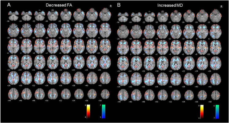
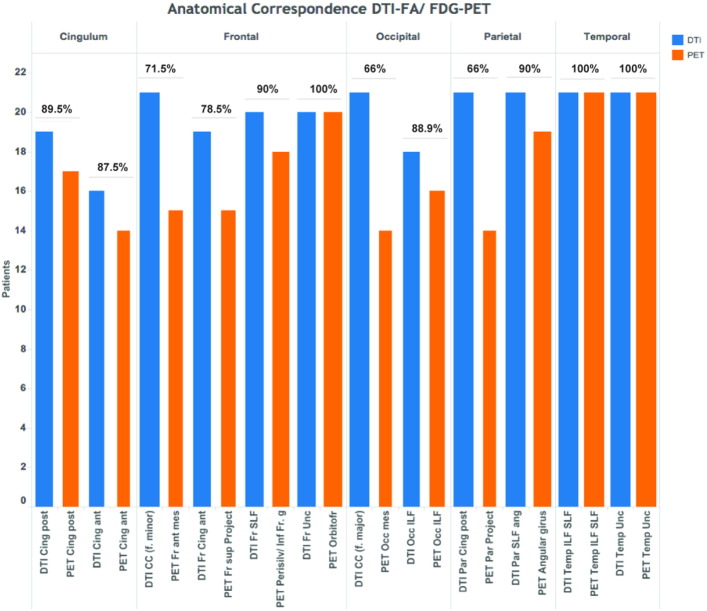
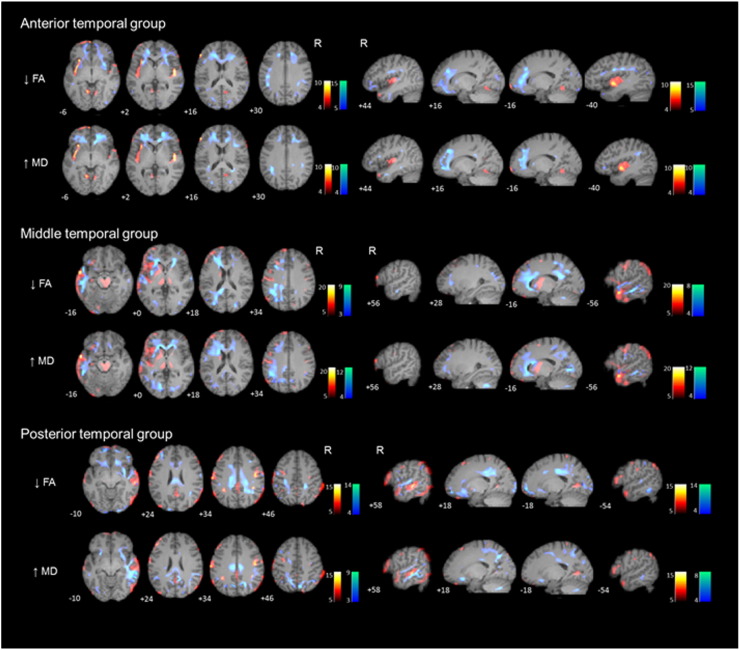
Results: We found widespread temporal and extratemporal 18F-FDG-PET and DTI abnormalities. Patterns of WM abnormalities and cortical glucose hypometabolism involved similar brain regions, being more extensive in the left than the right MTLE-HS. We classified patients into three groups according to temporal 18F-FDG-PET patterns: hypometabolism restricted to the anterior third (n = 7), hypometabolism extending to the middle third (n = 7), and hypometabolism extending to the posterior third (n = 7). Patients with anterior temporal hypometabolism showed DTI abnormalities in anterior association and commissural tracts while patients with posterior hypometabolism showed WM alterations in anterior and posterior tracts.

Conclusions: Patients with MTLE-HS have widespread metabolic and microstructural abnormalities that involve similar regions. The distribution patterns of these gray and white matter abnormalities differ between patients with left or right MTLE, but also with the extent of the 18F-FDG-PET hypometabolism along the epileptogenic temporal lobe. These findings suggest a variable network involvement among patients with MTLE-HS.

Keywords: DTI; Hippocampus; MTLE; Nerve net; Positron emission tomography.


Figures

Supplementary Fig. S1
Axial sections of images illustrating white matter alterations (cool colors), corresponding to decreased fractional anisotropy (A) and to increased mean diffusivity (B), and 18F-FDG-PET hypometabolism (warm colors) in patients with mesial temporal lobe epilepsy with hippocampal sclerosis (MTLE-HS) compared to controls. Numbers represent the corresponding slice. Right on image is patients' right.
Supplementary Fig. S2
Sum of values and proportion of anatomical correspondence for each DTI-WM fasciculus and 18F-FDG-PET cortical regions. (Abbreviations: Cing, cingulum; CC, corpus callosum; f., forceps; Fr, frontal; Par, parietal; Project, projections; SLF, Superior Longitudinal Fasciculus; Perisilv, perisilvian region; g, girus; Unc, uncinate fasciculus; Orbitofr, orbitofrontal cortex; Occ, occipital; ILF, Inferior Longitudinal Fasciculus; ant, anterior; post, posterior; sup, superior; mes, mesial).
Fig. 1
Fig. 1
(2-column fitting image). Axial sections of images illustrating white matter alterations (cool colors), corresponding to decreased fractional anisotropy (FA), and 18F-FDG-PET hypometabolism (warm colors) in patients with left (A) and right (B) mesial temporal lobe epilepsy with hippocampal sclerosis (MTLE-HS) compared to controls. Right on image is patients' right.
Fig. 2
Fig. 2
(2-column fitting image). Axial sections of images illustrating white matter alterations (cool colors), corresponding to increased mean diffusivity (MD), and 18F-FDG-PET hypometabolism (warm colors) in patients with left (A) and right (B) mesial temporal lobe epilepsy with hippocampal sclerosis (MTLE-HS) compared to controls. Right on image is patients' right.
Fig. 3
Fig. 3
(1.5-column fitting image). Axial (left) and sagittal (right) sections of images illustrating white matter alterations (cool colors) corresponding to decreased fractional anisotropy (FA) and to increased mean diffusivity (MD), and 18F-FDG-PET hypometabolism (warm colors) in patients with mesial temporal lobe epilepsy with hippocampal sclerosis (MTLE-HS) compared to controls, based on the extension of 18F-FDG-PET hypometabolism (anterior, middle and posterior groups). Numbers represent the corresponding slice. Axial images: right on image is patients' right. Sagittal images: left on image is patients' right.

References

    1. Ahmadi M.E., Hagler D.J., Jr., McDonald C.R., Tecoma E.S., Iragui V.J., Dale A.M., Halgren E. Side matters: diffusion tensor imaging tractography in left and right temporal lobe epilepsy. AJNR Am J Neuroradiol. 2009;30:1740–1747. - PMC - PubMed
    1. Archambaud F., Bouilleret V., Hertz-Pannier L., Chaumet-Riffaud P., Rodrigo S., Dulac O., Chassoux F., Chiron C. Optimizing statistical parametric mapping analysis of 18F-FDG PET in children. EJNMMI Res. 2013;3:2. - PMC - PubMed
    1. Asadi-Pooya A.A., Sharan A., Nei M., Sperling M.R. Corpus callosotomy. Epilepsy Behav. 2008;13:271–278. - PubMed
    1. Basser P.J., Pierpaoli C. Microstructural and physiological features of tissues elucidated by quantitative-diffusion-tensor MRI. J. Magn. Reson. B. 1996;111:209–219. - PubMed
    1. Bernhardt B.C., Bernasconi N., Concha L., Bernasconi A. Cortical thickness analysis in temporal lobe epilepsy: reproducibility and relation to outcome. Neurology. 2010;74:1776–1784. - PubMed

Publication types

MeSH terms

Substances

LinkOut - more resources