Review
. 2016 Nov 18;16(4):155-168.
doi: 10.1016/j.tjem.2016.09.008. eCollection 2016 Dec.

A systematic and technical guide on how to reduce a shoulder dislocation

Affiliations
Review

A systematic and technical guide on how to reduce a shoulder dislocation

H Alkaduhimi et al. Turk J Emerg Med. .

Abstract

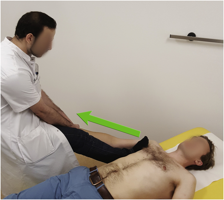
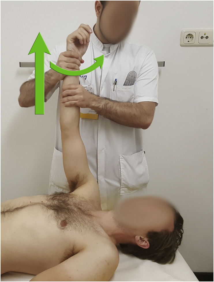
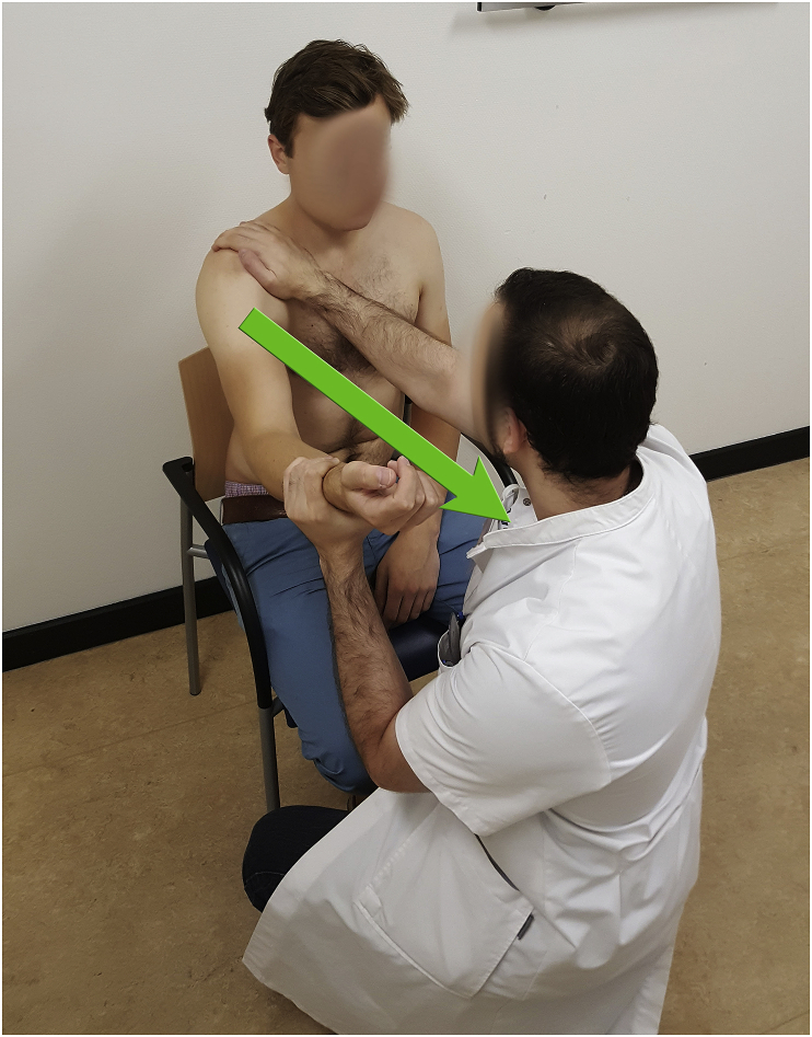
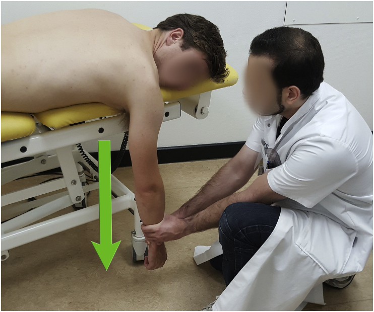
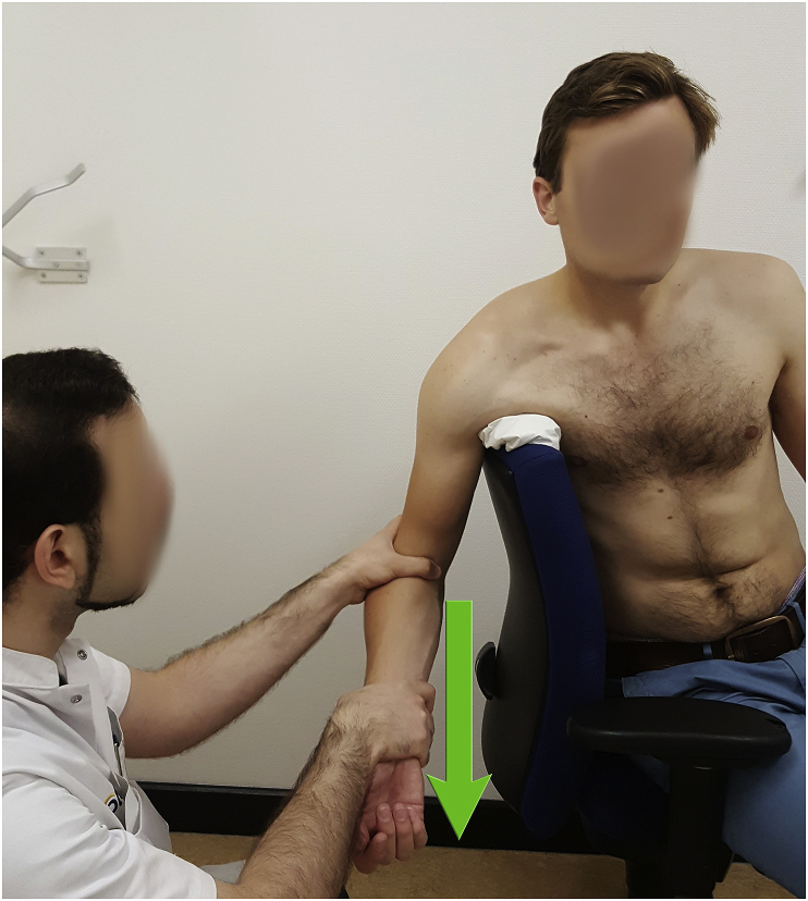
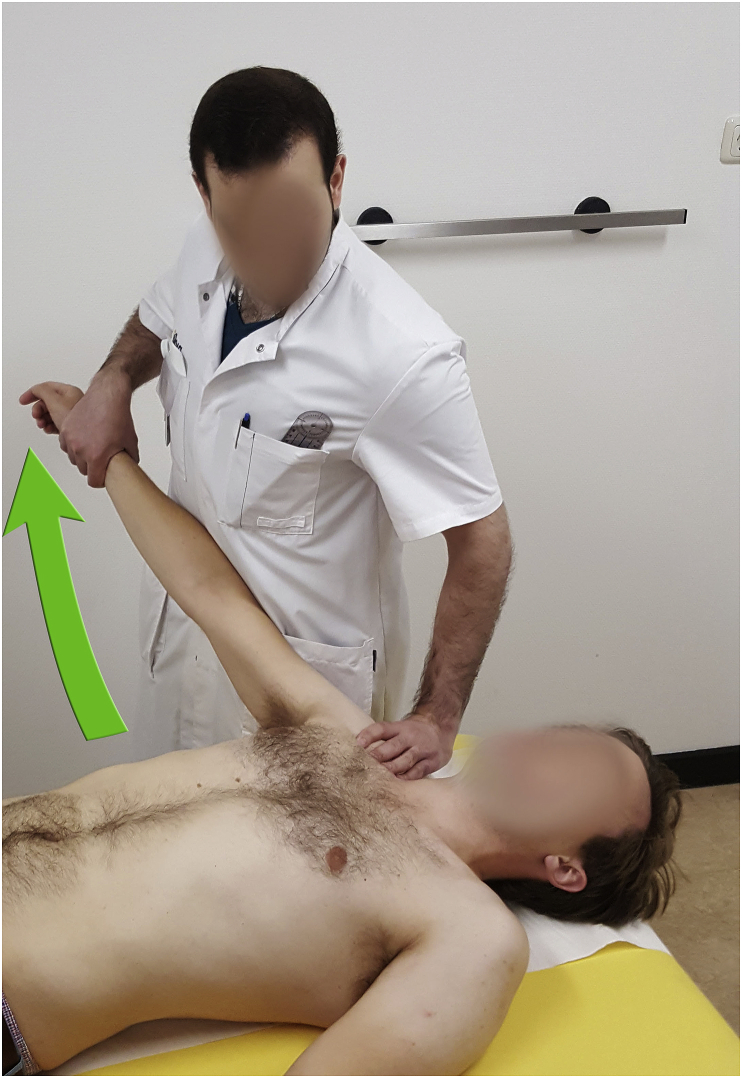
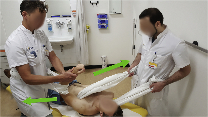
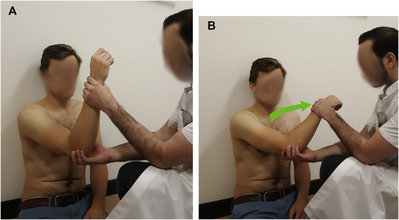
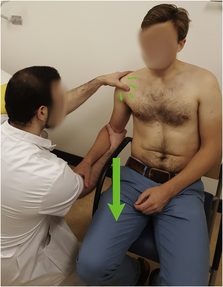
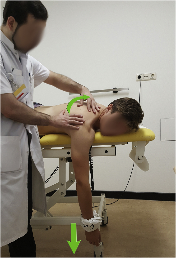
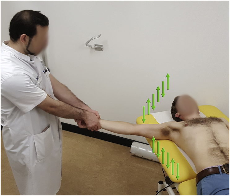
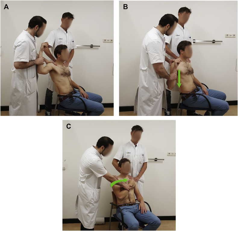
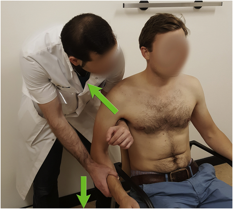
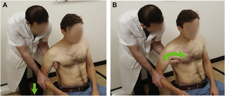
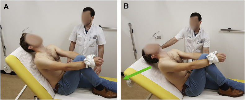
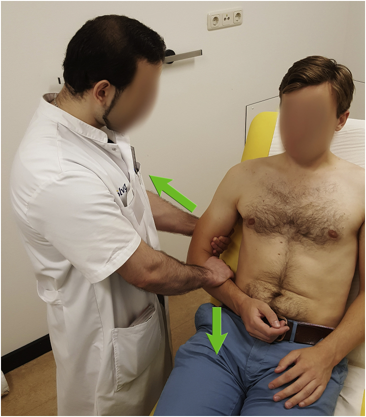
Objectives: Our objective is to provide a systematic and technical guide on how to reduce a shoulder dislocation, based on techniques that have been described in literature for patients with anterior and posterior shoulder instability.

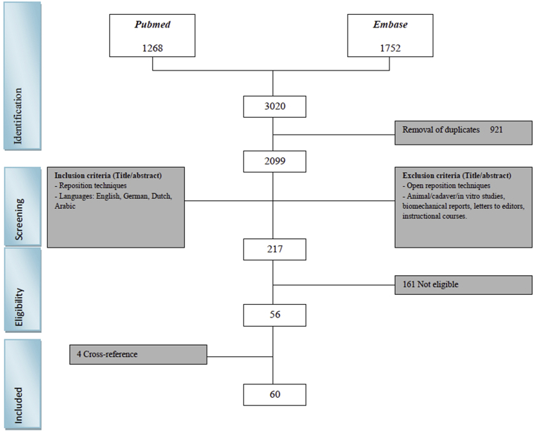
Materials and methods: A PubMed and EMBASE query was performed, screening all relevant literature on the closed reduction techniques. Studies regarding open reduction techniques and studies with fracture dislocations were excluded.

Results: In this study we give an overview of 23 different techniques for closed reduction and 17 modifications of these techniques.

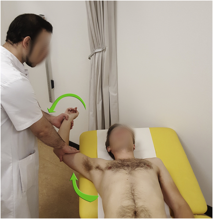
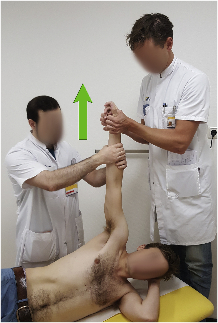
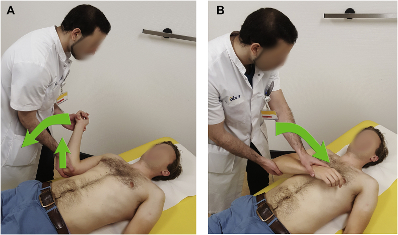
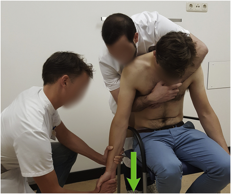
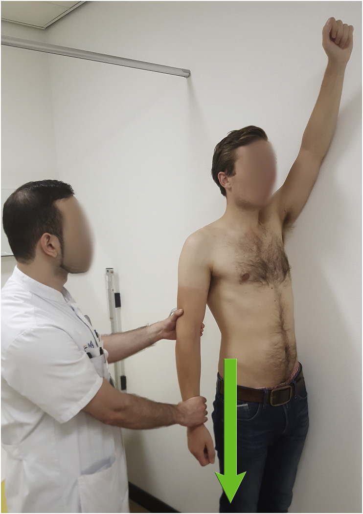
Discussion: In this review article we present a complete overview of the techniques, that have been described in the literature for closed reduction for shoulder dislocations. This manuscript can be regarded as a clinical guide how to perform a closed reduction maneuver, including several technical tips and tricks to optimize the success rate and to avoid complications.

Conclusion: There are 23 different reduction techniques with 17 modifications of these techniques. Knowledge of the different techniques is highly important for a good reduction.

Keywords: Glenohumeral; Instability; Maneuver; Reposition; Shoulder; Techniques.


Figures

Fig. 1
Fig. 1
Flow chart of study selection. This figure provides a flow chart of the study selection. 2099 titles and abstracts were screened, 217 studies were screened full-text and 60 studies were included.
Fig. 2
Fig. 2
Milch maneuver. The patient lies in a supine position. The practitioner holds the patient's arm at the wrist abducting it to an overhead position, externally rotating it to 90°. Subsequently, the humeral head is pushed into a superior lateral position.
Fig. 3
Fig. 3
Eskimo technique. The patient lies on the side of the unaffected shoulder. Two practitioners lift the patient by the dislocated arm, while the arm is abducted. If no reduction occurs, the practitioner can place his hand in the axilla and apply pressure on the humeral head to reposition it in the glenoid rim.
Fig. 4
Fig. 4
External rotation maneuver. The external rotation maneuver is performed with the patient in supine position with the arm of the patient adducted to the thorax, the elbow flexed at 90° and the shoulder flexed at 20°. (A) The practitioner then holds the hand of the patient in one arm and the elbow in the other arm and slowly externally rotates the arm of the patient till it is parallel to the body of the patient without providing traction. (B) After the reduction the arm should be rested on the patient in extended position of the shoulder and flexed position of the elbow. (C).
Fig. 5
Fig. 5
Kocher maneuver. During the Kocher maneuver the patient lies in a supine position with arm adducted and elbow flexed in 90°. The practitioner then provides external rotation in the shoulder till a resistance is felt. Hereafter, the shoulder is adducted and flexed in external rotation. (A) After the reduction the arm should be rested on the patient in extended position of the shoulder and flexed position of the elbow. (B).
Fig. 6
Fig. 6
Hippocratic maneuver. The Hippocratic method is performed with the patient lying in supine position. The practitioner holds the affected limb by the forearm and hand of the patient. The practitioner then places his heel in the axilla of the patient, acting as a fulcrum while the arm of the patient is adducted. This initiates the reduction.
Fig. 7
Fig. 7
Spaso. During the Spaso technique the patient lies in a supine position with arm extended and shoulder flexed to 90°. Longitudinal traction is applied. At the same time, the reduction is initiated by external rotation in the shoulder. (A) After the reduction the arm should be rested on the patient in extended position of the shoulder and flexed position of the elbow.
Fig. 7
Fig. 8
Sitting method. The patient is seated while facing the practitioner. The practitioner holds the forearm of the affected limb and flexes the shoulder to 90° while having the elbow of the patient slightly flexed. Then, the practitioner places his other arm on the anterior chest wall at the side of the affected limb, to control the glenoid tilt by manipulating a part of the scapula such as the acromion or coracoid process. Finally, the practitioner applies longitudinal traction to initiate the reduction.
Fig. 9
Fig. 9
Stimson's maneuver. During the Stimson maneuver the patient lies in prone position with his arm hanging from the examining table. A downward traction to the arm is applied for 10–20 min by the practitioner or by attaching weights to the wrist of the patient to fatigue the shoulder musculature. (B) Thereafter the weights are released allowing the humerus to fall back into its position.
Fig. 10
Fig. 10
Chair method. The patient sits sideways on a chair with the backrest of the chair in his axilla. The practitioner holds the hand of the patient in one hand and the elbow of the patient in the other while he ensures that the elbow of the patient is gently flexed. After calming the patient, downward traction is applied by the practitioner or by an attached sling with weights, facilitating reduction. A slight amount of external rotation in combination with gentle forward flexion can be applied by the right hand of the physician.
Fig. 11
Fig. 11
Aufmesser's method. The patient lies in supine position. The practitioner holds the hand of the patient in one hand and fixates the acromion of the patient with the other hand. The practitioner performs axial traction of the affected arm, while considering the muscular tension of the patient. Additionally, the practitioner can use his body as a fulcrum to add more lever force.
Fig. 12
Fig. 12
Matsen's traction-countertraction. The patient lies in supine position with a sheet around his thorax and also around the waist of the assistant standing at the contralateral side of the affected shoulder. Another sheet is wrapped around the waist and forearm of the practitioner standing on the side of the dislocated shoulder near the waist of the patient, while holding the elbow of the patient in 90° flexion and the shoulder in 90° abduction. The practitioner applies traction to the affected arm by leaning back while the assistant provides countertraction.
Fig. 13
Fig. 13
Bokor-Billmann's technique. The patients sits upright with his back against a firm surface to minimize movements of the upper body. The practitioner holds the wrist of the patient in one hand and the elbow in the other maintaining 90° flexion of the elbow and 90° flexion of the shoulder. The arm is then adducted until it reaches the midline of the thorax. Maintaining the arm in midline position the shoulder is internally rotated. At 25°–30° a resistance is felt, the practitioner should maintain constant pressure to overcome this resistance, inducing reduction.
Fig. 14
Fig. 14
Cunningham technique. The patient sits in a chair with his back straight and his arm adducted to the body with his arm in neutral position and his elbow in 90° flexion. The practitioner kneels on the side of the affected arm facing the opposite direction. The practitioner then slides his hand between the patients forearm and body having the hand of the patient resting on the upper arm of the practitioner. The practitioner applies gentle downward traction and massages the trapezius, deltoid and biceps muscle sequentially with his other hand initiating the reduction.
Fig. 15
Fig. 15
Scapular manipulation. The patient is lying in prone position with his shoulder flexed to 90° and his arm hanging in external rotated position from an examining table. A downward traction is applied by hanging weights on the wrist of the patient or by having an assistant apply traction. Thereafter the practitioner pushes the tip of the inferolateral scapular edge medially rotating upward, initiating the reduction.
Fig. 16
Fig. 16
FARES method. The patient lies in supine position with the practitioner standing at the side of the dislocated shoulder. The practitioner holds the wrist of the patient with both arms keeping the elbow of the patient extended and the forearm in neutral position. Then, the arm is slowly abducted in an oscillating movement (approximately 5 cm up- and downward movement) while constant longitudinal traction is applied. The arm is externally rotated when abducted beyond 90°. Reduction usually takes place at approximately 120°.
Fig. 17
Fig. 17
Legg maneuver. The patient is seated in a straight-backed chair to minimize movement while the assistant stabilizes the unaffected shoulder by applying downward pressure. The patient actively abducts his affected arm to 90°. Subsequently, the arm is externally rotated until the palm faces forward. The practitioner then flexes the elbow to 90° and stabilizes the affected arm behind the patient's head. (A) Then, the arm is adducted to the patients side while fully flexing the elbow. (B) The patient is then asked to internally rotate his arm across the chest. (C).
Fig. 18
Fig. 18
Manes' method. The patient is seated in a chair while the practitioner stands behind him and places his flexed forearm in the axilla of the affected shoulder of the patient. Thereafter, the practitioner presses the forearm in the axilla in a super lateral direction while applying gentle traction on the flexed forearm of the affected arm of the patient with his other hand.
Fig. 19
Fig. 19
Walz method. The patient sits straight-backed on a chair while the practitioner stands behind him. The practitioner then places one hand in the axilla making a fist and having his thumb facing upward, making contact with the humeral head. With the other hand, the practitioner holds the forearm of the patient and applies downward traction, usually initiating the reduction. (A) If no reduction occurs, the practitioner can rotate his hand externally in the axilla, pushing the humeral head laterally in the glenoid fossa.(B).
Fig. 20
Fig. 20
Boss-Holzach-Matter method. The patient sits on the examination table with his leg straight while his wrists are protected by cotton wool and bound together. The knee on the same side of the dislocated arm is then flexed to 90° and the patient places his forearms around this knee. (A) The head of the examination table is then lowered slowly and the patient is asked to lean back hyperextending his neck. (B).
Fig. 21
Fig. 21
DePalma's method. The affected arm is adducted and internally rotated. Then, caudal traction is performed while the medial side of the upper arm is pushed laterally.
Fig. 22
Fig. 22
Slump method. The patient sits in a chair leaning forward in a slump position letting the assistant support all of the body weight. When the patient is fully relaxed the assistant gives a sign to the practitioner to initiate the reduction maneuver. The practitioner then holds the elbow of the affected arm and applies longitudinal traction together with external rotation if necessary. Additionally, the inferior tip of the scapula can be pushed medially by the forearm of the assistant.
Fig. 23
Fig. 23
Forward pressure of humeral head. This method is performed under general anesthesia while the affected arm is flexed, adducted and internally rotated. The humeral head is gently pressed anteriorly while an assistant provides cross-body traction to the arm. (A) Reduction is then achieved by external rotation of the arm. (B).
Fig. 24
Fig. 24
Caudal traction. The patient is standing. The practitioner pulls the arm caudally.

References

    1. Schaider J.S.R. Clinical Practice of Emergency Medicine. Philadelphia Lippincott Williams & Wilkins; 2005. Shoulder injuries; p. 1033.
    1. Simon R.R.S.S., Koenigsknecht S.J. McGraw-Hill; New York: 2006. Emergency Orthopedics: The Extremities.
    1. Simonet W.T., Melton L.J., Cofield R.H., Ilstrup D.M. Incidence of anterior shoulder dislocation in Olmsted County, Minnesota. Clin Orthop Relat Res. 1984;1984(186):186–191. - PubMed
    1. Sineff S.S.R.E. Shoulder joint dislocation reduction. In: Reichman E.F.S.R., editor. Emergency Medicine Procedures. McGraw-Hill; New-York: 2004. p. 593.
    1. Zacchilli M. a, Owens BD. Epidemiology of shoulder dislocations presenting to emergency departments in the United States. J Bone Jt Surg Am. 2010;92(3):542–549. - PubMed

LinkOut - more resources