. 2016 Dec 20:6:38644.
doi: 10.1038/srep38644.

Engineering the surface properties of a human monoclonal antibody prevents self-association and rapid clearance in vivo

Affiliations

Engineering the surface properties of a human monoclonal antibody prevents self-association and rapid clearance in vivo

Claire L Dobson et al. Sci Rep. .

Abstract

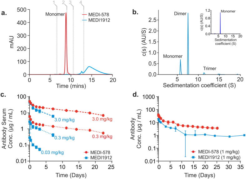
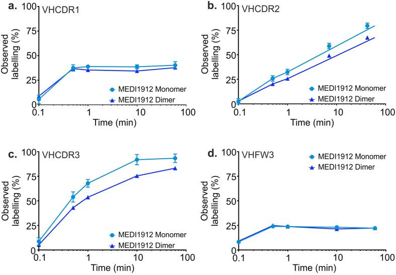
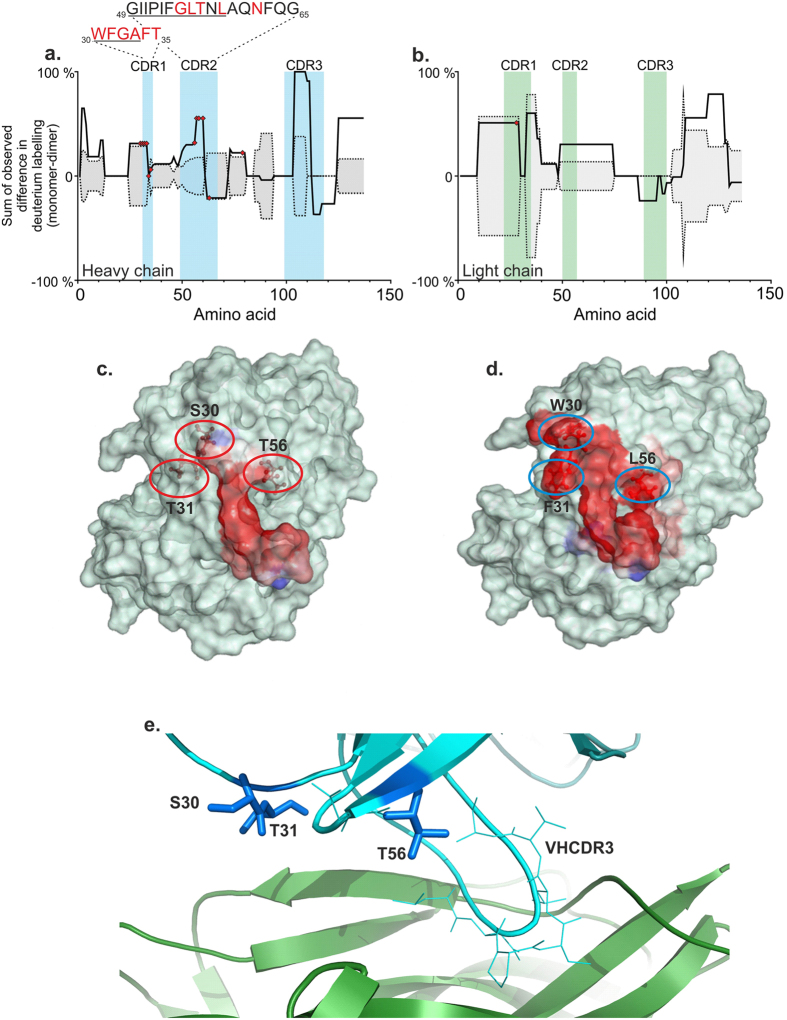
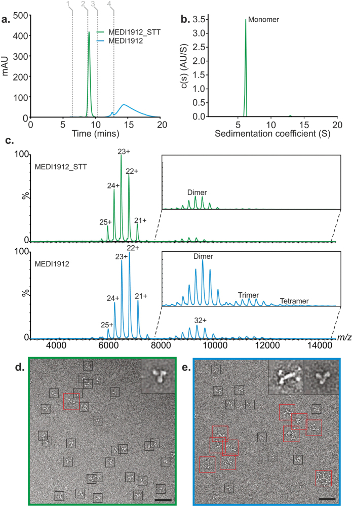
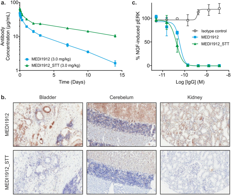
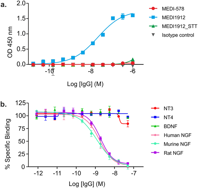
Uncontrolled self-association is a major challenge in the exploitation of proteins as therapeutics. Here we describe the development of a structural proteomics approach to identify the amino acids responsible for aberrant self-association of monoclonal antibodies and the design of a variant with reduced aggregation and increased serum persistence in vivo. We show that the human monoclonal antibody, MEDI1912, selected against nerve growth factor binds with picomolar affinity, but undergoes reversible self-association and has a poor pharmacokinetic profile in both rat and cynomolgus monkeys. Using hydrogen/deuterium exchange and cross-linking-mass spectrometry we map the residues responsible for self-association of MEDI1912 and show that disruption of the self-interaction interface by three mutations enhances its biophysical properties and serum persistence, whilst maintaining high affinity and potency. Immunohistochemistry suggests that this is achieved via reduction of non-specific tissue binding. The strategy developed represents a powerful and generic approach to improve the properties of therapeutic proteins.


Conflict of interest statement

The MedImmune authors are employees of the AstraZeneca Group and have stock/stock options in AstraZeneca. J.J.P. acknowledges financial support from MedImmune via The Beacon Project.

Figures

Figure 1
Figure 1. Biophysical and PK properties of MEDI-578 and MEDI1912.
(a) Size exclusion chromatography-HPLC (SEC-HPLC) elution profiles of MEDI-578 (red) and MEDI1912 (blue), each 1 mg/mL, at 280 nm (mAu) (0.1 M sodium phosphate, 0.1 M sodium sulphate, pH 6.8). Grey lines show calibrants used: Thyroglobulin (1), IgG (2), Ovalbumin (3) and Vitamin B12 (4). (b) Sedimentation-velocity analytical ultracentrifugation (SV-AUC) of MEDI1912 (1 mg/mL, 20 mM sodium succinate, 125 mM arginine, pH 6). (c) MEDI-578 and MEDI1912 PK profiles following intravenous administration of a 3.0 mg/kg, 0.3 mg/kg or 0.03 mg/kg dose to rats (n = 3). (d) MEDI-578 and MEDI1912 PK profiles following intravenous administration of a 1 mg/kg dose to cynomolgus monkeys (n = 3). Data points represent the mean ± SD.
Figure 2
Figure 2. MEDI1912 dimer shows site-specific protection against hydrogen exchange.
HDX-MS identified peptide regions within MEDI1912 that have a lower rate of labelling under conditions that stabilise the antibody dimer (1 mg/mL) versus the monomer (0.1 mg/mL). Significant differences in labelling (p < 0.01) were observed in VHCDR1 (a), VHCDR2 (b) and VHCDR3 (c), yet there was no difference, for example, in a peptide within VH framework region 3). This supports the hypothesis that there is a specific interface for MEDI1912 self-association involving the variable domains. Data are corrected for back-exchange and represent mean ±1 SD.
Figure 3
Figure 3. Mapping MEDI1912 self-interactions.
Site-localised protection against hydrogen-exchange in MEDI1912 heavy (a) and light (b) chain variable domains. For concentrations at which MEDI1912 is predominantly monomeric (M) and dimeric (D), the difference in uptake of deuterium label was calculated as ΔHDX(M-D). This was normalised per protein, with the region that showed maximum difference set to 100%. Protection due to self-association shows as a positive value on the y-axis (solid line – measured value; dashed line – 99% confidence limit). Amino acids altered in affinity maturation from MEDI-578 are marked in red circles. CDR sequences are listed for those that were affinity matured: red – mutated residue; underlined – significant HDX protection (p < 0.01). A spatial aggregation propensity (SAP) algorithm was applied to structural models of MEDI-578 (c) and MEDI1912 (d) revealing surface exposed hydrophobic patches (red). Three amino acids coincident with significant HDX protection in MEDI1912 dimer and SAP prediction: W30, F31 and L56 (blue circles) were chosen for reversion to the corresponding MEDI-578 amino acids: S30, T31, and T56 (red circles). The crystal structure of NGF (coloured green) in complex with the Fab of the MEDI1912 parental antibody, MEDI-578 (blue), (e) reveals that while the VHCDR3 (cyan lines) forms a loop that extends into a cavity within NGF, the amino acid residues 30 and 31 in the VHCDR1 and 56 in the VHCDR2 (blue sticks) are located at the periphery of the binding interface and do not make specific interactions with epitope residues.
Figure 4
Figure 4. Conformational properties of MEDI1912 and MEDI1912_STT using nESI-IMS-MS and negative stain TEM.
(a) High performance size exclusion chromatography-HPLC (HP-SEC-HPLC) elution profiles of MEDI1912 (blue) and MEDI1912_STT (green) at 280 nm (mAU). Grey lines show calibrants used: Thyroglobulin (1), IgG (2), Ovalbumin (3) and Vitamin B12 (4). (b) SV-AUC of MEDI1912_STT showing the distribution of species formed. (c) Nano electrospray ionisation (nESI) mass spectra of MEDI1912 (blue, lower) and MEDI1912_STT (green, upper) showing the different oligomers observed for the two proteins. The insets highlight the higher-order species formed from each sample. (d) Negative stain TEM images of MEDI1912_STT (green, left) and MEDI1912 (blue, right) reveal a distribution of oligomeric species only for the latter protein. Black boxes, monomer; red boxes, dimers/oligomers. Scale bar = 25 nm. Inset shows averaged particle images for monomer and dimer species observed (31 nm (square)).
Figure 5
Figure 5. Effect of STT mutations on MEDI1912 pharmacokinetics, tissue specificity and functional potency.
(a) MEDI1912 and MEDI1912_STT PK profiles following intravenous administration of a 3 mg/kg dose to rats (n = 3). (b) Human bladder, cerebellum and kidney tissue immunohistochemically stained with MEDI1912 and MEDI1912_STT at 0.18 μg/mL. Significant staining was demonstrated by MEDI1912, showing strong staining in connective tissue, smooth muscle (around blood vessels) and other areas that is consistent with non-specific staining. MEDI1912_STT showed no evidence of staining in any tissues evaluated. (c) MEDI1912_STT retains functional potency in vitro compared with MEDI1912 in a phospho-ERK activation assay. Data points represent the mean ± SD.
Figure 6
Figure 6. In vitro characterisation of MEDI1912 and MEDI1912_STT.
(a) Baculovirus ELISA and (b) specificity of MEDI1912 for human, mouse and rat NGF, and a panel of structurally related proteins determined using a homogeneous time resolved fluorescence assay (HTRF).

References

    1. Reichert J. M. Antibodies to watch in 2016. MAbs 8, 197–204 (2016). - PMC - PubMed
    1. Vugmeyster Y. et al.. Complex pharmacokinetics of a humanized antibody against human amyloid beta peptide, anti-abeta Ab2, in nonclinical species. Pharm. Res. 28, 1696–1706 (2011). - PubMed
    1. Wu H. et al.. Development of motavizumab, an ultra-potent antibody for the prevention of respiratory syncytial virus infection in the upper and lower respiratory tract. J. Mol. Biol. 368, 652–665 (2007). - PubMed
    1. Deng R. et al.. Projecting human pharmacokinetics of therapeutic antibodies from nonclinical data: what have we learned? MAbs 3, 61–66 (2011). - PMC - PubMed
    1. Hotzel I. et al.. A strategy for risk mitigation of antibodies with fast clearance. MAbs 4, 753–760 (2012). - PMC - PubMed

Publication types

MeSH terms