. 2016 Dec 20:6:39311.
doi: 10.1038/srep39311.

RNA activation of haploinsufficient Foxg1 gene in murine neocortex

Affiliations

RNA activation of haploinsufficient Foxg1 gene in murine neocortex

Cristina Fimiani et al. Sci Rep. .

Abstract

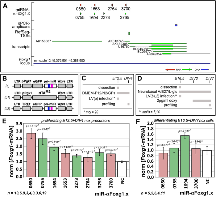
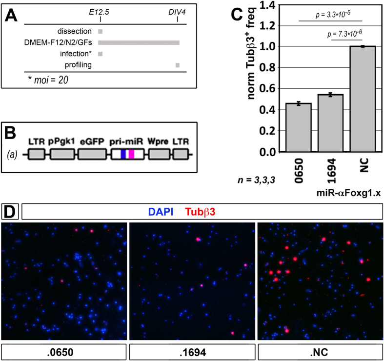
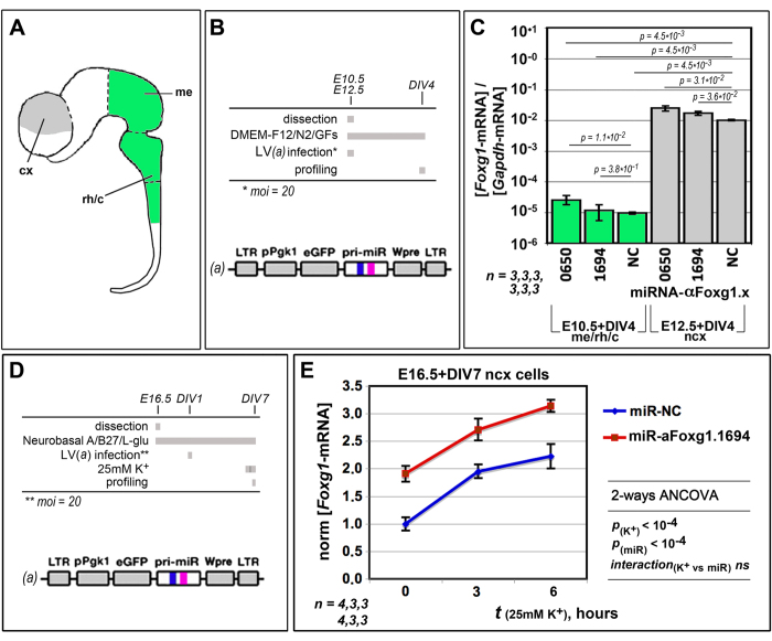
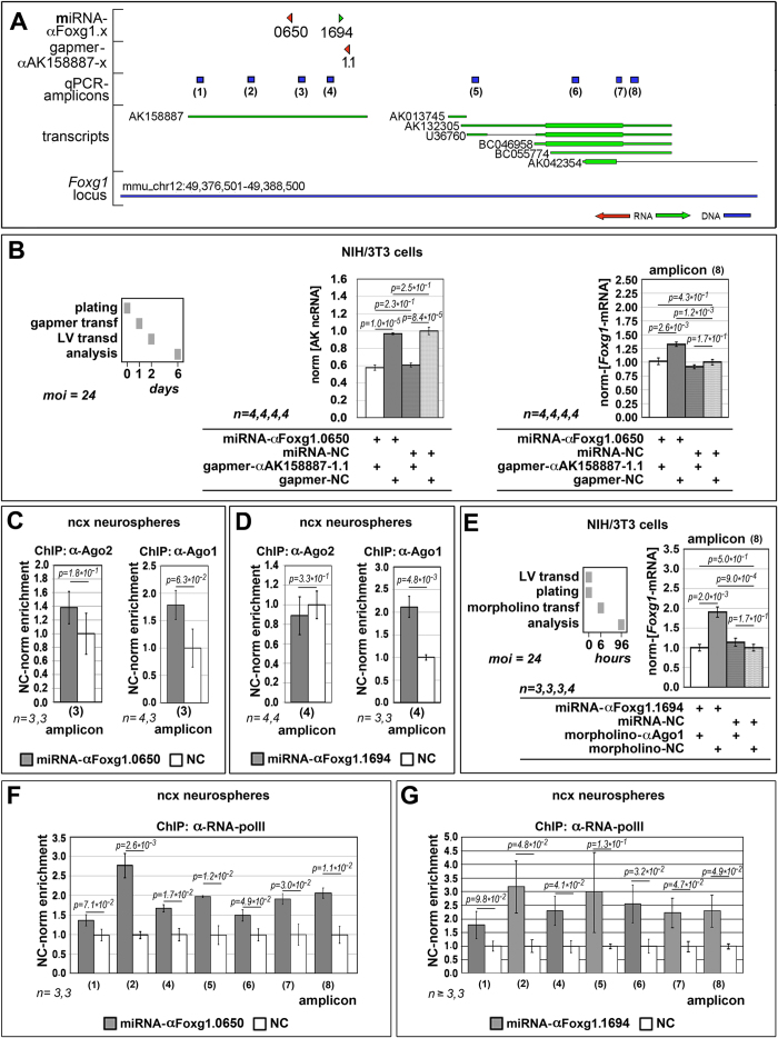
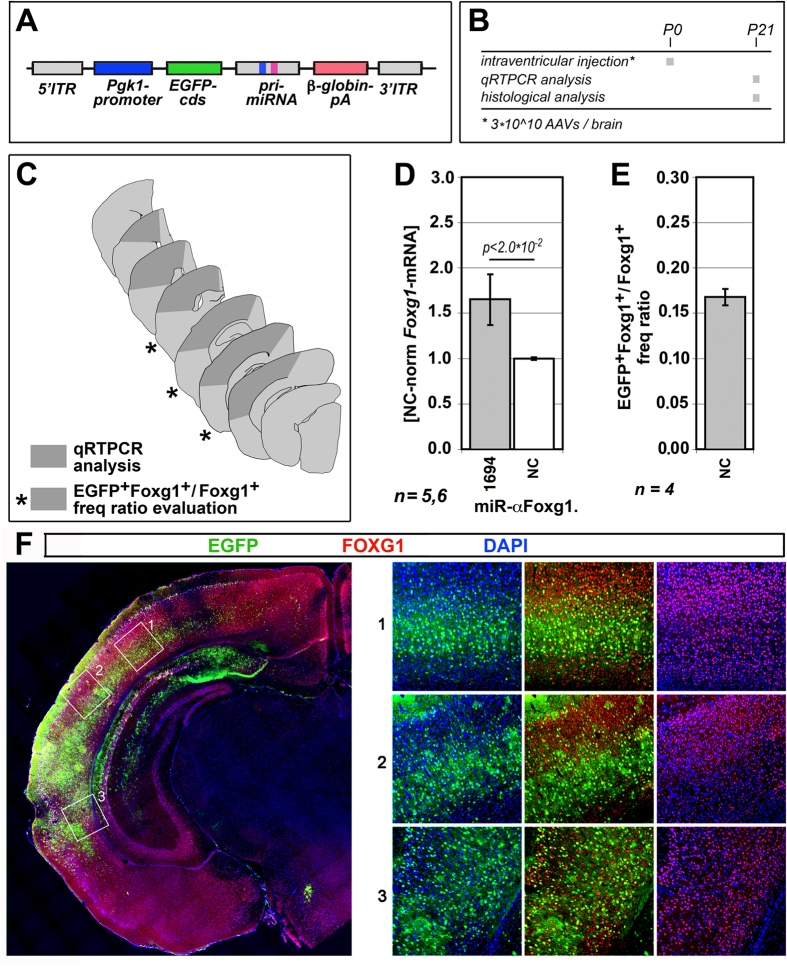
More than one hundred distinct gene hemizygosities are specifically linked to epilepsy, mental retardation, autism, schizophrenia and neuro-degeneration. Radical repair of these gene deficits via genome engineering is hardly feasible. The same applies to therapeutic stimulation of the spared allele by artificial transactivators. Small activating RNAs (saRNAs) offer an alternative, appealing approach. As a proof-of-principle, here we tested this approach on the Rett syndrome-linked, haploinsufficient, Foxg1 brain patterning gene. We selected a set of artificial small activating RNAs (saRNAs) upregulating it in neocortical precursors and their derivatives. Expression of these effectors achieved a robust biological outcome. saRNA-driven activation (RNAa) was limited to neural cells which normally express Foxg1 and did not hide endogenous gene tuning. saRNAs recognized target chromatin through a ncRNA stemming from it. Gene upregulation required Ago1 and was associated to RNApolII enrichment throughout the Foxg1 locus. Finally, saRNA delivery to murine neonatal brain replicated Foxg1-RNAa in vivo.


Conflict of interest statement

C.F., E.G., Q.S. and A.M. do not declare any conflicts. G.G. is a co-founder of Voyager Therapeutics specialized in rAAV-based gene therapy, and holds equity in the company. Moreover, G.G. is also an inventor on patents with potential royalties licensed to Voyager Therapeutics and other biopharmaceutical companies.

Figures

Figure 1
Figure 1. Screening for miRNA-like, small RNAs activating Foxg1-mRNA (Foxg1-saRNAs) in murine neocortical precursors and derivatives.
(A) Schematics of the Foxg1 locus including saRNA positions and orientations as well as the diagnostic qRTPCR amplicon. (B–D) Lentiviral reagents and protocols employed for this screening. (E,F) Foxg1-mRNA levels in proliferating neocortical precursors and their differentiating derivatives, manipulated as in (C) and (D), respectively. Values double normalized, against Gapdh and control (NC). E, embryonic day. DIV, days in vitro. Bars represent sem’s. n = number of biological replicates. p-values were calculated by the t-Student algoritm (one-tail, unpaired). All results with p < 0.05 further passed Benjamini-Hochberg filtering, with FDR < 1/m.
Figure 2
Figure 2. Histogenetic outcome of Foxg1-RNAa.
(A,B) Protocols and lentiviral reagents employed for this assay. (C) Quantification of cells immunopositive for the neuron-specific Tubβ3 marker, in cultures of neocortical precursors expressing Foxg1-saRNAs. (D) Examples of Tubβ3+ immuno-fluorescences referred to in (C). E, embryonic day. DIV, days in vitro. Bars represent sem’s. n = number of biological replicates. Statistical significance of results evaluated by t-Student assay (one-tail, unpaired). Absolute average frequency of Tubβ3+ cells in NC samples was (27.25 ± 0.16)%.
Figure 3
Figure 3. Compliance of Foxg1-RNAa with endogenous gene regulation.
(A) Idealized representation of the murine early neural tube, including cortical (cx), mesencephalic (me) and rhombo-cervical (rh/c) domains. (B) Protocols and lentiviral reagent employed for the assay referred to in (C). (C) Impact of miR-αFoxg1.0650 and 0.1694 on Foxg1-mRNA levels in proliferating precursors from the me/rh/c and cx domains. (D) Protocols and lentiviral reagent employed for the assay referred to in (E). (E) Foxg1-mRNA modulation by miR-αFoxg1.1694 in differentiating neocortical derivatives upon their timed terminal exposure to 25 mM K+. E, embryonic day. DIV, days in vitro. Bars represent sem’s. n = number of biological replicates. Statistical significance of results evaluated by t-Student (one-tail, unpaired) (C) and ANCOVA (two-ways, unpaired) (E) assays. ns, not significant.
Figure 4
Figure 4. Molecular mechanisms underlying Foxg1-RNAa.
(A) Schematics of the Foxg1 locus including miRNA and gapmer positions and orientations, as well as diagnostic qPCR amplicons. (B) AK158887-ncRNA and Foxg1-mRNA levels in NIH/3T3 cells upon combined delivery of miR-αFoxg1.0650 and gapmer-αAK158887-1.1. Values double normalized, against Gapdh and control (NC). (C,D) qPCR quantification of Foxg1 chromatin enrichment, upon immunoprecipitation (ChIP) by antibodies against Argonaute 2 (α-Ago2) and Argonaute 1 (α-Ago1). Evaluation performed in neocortical precursors challenged by miR-αFoxg1.0650 (C) and miR-aFoxg1.1694 (D), according to the protocol shown in Fig. 1B,C. Values double normalized against input chromatin and control (NC). (E) Foxg1-mRNA levels in NIH/3T3 cells upon combined delivery of miR-αFoxg1.1694 and morpholino-αAgo1. Values double normalized, against Gapdh and control (NC). (F,G) qPCR quantification of Foxg1 chromatin enrichment, upon ChIP by antibodies against RNA polymerase II (α-RNA-polII). Evaluation performed in neocortical precursors challenged by miR-αFoxg1.0650 (F) and miR-aFoxg1.1694 (G), according to the protocol shown in Fig. 1B,C. Values double normalized against input chromatin and control (NC). Bars represent sem’s. n = number of biological replicates. p-values were calculated by the t-Student algoritm (one-tail, unpaired). All panel 4 F results with p < 0.05 further passed Benjamini-Hochberg filtering, with FDR < 1 /m. The same applies to panel 4 G, except amplicon results.
Figure 5
Figure 5. Foxg1-RNAa in murine neocortex.
(A) Schematics of AAV9-pseudotyped, self-complementary AAV2-derivative, adeno-associated viral vector, driving constitutive expression of Foxg1-activating miRNAs. (B) Protocol employed for the assays referred to in (CF). (C) Location of neocortical sectors subject of the analyses shown in (DF). (D) Quantification of Foxg1-mRNA levels in neocortex of juvenile mice previously injected by scAAVs encoding for miR-αFoxg1.1694. (E) Evaluation of frequency of Foxg1+ cells transduced by EGFP-encoding control virus (NC). (F) Examples of αFoxg1/αEGFP-immunoprofiled sections referred to in (C,E). P, post-natal day. Bars represent sem’s. n = number of biological replicates (i.e. brains). Statistical significance of results evaluated by t-Student assay (one-tail, unpaired).

References

    1. Barøy T. et al.. Haploinsufficiency of two histone modifier genes on 6p22.3, ATXN1 and JARID2, is associated with intellectual disability. Orphanet Journal of Rare Diseases 8, 3 (2013). - PMC - PubMed
    1. Carvill G. L. & Mefford H. C. Microdeletion syndromes. Current Opinion in Genetics & Development 23, 232–239 (2013). - PubMed
    1. Lal D. et al.. Burden Analysis of Rare Microdeletions Suggests a Strong Impact of Neurodevelopmental Genes in Genetic Generalised Epilepsies. PLOS Genetics 11, e1005226 (2015). - PMC - PubMed
    1. Lee B. H., Smith T. & Paciorkowski A. R. Autism spectrum disorder and epilepsy: Disorders with a shared biology. Epilepsy & Behavior 47, 191–201 (2015). - PMC - PubMed
    1. Silva A. M., Brown J. M., Buckle V. J., Wade-Martins R. & Lufino M. M. P. Expanded GAA repeats impair FXN gene expression and reposition the FXN locus to the nuclear lamina in single cells. Hum. Mol. Genet. 24, 3457–3471 (2015). - PMC - PubMed

Publication types

MeSH terms