Effect of NR5A2 inhibition on pancreatic cancer stem cell (CSC) properties and epithelial-mesenchymal transition (EMT) markers
- PMID: 27996162
- PMCID: PMC5392129
- DOI: 10.1002/mc.22604
Effect of NR5A2 inhibition on pancreatic cancer stem cell (CSC) properties and epithelial-mesenchymal transition (EMT) markers
Erratum in
-
Corrigendum.Mol Carcinog. 2018 Jan;57(1):142. doi: 10.1002/mc.22766. Mol Carcinog. 2018. PMID: 29205538 No abstract available.
Abstract
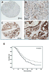
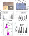
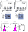
NR5A2 (aka LRH-1) has been identified as a pancreatic cancer susceptibility gene with missing biological link. This study aims to demonstrate expression and potential role of NR5A2 in pancreatic cancer. NR5A2 expression was quantified in resected pancreatic ductal adenocarcinomas and the normal adjacent tissues of 134 patients by immunohistochemistry. The intensity and extent of NR5A2 staining was quantified and analyzed in association with overall survival (OS). The impact of NR5A2 knockdown on pancreatic cancer stem cell (CSC) properties and epithelial-mesenchymal transition (EMT) markers was examined in cancer cells using RT-PCR and Western Blot. NR5A2 was overexpressed in pancreatic tumors, the IHC-staining H score (mean ± SE) was 96.4 ± 8.3 in normal versus 137.9 ± 8.2 in tumor tissues (P < 0.0001). Patients with a higher NR5A2 expression had a median survival time 18.4 months compared to 23.7 months for those with low IHC H scores (P = 0.019). The hazard ratio of death (95% confidence interval) was 1.60 (1.07-2.41) after adjusting for disease stage and tumor grade (P = 0.023). NR5A2 was highly expressed in pancreatic cancer sphere forming cells. NR5A2-inhibition by siRNA was associated with reduced sphere formation and decreased levels of CSCs markers NANOG, OCT4, LIN28B, and NOTCH1. NR5A2 knockdown also resulted in reduced expression of FGB, MMP2, MMP3, MMMP9, SNAIL, and TWIST, increased expression of epithelial markers E-cadherin and β-catenin, and a lower expression of mesenchymal marker Vimentin. Taken together, our findings suggest that NR5A2 could play a role in CSC stemness and EMT in pancreatic cancer, which may contribute to the worse clinical outcome.
Keywords: EMT; expression; stem cell-like cancer cell; survival.
© 2017 Wiley Periodicals, Inc.
Figures


Similar articles
-
LGR5 Is a Gastric Cancer Stem Cell Marker Associated with Stemness and the EMT Signature Genes NANOG, NANOGP8, PRRX1, TWIST1, and BMI1.PLoS One. 2016 Dec 29;11(12):e0168904. doi: 10.1371/journal.pone.0168904. eCollection 2016. PLoS One. 2016. PMID: 28033430 Free PMC article.
-
Nr5a2 promotes cancer stem cell properties and tumorigenesis in nonsmall cell lung cancer by regulating Nanog.Cancer Med. 2019 Mar;8(3):1232-1245. doi: 10.1002/cam4.1992. Epub 2019 Feb 10. Cancer Med. 2019. PMID: 30740909 Free PMC article.
-
CCL21/CCR7 Axis Contributed to CD133+ Pancreatic Cancer Stem-Like Cell Metastasis via EMT and Erk/NF-κB Pathway.PLoS One. 2016 Aug 9;11(8):e0158529. doi: 10.1371/journal.pone.0158529. eCollection 2016. PLoS One. 2016. PMID: 27505247 Free PMC article.
-
The epithelial to mesenchymal transition (EMT) and cancer stem cells: implication for treatment resistance in pancreatic cancer.Mol Cancer. 2017 Feb 28;16(1):52. doi: 10.1186/s12943-017-0624-9. Mol Cancer. 2017. PMID: 28245823 Free PMC article. Review.
-
Pancreatic cancer stem cell markers and exosomes - the incentive push.World J Gastroenterol. 2016 Jul 14;22(26):5971-6007. doi: 10.3748/wjg.v22.i26.5971. World J Gastroenterol. 2016. PMID: 27468191 Free PMC article. Review.
Cited by
-
ITGA7 functions as a tumor suppressor and regulates migration and invasion in breast cancer.Cancer Manag Res. 2018 May 1;10:969-976. doi: 10.2147/CMAR.S160379. eCollection 2018. Cancer Manag Res. 2018. PMID: 29760566 Free PMC article.
-
Curcumin reversed chronic tobacco smoke exposure induced urocystic EMT and acquisition of cancer stem cells properties via Wnt/β-catenin.Cell Death Dis. 2017 Oct 5;8(10):e3066. doi: 10.1038/cddis.2017.452. Cell Death Dis. 2017. PMID: 28981096 Free PMC article.
-
NR5A2 promotes malignancy progression and mediates the effect of cisplatin in cutaneous squamous cell carcinoma.Immun Inflamm Dis. 2024 Feb;12(2):e1172. doi: 10.1002/iid3.1172. Immun Inflamm Dis. 2024. PMID: 38358044 Free PMC article.
-
Activation of Glucocorticoid Receptor Inhibits the Stem-Like Properties of Bladder Cancer via Inactivating the β-Catenin Pathway.Front Oncol. 2020 Aug 5;10:1332. doi: 10.3389/fonc.2020.01332. eCollection 2020. Front Oncol. 2020. PMID: 32850423 Free PMC article.
-
LRH1 Acts as an Oncogenic Driver in Human Osteosarcoma and Pan-Cancer.Front Cell Dev Biol. 2021 Mar 15;9:643522. doi: 10.3389/fcell.2021.643522. eCollection 2021. Front Cell Dev Biol. 2021. PMID: 33791301 Free PMC article.
References
-
- Fayard E, Auwerx J, Schoonjans K. LRH-1: an orphan nuclear receptor involved in development, metabolism and steroidogenesis. Trends Cell Biol. 2004;14(5):250–260. - PubMed
-
- Botrugno OA, Fayard E, Annicotte JS, et al. Synergy between LRH-1 and beta-catenin induces G1 cyclin-mediated cell proliferation. Mol Cell. 2004;15(4):499–509. - PubMed
-
- Wang SL, Zheng DZ, Lan FH, et al. Increased expression of hLRH-1 in human gastric cancer and its implication in tumorigenesis. Mol Cell Biochem. 2008;308(1-2):93–100. - PubMed
Publication types
MeSH terms
Substances
Grants and funding
LinkOut - more resources
Full Text Sources
Other Literature Sources
Medical
Research Materials
Miscellaneous

