. 2017 Jan 18;139(2):849-855.
doi: 10.1021/jacs.6b10894. Epub 2017 Jan 5.

Three-Dimensional Structure Determination of Surface Sites

Affiliations

Three-Dimensional Structure Determination of Surface Sites

Pierrick Berruyer et al. J Am Chem Soc. .

Abstract

The spatial arrangement of atoms is directly linked to chemical function. A fundamental challenge in surface chemistry and catalysis relates to the determination of three-dimensional structures with atomic-level precision. Here we determine the three-dimensional structure of an organometallic complex on an amorphous silica surface using solid-state NMR measurements, enabled through a dynamic nuclear polarization surface enhanced NMR spectroscopy approach that induces a 200-fold increase in the NMR sensitivity for the surface species. The result, in combination with EXAFS, is a detailed structure for the surface complex determined with a precision of 0.7 Å. We observe a single well-defined conformation that is folded toward the surface in such a way as to include an interaction between the platinum metal center and the surface oxygen atoms.


Conflict of interest statement

The authors declare no competing financial interest.

Figures
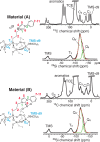
Figure 1
Figure 1
Chemical structure and NMR signal assignments. Left: the chemical structure of material A together with the 13C and 29Si CPMAS DNP SENS spectra for A-N1. Right: the same for material B. Symbol * denotes spinning side bands, and § denotes pentachloroethane impurity found in commercial 1,1,2,2 tetra-chloroethane used as the impregnating solvent (see Supporting Information Figure S7 for details). For the 29Si spectra, the deconvolution of the SiQ4/SiQ4′ peaks is shown, where the component Gaussian peaks are drawn with solid green lines and the sum is drawn with a dashed red line.
Figure 2
Figure 2
Three-dimensional structure determination. Possible 3D structures for the system were generated in silico, as visualized here with a simple “ball and stick” model, by rotating atoms around the 4 axes (α1, α3, α4, and α5) in 15° steps for each of the four selected SiT3–SiQ4′ distances. Analytical REDOR curves were then calculated for each structure and compared to the ensemble of experimental REDOR curves to determine (i) the single 3D structure in best agreement with the experimental data, and (ii) the distribution of conformers that agree with the data to within the estimated experimental error.
Figure 3
Figure 3
Three-dimensional structures of materials A and B. The experimental DNP SENS REDOR data are shown on the left for A (above) and for the Pt-complex B (below) with black dots for the 8 different spin pairs which generate nontrivial constraints. The solid red and green lines are the calculated REDOR curves for the structures in best agreement with the experimental data. The internuclear distances for each spin pair in the structure in best agreement with the experimental data are reported for each curve. The corresponding best fit structures are shown on the right, and coordinates are given in the SI. The dashed lines shown on the structures correspond to the REDOR constraints used for structure determination.
Figure 4
Figure 4
Ensemble of conformers for the Pt-complex B. There are 14 structures of B with dSiT3-SiQ4′ = 4.50 Å, 25 with dSiT3-SiQ4′ = 5.59 Å, and 23 with dSiT3-SiQ4′ = 5.83 Å shown. The RMSD among the ensemble is 0.62 Å for dSiT3-SiQ4′ = 4.50 Å, 0.71 Å for dSiT3-SiQ4′ = 5.59 Å, and 0.70 Å for dSiT3-SiQ4′ = 5.83 Å. The structures are superimposed by aligning SiT3, SiQ4′, and O.

References

    1. Tsubogo T.; Oyamada H.; Kobayashi S. Nature 2015, 520, 329–332. 10.1038/nature14343. - DOI - PubMed
    1. Fu Q.; Li W.-X.; Yao Y.; Liu H.; Su H.-Y.; Ma D.; Gu X.-K.; Chen L.; Wang Z.; Zhang H.; Wang B.; Bao X. Science 2010, 328, 1141–1144. 10.1126/science.1188267. - DOI - PubMed
    1. Kim Y.; Macfarlane R. J.; Jones M. R.; Mirkin C. A. Science 2016, 351, 579–582. 10.1126/science.aad2212. - DOI - PubMed
    1. Blanc F.; Berthoud R.; Coperet C.; Lesage A.; Emsley L.; Singh R.; Kreickmann T.; Schrock R. R. Proc. Natl. Acad. Sci. U. S. A. 2008, 105, 12123–12127. 10.1073/pnas.0802147105. - DOI - PMC - PubMed
    1. Hughes M. D.; Xu Y.-J.; Jenkins P.; McMorn P.; Landon P.; Enache D. I.; Carley A. F.; Attard G. a; Hutchings G. J.; King F.; Stitt E. H.; Johnston P.; Griffin K.; Kiely C. J. Nature 2005, 437, 1132–1135. 10.1038/nature04190. - DOI - PubMed

Publication types

LinkOut - more resources