. 2016 Dec 20;12(12):e1006486.
doi: 10.1371/journal.pgen.1006486. eCollection 2016 Dec.

Phosphatidylserine Ameliorates Neurodegenerative Symptoms and Enhances Axonal Transport in a Mouse Model of Familial Dysautonomia

Affiliations

Phosphatidylserine Ameliorates Neurodegenerative Symptoms and Enhances Axonal Transport in a Mouse Model of Familial Dysautonomia

Shiran Naftelberg et al. PLoS Genet. .

Abstract

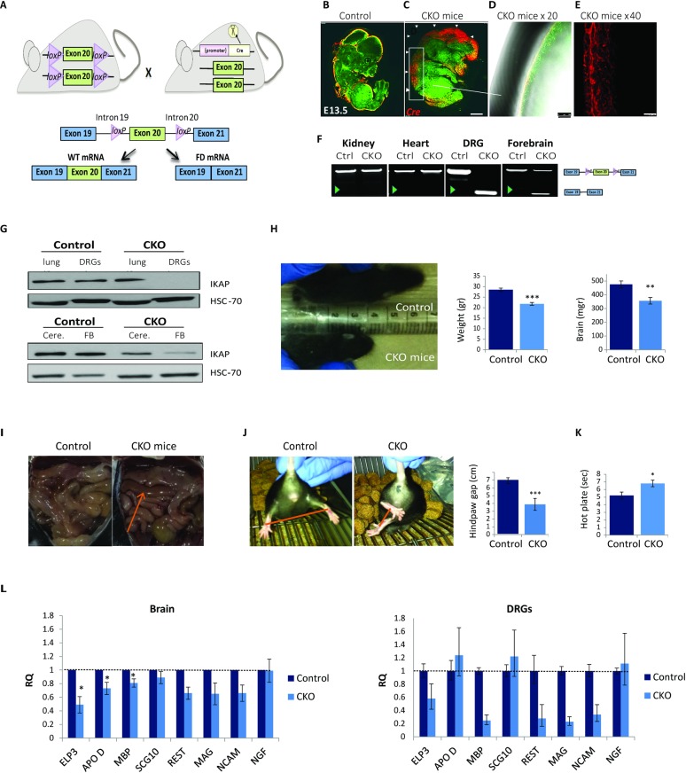
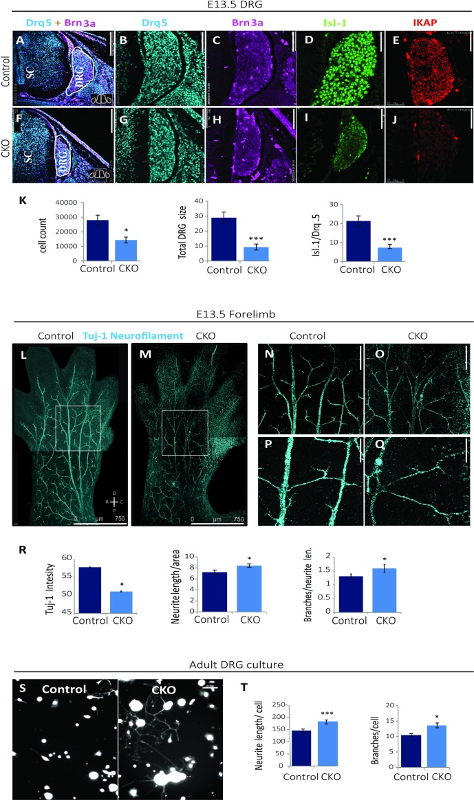
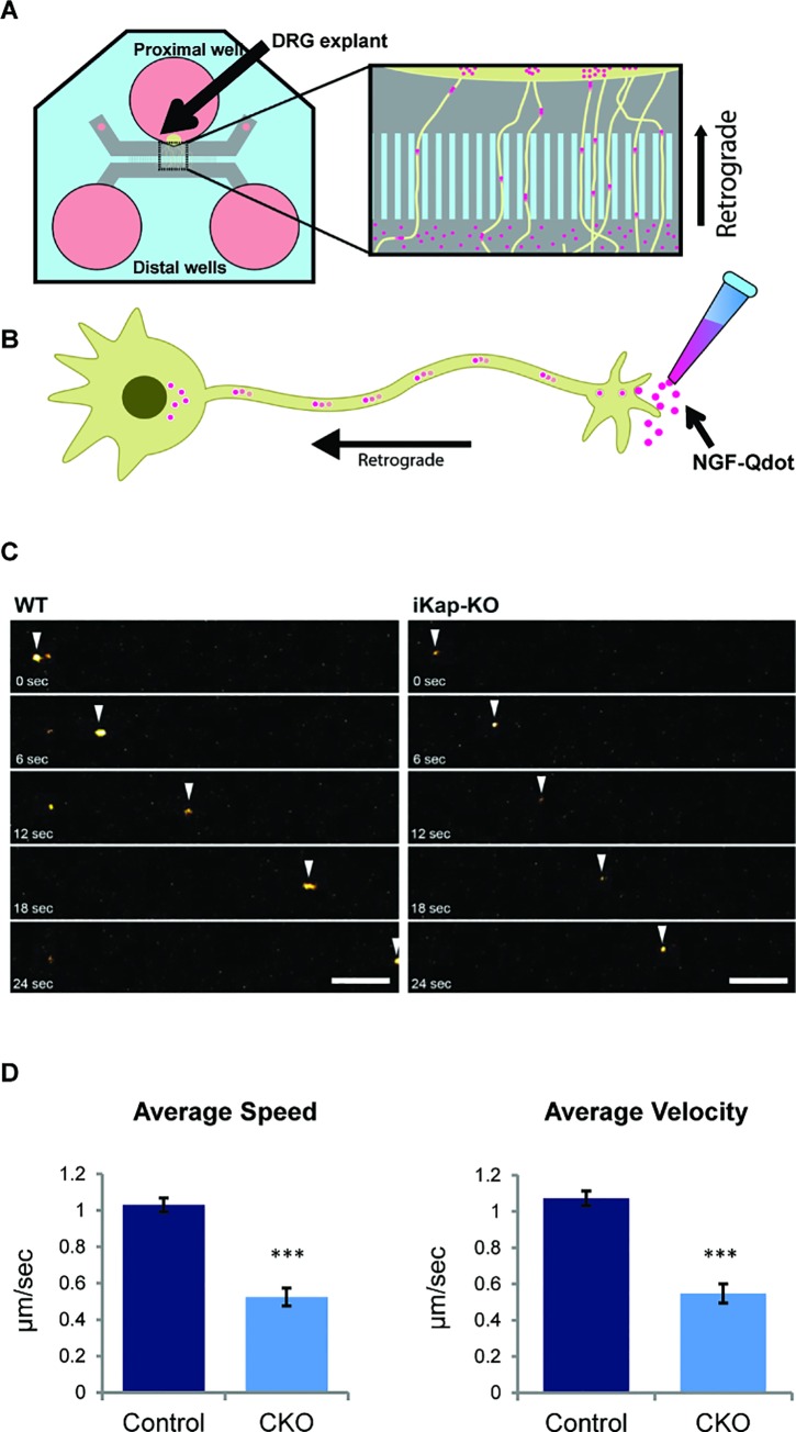
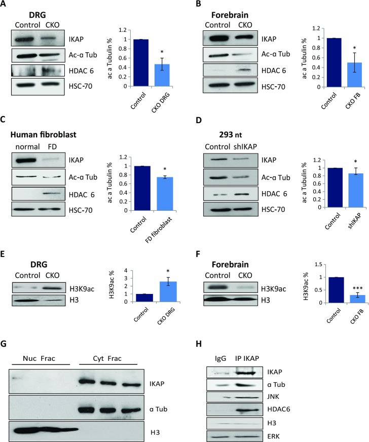
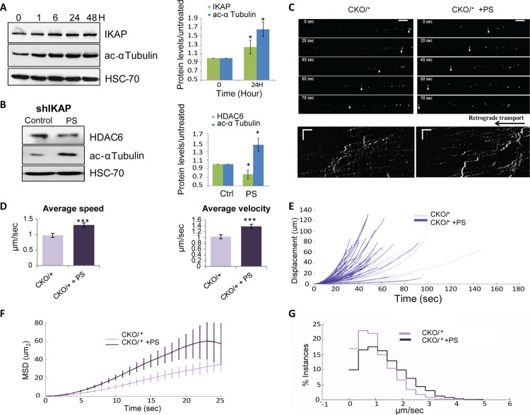
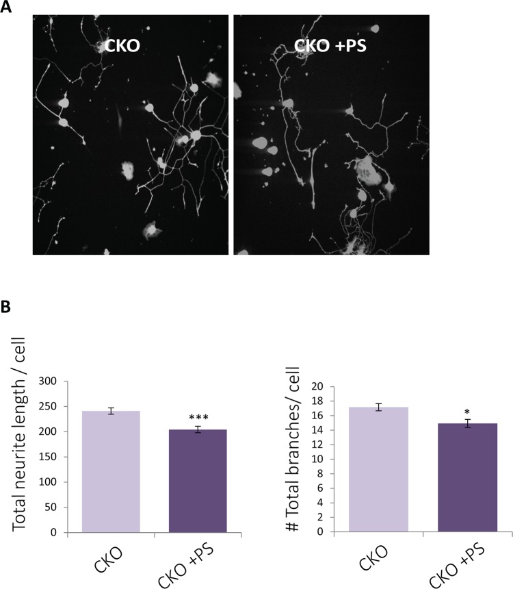
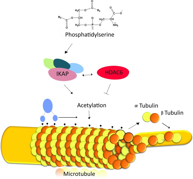
Familial Dysautonomia (FD) is a neurodegenerative disease in which aberrant tissue-specific splicing of IKBKAP exon 20 leads to reduction of IKAP protein levels in neuronal tissues. Here we generated a conditional knockout (CKO) mouse in which exon 20 of IKBKAP is deleted in the nervous system. The CKO FD mice exhibit developmental delays, sensory abnormalities, and less organized dorsal root ganglia (DRGs) with attenuated axons compared to wild-type mice. Furthermore, the CKO FD DRGs show elevated HDAC6 levels, reduced acetylated α-tubulin, unstable microtubules, and impairment of axonal retrograde transport of nerve growth factor (NGF). These abnormalities in DRG properties underlie neuronal degeneration and FD symptoms. Phosphatidylserine treatment decreased HDAC6 levels and thus increased acetylation of α-tubulin. Further PS treatment resulted in recovery of axonal outgrowth and enhanced retrograde axonal transport by decreasing histone deacetylase 6 (HDAC6) levels and thus increasing acetylation of α-tubulin levels. Thus, we have identified the molecular pathway that leads to neurodegeneration in FD and have demonstrated that phosphatidylserine treatment has the potential to slow progression of neurodegeneration.


Conflict of interest statement

The authors have declared that no competing interests exist.

Figures

Fig 1
Fig 1. Generation of Tyrp2-Cre;IKBKAPFDloxP/FDloxP (CKOTyrp2 FD) mice.
(A) Two loxP sequences were inserted in the introns flanking exon 20 of the IKBKAP gene (IKBAKPFDloxP/FDloxP mouse, the mouse shown on the left). IKBAKPFDloxP/FDloxP mice were mated with Tyrp2-Cre mice (the mouse on the right). The lower panel shows a schematic representation of the IKBKAPFDloxP/FDloxP construct. Cre activation leads to exon 20 deletion in targeted tissues. (B-E) Whole-mount immunostaining using Cre (red) and Tuj-1 (green) antibodies of (B) control and (C-E) CKOTyrp2 FD mice; enlargements are shown in D and E. (F) DNA from the indicated organs was extracted and analyzed to examine exon 20 deletion. Green arrowhead indicates removal of IKBKAP exon 20. (G) Western blot of IKAP in the lungs, DRGs, cerebellums (Cere.), and forebrains (FB) of control and CKOTyrp2 FD mice. (H) Left panel: Photographs of CKOTyrp2 FD and control littermates 10 days after birth (P10). Middle panel: Weights of CKOTyrp2 FD and control mice (n = 40 per group, ***p<0.001). Right panel: Weights of brains from CKOTyrp2 FD and control littermates (n = 10 per group, **p<0.01). (I) CKOTyrp2 FD mice have brownish and swollen intestines (indicated by arrow). (J) Tail hanging test of three-month old CKOTyrp2 FD and control littermates and plots of hindpaw gaps during tail hanging (n = 5 per group, ***p<0.001). (K) Hot-plate analgesia evaluation of thermal sensation and peripheral sensory nerve function of CKOTyrp2 FD and control mice (n = 20 per group, *p<0.05). (L) qRT-PCR analysis of genes, known to be abnormally regulated in FD patients in brains (n = 3 of each group) and DRGs (pool of four mice for each group) of CKOTyrp2 FD and control mice. Error bars represent ± SEM and for DRGs error bars represent technical standard deviation of three repeats.
Fig 2
Fig 2. Deletion of IKBKAP exon 20 results in grossly reduced DRG size and interruption of peripheral projections.
(A-J) Frozen cryo cross-sections of E13.5 CKOTyrp2 FD and control littermate embryos were immunostained for DNA marker Drq5 (blue), DRG markers Brn3a (pink) and Isl-1 (green), and IKAP (red). Lumbar DRG cross-sections show decreases in DRG size as indicated by the gross morphology and decreased numbers of cells expressing Brn3a and Isl-1 in CKOTyrp2 FD mice compared to controls. Scale bars 250 μm for panels A and F and 50 μm for panels B-E and G-J. (K) Quantification of average cell counts based on Drq5 immunostaining (n = 5 per group,*p<0.05), of total DRG size (in cm3, ***p<0.001), and of the Isl-1/Drq5 ratio for the CKOTyrp2 FD DRGs compared to controls at E13.5 (***p<0.001). (L-Q) Embryos at E13.5 were whole-mount stained for neuronal marker Tuj-1 (blue). Scale bars: panels L and M, 750 μm; panels N and O, 250 μm; panels P and Q, 100 μm. (R) Quantification of Tuj-1 intensity (mean ROI intensity ± SEM,), average neurite length (mean ROI length/forelimb ROI size ± SEM), and numbers of branches (mean of total branches/neurite length ± SEM) based on ImageJ analysis of whole-mount staining (n = 40 per group, *p<0.05). (S) DRGs from 3 month-old CKOTyrp2 FD and control mice were cultured for 24 h and stained using calcein to detect neurites and Hoechst dye to detect cell bodies. (T) Plots of neurite lengths and total numbers of branches normalized to cells number of DRGs cultured from 3 month-old CKOTyrp2 FD and control mice (n = 300 per group, ***p<0.001 and *p<0.05, respectively). Error bars represent ± SEM.
Fig 3
Fig 3. Deletion of IKBKAP exon 20 results in disrupted NGF axonal transport.
(A) Schematic for the microfluidic system to track retrograde transport in DRG neurons. (B) Representation of retrograde axonal transport after addition of NGF-Qdot (pink). (C) DRG E13.5 explants were grown in microfluidic chambers, labeled NGF was added to the distal side, and bright field and fluorescent images were taken 24 h after plating. Arrows indicate transported particles. Scale bars: horizontal, 10 μm; vertical, 50 s. (D) The average velocities and speeds of labeled NGF were lower in CKOTyrp2 FD DRGs than control DRGs (***p<0.001). Error bars represent ± SEM.
Fig 4
Fig 4. IKAP is a cytoplasmic protein that interacts with α-tubulin and HDAC6.
(A-D) Right panels: Representative western blots for IKAP, acetylated α-tubulin, and HDAC6. Left panels: Relative levels of acetylated α-tubulin in control (set to 1) and IKAP-deficient samples from (A) control and CKOTyrp2 FD DRG extracts, (B) control and CKOTyrp2 FD forebrains, (C) extracts generated from fibroblasts from normal controls and an FD patient, and (D) HEK 293nt controls and shIKAP cells. HSC-70 levels were used as a protein loading control. Quantifications are of three biological replicates (*p<0.05). (E-F) Western blots and quantification of histone H3K9ac levels in (E) DRGs (*p<0.05) and (F) forebrain CKOTyrp2 FD DRGs and control DRGs (***p<0.001). (G) Western blots of nuclear (Nuc) and cytoplasmic (Cyt) fractions of HEK 293nt lysates. Histone H3 was present only in the nuclear fraction and α-tubulin only in the cytoplasmic fraction. (H) HEK 293nt cell lysate was immunoprecipitated with anti-IKAP antibody followed by immunoblot analysis for indicated proteins. Error bars represent SEM.
Fig 5
Fig 5. Phosphatidylserine elevates acetylated α-tubulin levels and rescues axonal transport.
(A) HEK 293nt cells were treated with 200 μg/μl PS for the indicated time. Left panel: Proteins were extracted, and IKAP, acetylated α-tubulin, and HSC-70 levels were analyzed by western blot. Right panel: Quantification of 24-h data with control levels set to 1 (n = 3, *p<0.05). (B) Western blot of extract of HEK 293nt shIKAP cells treated with 200 μg/μl PS for 24 h. (C-G) PS alters NGF transport in DRG explants culture from CKO/+ embryos. Labeled NGF was added to the distal side of the culture, and bright field and fluorescent images were taken 24 hours after addition of PS or vehicle. (C) NGF-Qdot transport was imaged in DRG neurons upon PS treatment. The arrowheads track representative faster Q-dots along the axon of PS treatment neurons. Below is a representative kymograph demonstrating faster NGF-Qdot transport in PS-treated compared to control cells. (D) Mean average velocities and speeds (***p<0.001), (E) displacement, and (F) mean square displacement plotted vs. time of labeled NGF in CKO/+ DRG cultures treated with PS or vehicle. Error bars represent SEM. (G) Comparisons of the distribution profiles for instantaneous velocities show that PS treatment induces an overall shift toward faster transport velocities.
Fig 6
Fig 6. Phosphatidylserine improves neuritis outgrowth.
(A) DRGs were extracted from 3 month-old CKOTyrp2 FD mice and grown in culture. Cells were treated with 200 μg/ul PS for 24 hrs. Cultures were stained with calcein to detect neurites and Hoechst dye to detect cell bodies. (B). Neurite length (***p<0.001) and total branches (*p<0.05) were compared to untreated and vehicle only controls and normalized to number of DRGs. Error bars represent ± SEM.
Fig 7
Fig 7. A suggested model for the effect of PS on impaired axonal transport in FD.
Phosphatidylserine affects axonal transport and microtubule stabilization by balancing the interplay of IKAP and HDAC6 levels. IKAP is part of the Elongator complex that contains the catalytic acetyltransferase subunit of ELP3. The Elongator complex acetylates α-tubulin, which is crucial for dynein movement and polymerization of microtubules. HDAC6 destabilizes acetylated α-tubulin, and its levels are influenced by levels of IKAP and other Elongator components. Phosphatidylserine elevates IKAP levels and downregulates HDAC6 levels and thus facilitates axonal transport and microtubule stabilization.

Similar articles

Cited by

References

    1. Lehavi O, Aizenstein O, Bercovich D, Pavzner D, Shomrat R, et al. (2003) Screening for familial dysautonomia in Israel: evidence for higher carrier rate among Polish Ashkenazi Jews. Genet Test 7: 139–142. 10.1089/109065703322146830 - DOI - PubMed
    1. Axelrod FB, Goldberg JD, Ye XY, Maayan C (2002) Survival in familial dysautonomia: Impact of early intervention. J Pediatr 141: 518–523. 10.1067/mpd.2002.127088 - DOI - PubMed
    1. Riley CM, Day RL, et al. (1949) Central autonomic dysfunction with defective lacrimation; report of five cases. Pediatrics 3: 468–478. - PubMed
    1. Mahloudji M, Brunt PW, McKusick VA (1970) Clinical neurological aspects of familial dysautonomia. J Neurol Sci 11: 383–395. - PubMed
    1. Palma JA, Norcliffe-Kaufmann L, Fuente-Mora C, Percival L, Mendoza-Santiesteban C, et al. (2014) Current treatments in familial dysautonomia. Expert Opin Pharmacother 15: 2653–2671. 10.1517/14656566.2014.970530 - DOI - PMC - PubMed

MeSH terms

LinkOut - more resources