. 2016 Dec 20;13(12):e1002200.
doi: 10.1371/journal.pmed.1002200. eCollection 2016 Dec.

IL-7 Receptor Mutations and Steroid Resistance in Pediatric T cell Acute Lymphoblastic Leukemia: A Genome Sequencing Study

Affiliations

IL-7 Receptor Mutations and Steroid Resistance in Pediatric T cell Acute Lymphoblastic Leukemia: A Genome Sequencing Study

Yunlei Li et al. PLoS Med. .

Abstract

Background: Pediatric acute lymphoblastic leukemia (ALL) is the most common childhood cancer and the leading cause of cancer-related mortality in children. T cell ALL (T-ALL) represents about 15% of pediatric ALL cases and is considered a high-risk disease. T-ALL is often associated with resistance to treatment, including steroids, which are currently the cornerstone for treating ALL; moreover, initial steroid response strongly predicts survival and cure. However, the cellular mechanisms underlying steroid resistance in T-ALL patients are poorly understood. In this study, we combined various genomic datasets in order to identify candidate genetic mechanisms underlying steroid resistance in children undergoing T-ALL treatment.

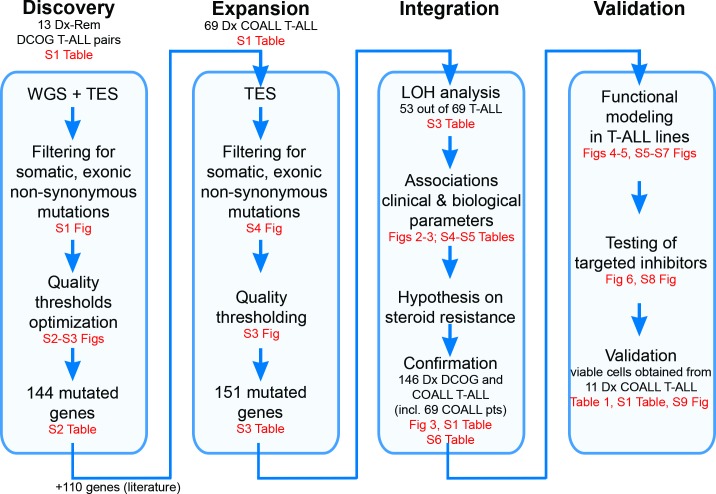
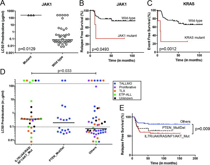
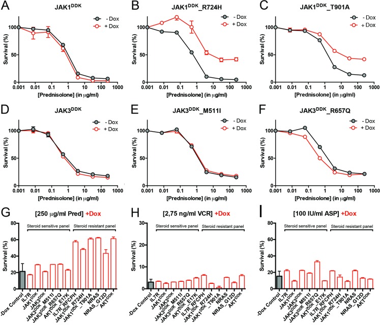
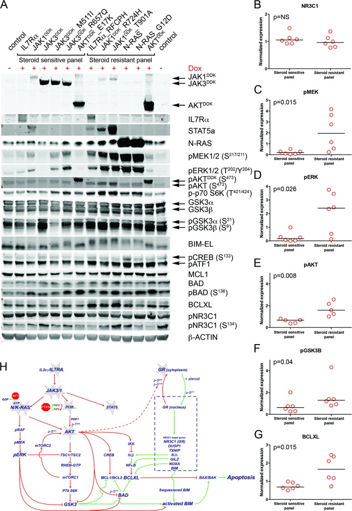
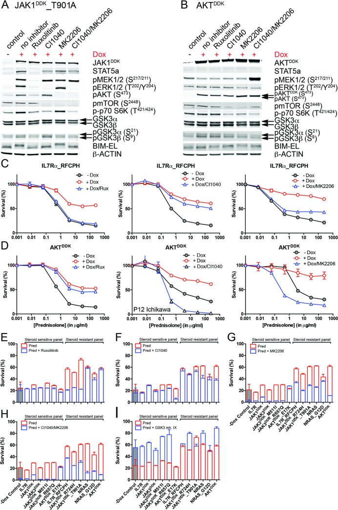
Methods and findings: We performed whole genome sequencing on paired pre-treatment (diagnostic) and post-treatment (remission) samples from 13 patients, and targeted exome sequencing of pre-treatment samples from 69 additional T-ALL patients. We then integrated mutation data with copy number data for 151 mutated genes, and this integrated dataset was tested for associations of mutations with clinical outcomes and in vitro drug response. Our analysis revealed that mutations in JAK1 and KRAS, two genes encoding components of the interleukin 7 receptor (IL7R) signaling pathway, were associated with steroid resistance and poor outcome. We then sequenced JAK1, KRAS, and other genes in this pathway, including IL7R, JAK3, NF1, NRAS, and AKT, in these 69 T-ALL patients and a further 77 T-ALL patients. We identified mutations in 32% (47/146) of patients, the majority of whom had a specific T-ALL subtype (early thymic progenitor ALL or TLX). Based on the outcomes of these patients and their prednisolone responsiveness measured in vitro, we then confirmed that these mutations were associated with both steroid resistance and poor outcome. To explore how these mutations in IL7R signaling pathway genes cause steroid resistance and subsequent poor outcome, we expressed wild-type and mutant IL7R signaling molecules in two steroid-sensitive T-ALL cell lines (SUPT1 and P12 Ichikawa cells) using inducible lentiviral expression constructs. We found that expressing mutant IL7R, JAK1, or NRAS, or wild-type NRAS or AKT, specifically induced steroid resistance without affecting sensitivity to vincristine or L-asparaginase. In contrast, wild-type IL7R, JAK1, and JAK3, as well as mutant JAK3 and mutant AKT, had no effect. We then performed a functional study to examine the mechanisms underlying steroid resistance and found that, rather than changing the steroid receptor's ability to activate downstream targets, steroid resistance was associated with strong activation of MEK-ERK and AKT, downstream components of the IL7R signaling pathway, thereby inducing a robust antiapoptotic response by upregulating MCL1 and BCLXL expression. Both the MEK-ERK and AKT pathways also inactivate BIM, an essential molecule for steroid-induced cell death, and inhibit GSK3B, an important regulator of proapoptotic BIM. Importantly, treating our cell lines with IL7R signaling inhibitors restored steroid sensitivity. To address clinical relevance, we treated primary T-ALL cells obtained from 11 patients with steroids either alone or in combination with IL7R signaling inhibitors; we found that including a MEK, AKT, mTOR, or dual PI3K/mTOR inhibitor strongly increased steroid-induced cell death. Therefore, combining these inhibitors with steroid treatment may enhance steroid sensitivity in patients with ALL. The main limitation of our study was the modest cohort size, owing to the very low incidence of T-ALL.

Conclusions: Using an unbiased sequencing approach, we found that specific mutations in IL7R signaling molecules underlie steroid resistance in T-ALL. Future prospective clinical studies should test the ability of inhibitors of MEK, AKT, mTOR, or PI3K/mTOR to restore or enhance steroid sensitivity and improve clinical outcome.


Conflict of interest statement

RCB and GJRZ are founders and shareholders of Netherlands Translational Research Center B.V. The other authors have declared that no competing interests exist.

Figures

Fig 1
Fig 1. Study overview.
Discovery phase: Whole genome sequencing (WGS) followed by targeted exome sequencing (TES) and prioritization of high-confidence mutations in 13 paired diagnostic (Dx)–remission (Rem) T cell acute lymphoblastic leukemia (T-ALL) patient samples. Expansion phase: TES for 254 genes on diagnostic materials of 69 T-ALL patients. Integration phase: Integration of high-confidence TES mutation and array comparative genomic hybridization loss of heterozygosity (LOH) datasets and associations with clinical and biological data. Confirmation of findings was done using an extended cohort of 146 diagnostic T-ALL patient samples, including the 69 patients (pts) in the expansion phase and 77 additional patients. Validation phase: Functional modeling in T-ALL cell lines, determination of efficacy of targeted inhibitors to revert phenotype, and testing in primary T-ALL patient samples. COALL, Co-operative Study Group for Childhood Acute Lymphoblastic Leukemia; DCOG, Dutch Childhood Oncology Group.
Fig 2
Fig 2. Whole genome and targeted exome sequencing results for pediatric T-ALL patients at diagnosis.
(A and B) Visualization of chromosomal breakpoint junctions in diagnostic leukemia cells of ETP-ALL patient #10793. (A) Circos plot of somatic structural variations as detected in the diagnostic sample (Dx) along with SNV densities (1-Mb window) and predicted LOH data as explained in the accompanying legend. Interchromosomal junctions are displayed as red lines, and intrachromosomal junctions are displayed as grey lines. (B) Allele-specific copy number variations as determined by Affymetric SNP Array analysis and multiple WGS-predicted chromosomal breakpoints as a consequence of chromothripsis affecting Chromosomes 7 and 14 are displayed. In the copy number plots, black dots indicate the unsmoothed allele-specific copy numbers. Red dots are the smoothed maximal allele-specific copy numbers using a sliding window of 30 SNP probes, while the green dots reflect the smoothed minimal allele-specific copy numbers. Chromosomal breakpoint junctions are displayed for translocations (red arrows), inversions (blue arrows), deletions (green arrows), duplications (purple arrows), and complex rearrangements (grey arrows). Affected (in black) and flanking (in grey) genes for the interchromosomal translocations are indicated. (C) Overview of most deregulated cellular processes among 127 genes carrying mutations/aberrations in the diagnostic material of two or more patients in the expansion cohort of 69 T-ALL patients. The number of patients with each T-ALL subtype is indicated. The p-value for each process represents the significance level for enrichment of mutations/aberrations that affect this pathway in ETP-ALL patients compared to other T-ALL subtypes, and was calculated by two-sided Fisher’s exact test. See also S4 Table. ETP-ALL, early thymic progenitor acute lymphoblastic leukemia; LOH, loss of heterozygosity; SNV, single nucleotide variant; T-ALL, T cell acute lymphoblastic leukemia.
Fig 3
Fig 3. Mutations/aberrations affecting the IL7R signaling pathway in pediatric T-ALL patients at diagnosis predict diminished steroid response and poor outcome.
Mutations in (A and B) JAK1 or (C) KRAS detected by TES in diagnostic samples from 69 T-ALL patients are associated with diminished steroid response and/or poor survival. IL7R signaling mutations in diagnostic samples from 146 T-ALL patients are associated with reduced (D) in vitro steroid sensitivity and (E) relapse-free survival. Patients harboring NR3C1 deletion as a consequence of a chromosomal 5q deletion were excluded from these analyses. See also S6 Table. ETP-ALL, early thymic progenitor acute lymphoblastic leukemia; T-ALL, T cell acute lymphoblastic leukemia; TES, targeted exome sequencing.
Fig 4
Fig 4. Activating IL7R signaling mutations can confer resistance to steroid treatment.
(A–F) Steroid response curves (from triplicate experiments ± standard deviation) for SUPT1 cells that express (A) JAK1, (B) JAK1R724H, (C) JAK1T901A, (D) JAK3, (E) JAK3M511I, or (F) JAK3R657Q from doxycycline-inducible lentiviral expression constructs. Steroid response curves are shown for induced (+Dox) and non-induced (−Dox) cells that have been exposed to serial dilutions of prednisolone for 72 h. (G–I) Mean survival of SUPT1 cells (triplicate experiments ± standard deviation) expressing wild-type or mutant IL7R signaling molecules (+Dox: open red bars) following a 72-h exposure to (G) prednisolone (Pred), (H) vincristine (VCR), or (I) L-asparaginase (ASP). Black bars represent the mean survival of all SUPT1 lines under non-induced conditions following exposure to prednisolone, vincristine, or L-asparaginase (−Dox control). The steroid-sensitive panel refers to SUPT1 lines that retain a similarly sensitive steroid response following expression of IL7R, JAK1, JAK3, JAK3M511I, JAK3R657Q, or AKTE17K compared to non-induced control conditions. The steroid-resistant panel refers to lines that acquire steroid resistance following expression of IL7RRFCPH, JAK1R724H, JAK1T901A, NRAS, NRASG12D, or AKT. See also S5 Fig.
Fig 5
Fig 5. Steroid resistance induced by wild-type or mutant IL7R signaling molecules is associated with activation of MEK-ERK and/or AKT.
(A) Western blot results for total and/or phosphorylated levels of IL7R signaling molecules following doxycycline induction (+Dox) of wild-type or mutant forms of IL7R, JAK1, JAK3, NRAS, or AKT molecules in SUPT1 cells. The steroid-sensitive and -resistant panels are indicated. Cellular lysate of parental SUPT1 cells was used as a control. (B–G) β-actin-normalized protein concentrations for (B) NR3C1, (C) pMEK, (D) pERK, (E) pAKT, (F) pGSK3B, and (G) BCLXL in doxycycline-induced steroid-sensitive and -resistant SUPT1 lines. Significance levels were calculated using the Mann-Whitney U test. In (E), phospho-AKT levels are shown for all lines except for lines induced to express construct-driven AKT and AKTE17K. (H) Schematic overview of crosstalk between the proapoptotic NR3C1 response following steroid exposure and activation of MEK-ERK and AKT pathways downstream of IL7R signaling mutations. Green vectors indicate molecules that drive a proapoptotic, steroid-sensitive response, whereas red vectors indicate molecules that drive an antiapoptotic, steroid-resistant response. See also S7 Fig.
Fig 6
Fig 6. Reversal of steroid resistance by IL7R signaling inhibitors.
Activity status of IL7R signaling molecules by Western blot analysis in SUPT1 cells expressing (A) JAK1T901A or (B) wild-type AKT that are exposed to ruxolitinib (2 μM), CI1040 (10 μM), MK2206 (2 μM), or the CI1040/MK2206 combination for 24 h compared to non-induced and doxycycline-induced controls. (C and D) SUPT1 or P12 Ichikawa (D, middle panel) T cell acute lymphoblastic leukemia cell response curves following 72-h exposure to serial dilutions of prednisolone (triplicate experiments ± standard deviation) without (−Dox, grey circles) or with (+Dox, open red circles) induction of (C) IL7RRFCPH or (D) AKT. The effect of 2 μM ruxolitinib (left panels), 10 μM CI1040 (middle panels), and 2 μM MK2206 (right panels) on the steroid response under doxycycline-induced conditions are shown (open blue triangles). (E–I) Mean survival (triplicate experiments ± standard deviation) of SUPT1 lines expressing wild-type or mutant IL7R signaling molecules following 72-h treatment with prednisolone (250 μg/ml) in the absence (open red bars) or presence (open blue bars) of (E) ruxolitinib, (F) CI1040, (G) MK2206, (H) the combination of CI1040/MK2206, or (I) GSK3 inhibitor IX. Steroid-sensitive and -resistant panels are indicated. For each experiment, the mean survival for all non-induced (−Dox) SUPT1 lines that are exposed to prednisolone (red, grey-filled bar) or prednisolone plus inhibitor (blue, grey-filled bar) is shown as a control. See also S8 Fig. Dox, doxycycline; inh., inhibitor; Pred, prednisolone; Rux, ruxolitinib.

References

    1. Inaba H, Pui CH. Glucocorticoid use in acute lymphoblastic leukaemia. Lancet Oncol. 2010;11(11):1096–106. 10.1016/S1470-2045(10)70114-5 - DOI - PMC - PubMed
    1. Lauten M, Stanulla M, Zimmermann M, Welte K, Riehm H, Schrappe M. Clinical outcome of patients with childhood acute lymphoblastic leukaemia and an initial leukaemic blood blast count of less than 1000 per microliter. Klin Padiatr. 2001;213(4):169–74. 10.1055/s-2001-16848 - DOI - PubMed
    1. Pieters R, Huismans DR, Loonen AH, Hahlen K, van der Does-van den Berg A, van Wering ER, et al. Relation of cellular drug resistance to long-term clinical outcome in childhood acute lymphoblastic leukaemia. Lancet. 1991;338(8764):399–403. - PubMed
    1. Schlossmacher G, Stevens A, White A. Glucocorticoid receptor-mediated apoptosis: mechanisms of resistance in cancer cells. J Endocrinol. 2011;211(1):17–25. 10.1530/JOE-11-0135 - DOI - PubMed
    1. Irving JA, Minto L, Bailey S, Hall AG. Loss of heterozygosity and somatic mutations of the glucocorticoid receptor gene are rarely found at relapse in pediatric acute lymphoblastic leukemia but may occur in a subpopulation early in the disease course. Cancer Res. 2005;65(21):9712–8. 10.1158/0008-5472.CAN-05-1227 - DOI - PubMed

MeSH terms