. 2016 Dec 20;11(12):e0168436.
doi: 10.1371/journal.pone.0168436. eCollection 2016.

Identification and Characterisation of a Novel Protein FIP-sch3 from Stachybotrys chartarum

Affiliations

Identification and Characterisation of a Novel Protein FIP-sch3 from Stachybotrys chartarum

Shuying Li et al. PLoS One. .

Abstract

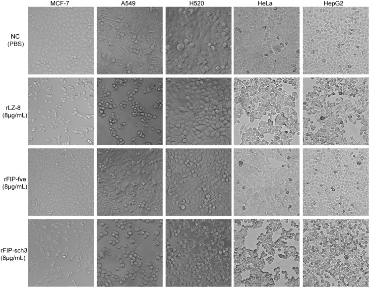
In this study, a novel FIP named FIP-sch3 has been identified and characterised. FIP-sch3 was identified in the ascomycete Stachybotrys chartarum, making it the second FIP to be identified outside the order of Basidiomycota. Recombinant FIP-sch3 (rFIP-shc3) was produced in Escherichia coli and purified using GST-affinity magnetic beads. The bioactive characteristics of FIP-sch3 were compared to those of well-known FIPs LZ-8 from Ganoderma lucidum and FIP-fve from Flammulina velutipes, which were produced and purified using the same method. The purified rFIP-sch3 exhibited a broad spectrum of anti-tumour activity in several types of tumour cells but had no cytotoxicity in normal human embryonic kidney 293 cells. Assays that were implemented to study these properties indicated that rFIP-sch3 significantly suppressed cell proliferation, induced apoptosis and inhibited cell migration in human lung adenocarcinoma A549 cells. The anti-tumour effects of rFIP-sch3 in A549 cells were comparable to those of rLZ-8, but they were significantly greater than those of rFIP-fve. Molecular assays that were built on real-time PCR further revealed potential mechanisms related to apoptosis and migration and that underlie phenotypic effects. These results indicate that FIP-shc3 has a unique anti-tumour bioactive profile, as do other FIPs, which provide a foundation for further studies on anti-tumour mechanisms. Importantly, this study also had convenient access to FIP-sch3 with potential human therapeutic applications.


Conflict of interest statement

The authors have declared that no competing interests exist.

Figures

Fig 1
Fig 1. Alignment of FIP-sch3 with other FIPs.
The LZ-8, FIP-gat, FIP-gja, FIP-gmi, FIP-gts, FIP-gap, FIP-cru, FIP-tvc, FIP-fve, FIP-vvo, FIP-nha, and FIP-ppl sequences came from G. lucidium, G. atrum, G. japonicum, G. microsporum, G. tsugae, G, applanatum, C. rutilus, T. versicolor, F. velutipes, V. volvacea, N. haematococca, and P. placenta, respectively. Shaded (solid bright yellow) residues match the consensus sequence exactly.
Fig 2
Fig 2. Evolutionary relationships of FIPs.
The evolutionary history was inferred using the Neighbour-Joining method [21]. The optimal tree with a sum of branch length = 2.61058163 is shown. The percentage of replicate trees in which the associated FIPs clustered together in the bootstrap test (1000 replicates) are shown next to the branches [22]. The tree is drawn to scale with branch lengths in the same units as those of the evolutionary distances that were used to infer the phylogenetic tree. The evolutionary distances were computed using the JTT matrix-based method [23] and have units of the number of amino acid substitutions per site. The analysis involved 13 amino acid sequences. All positions containing gaps and missing data were eliminated. There were a total of 110 positions in the final dataset. Evolutionary analyses were conducted in MEGA7.
Fig 3
Fig 3. Analysis of purified rFIPs.
(a) Gel filtration chromatography of rFIPs after the GST tag was removed. The standard markers noted in the figure are 66, 43, 29 and 17 kDa. Native LZ-8 and FIP-fve are dimers. The calculated molecular weight of FIP-sch3 as a dimer is 28.3 kDa. (b) SDS-PAGE results of purified rFIPs. The samples were purified using gel filtration chromatography. Purified proteins were resolved by 15% SDS-PAGE, stained with Coomassie brilliant blue R-250, and then destained with ethanol-acetic acid-water.
Fig 4
Fig 4. Tumour cytotoxicity assay after rFIP treatment.
MCF-7, A549, H520, HeLa and HepG2 cells were treated with 8 μg/mL rLZ-8, rFIP-fve or rFIP-sch3 for 24 h; NC is the control cells after PBS treatment.
Fig 5
Fig 5. Effect of rFIP-sch3 treatment on A549 and 293 cell viability.
(a) A549 and 293 cells were treated with different concentrations of rFIP-sch3 (1, 2, 4, 8, 16, 32 and 64 μg/mL) for 24 h. Cell viability was measured using a CCK assay, and the half-maximal inhibitory concentration (IC50) was calculated using SPSS 19 software. (b) Cytotoxicity comparison between rFIPs in A549 cells. A549 cells were treated with 8 μg/mL rFIPs for 24 h. * p < 0.05, and *** p < 0.001.
Fig 6
Fig 6. FACS analysis of apoptosis induced by rFIPs.
A549 cells were subjected to treatments including PBS (negative control, NC), rLZ-8 (8 μg/mL), rFIP-fve (8 μg/mL), or rFIP-sch3 (8 μg/mL) at 37°C in a CO2 incubator for 24 h. The treated cells were then trypsinized, counted, and stained with annexin V-EGFP and PI, and the data were analysed using FACS. All experiments were performed in duplicate. (a) The data of one representative experiment are presented. The upper right quadrant (UR) represents late apoptotic cells that were stained with Annexin V-EGFP and PI, and the lower right quadrant (LR) represents early apoptotic cells that were stained with Annexin V-EGFP. (b) Phase percentages for the UR quadrant and LR quadrant are depicted on the bar graph. Each bar represents the mean ± SD (n = 3). *p < 0.05; ***p < 0.001, compared to the NC.
Fig 7
Fig 7. Effects of rFIPs on cell migration.
A549 cells were cultured overnight and then treated with PBS (NC), rLZ-8 (8 μg/mL), rFIP-fve (8 μg/mL), or rFIP-sch3 (8 μg/mL). After incubation for 24 h, cell migration (wound healing) was monitored by microscopy.
Fig 8
Fig 8. Differentially expressed gene detection resulting from rFIP-sch3 treatment.
A549 cells were treated with 8 μg/mL rFIP-sch3 for 24 h. Relative mRNA levels of the ten genes (p53, BCL-2, Bax, CCR10, DRD1, DUSP1, ITPR1, TNFRSF6, JAK2 and SMPD1) were detected using qPCR, described as the means (error bars indicate SD) and represented as fold changes from controls.

References

    1. Wang XF, Su KQ, Bao TW, Cong WR, Chen YF, Li QZ, et al. Immunomodulatory effects of fungal proteins. Curr Top Nutraceutical Res. 2012;10:1–11.
    1. Li QZ, Wang XF, Zhou XW. Recent status and prospects of the fungal immunomodulatory protein family. Crit Rev Biotechnol. 2011;31(4):365–75. 10.3109/07388551.2010.543967 - DOI - PubMed
    1. Paaventhan P, Joseph JS, Seow SV, Vaday S, Robinson H, Chua KY, et al. A 1.7Å structure of Fve, a member of the new fungal immunomodulatory protein family. J Mol Biol. 2003;332(2):461–70. - PubMed
    1. Huang L, Sun F, Liang C, He YX, Bao R, Liu L, et al. Crystal structure of LZ-8 from the medicinal fungus Ganoderma lucidum. Proteins. 2008;75(2):524–7. - PubMed
    1. Ko JL, Hsu CI, Lin RH, Kao CL, Lin JY. A new fungal immunomodulatory protein, FIP-fve isolated from the edible mushroom, Flammulina velutipes and its complete amino acid sequence. Eur J Biochem. 1995;228(2):244–9. - PubMed

MeSH terms