. 2016 Dec 20;11(12):e0168617.
doi: 10.1371/journal.pone.0168617. eCollection 2016.

The C-Terminal SynMuv/DdDUF926 Domain Regulates the Function of the N-Terminal Domain of DdNKAP

Affiliations

The C-Terminal SynMuv/DdDUF926 Domain Regulates the Function of the N-Terminal Domain of DdNKAP

Bhagyashri D Burgute et al. PLoS One. .

Abstract

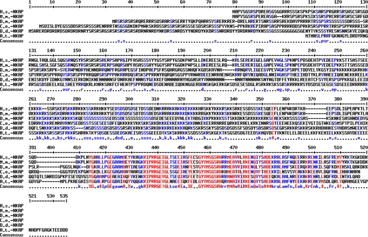
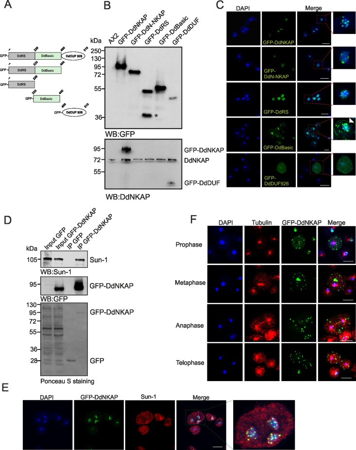
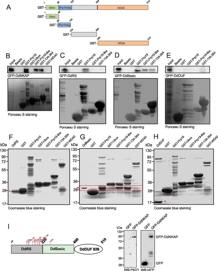
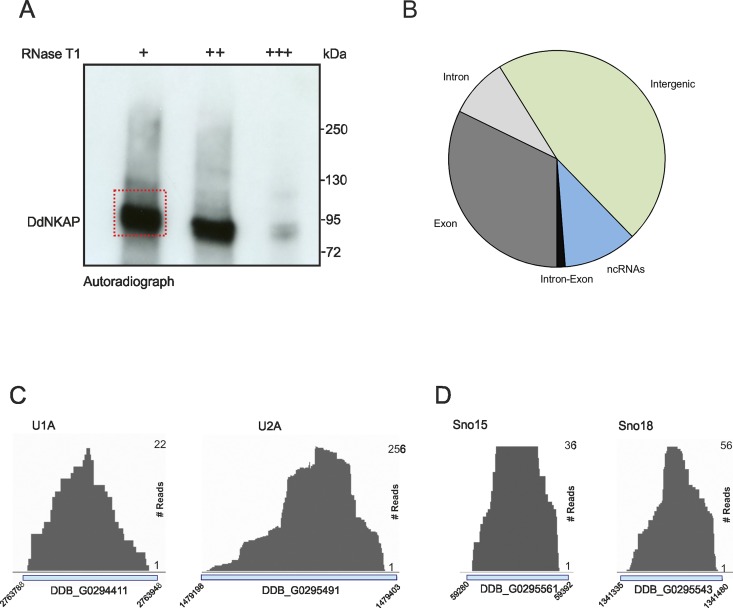
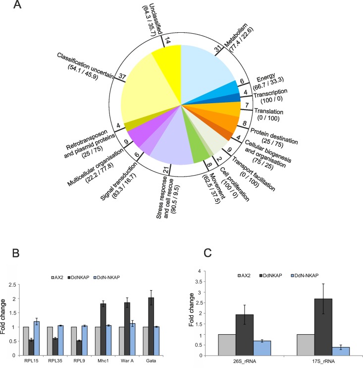
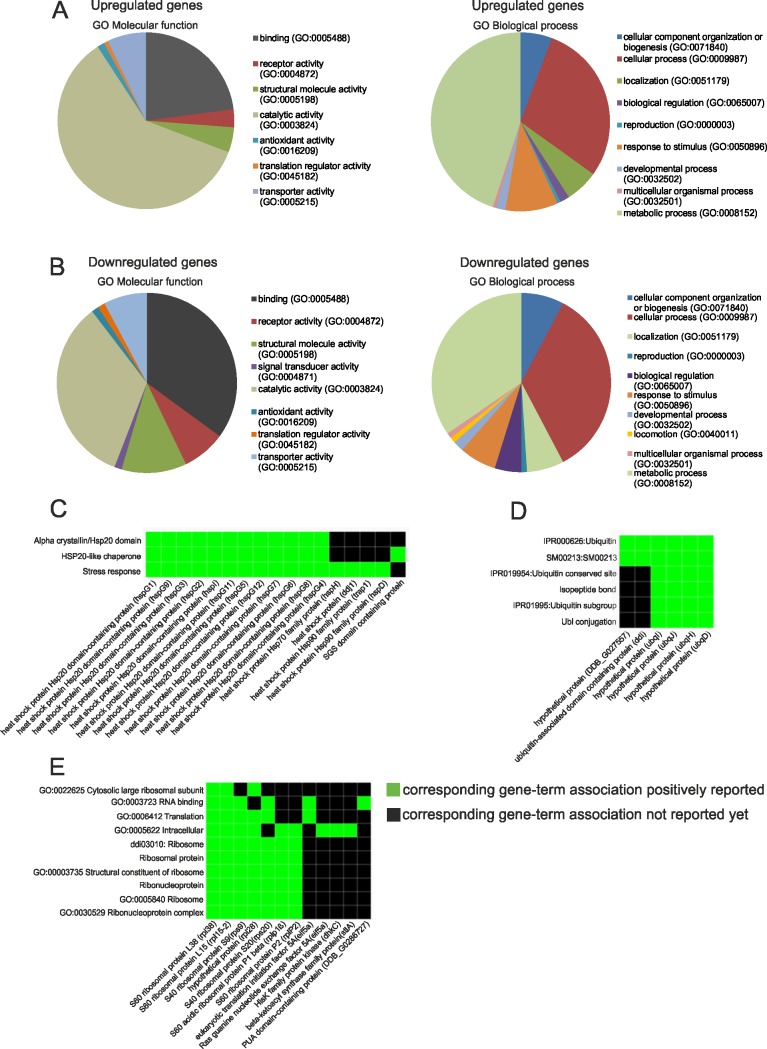
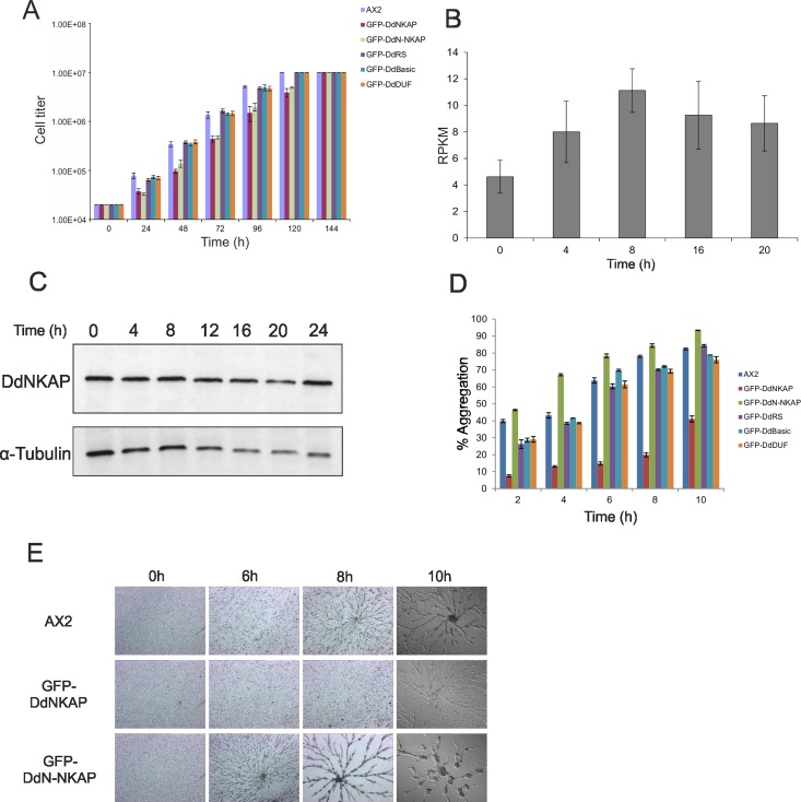
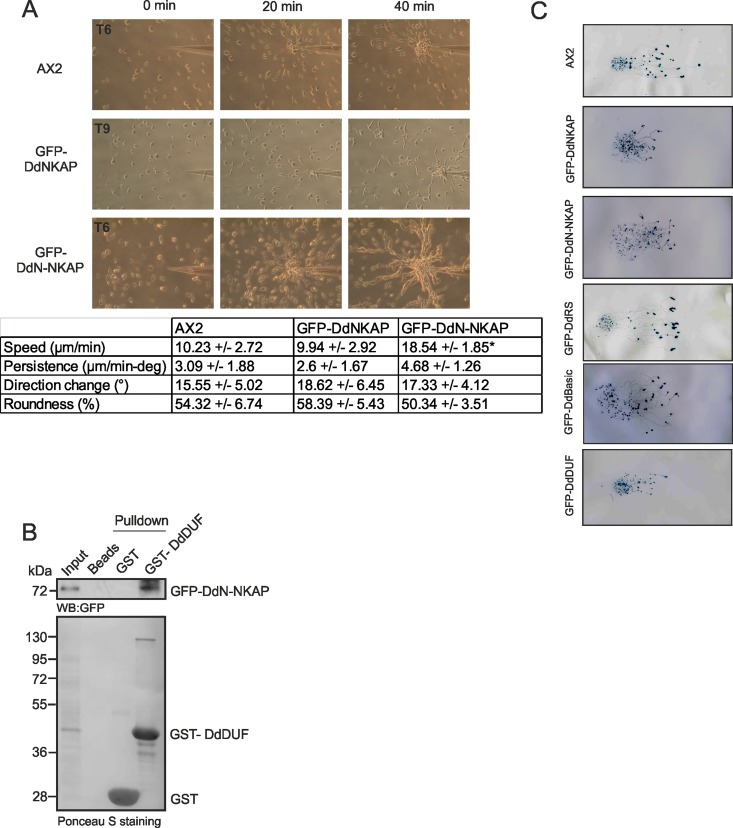
NKAP (NF-κB activating protein) is a highly conserved SR (serine/arginine-rich) protein involved in transcriptional control and splicing in mammals. We identified DdNKAP, the Dictyostelium discoideum ortholog of mammalian NKAP, as interacting partner of the nuclear envelope protein SUN-1. DdNKAP harbors a number of basic RDR/RDRS repeats in its N-terminal domain and the SynMuv/DUF926 domain at its C-terminus. We describe a novel and direct interaction between DdNKAP and Prp19 (Pre mRNA processing factor 19) which might be relevant for the observed DdNKAP ubiquitination. Genome wide analysis using cross-linking immunoprecipitation-high-throughput sequencing (CLIP-seq) revealed DdNKAP association with intergenic regions, exons, introns and non-coding RNAs. Ectopic expression of DdNKAP and its domains affects several developmental aspects like stream formation, aggregation, and chemotaxis. We conclude that DdNKAP is a multifunctional protein, which might influence Dictyostelium development through its interaction with RNA and RNA binding proteins. Mutants overexpressing full length DdNKAP and the N-terminal domain alone (DdN-NKAP) showed opposite phenotypes in development and opposite expression profiles of several genes and rRNAs. The observed interaction between DdN-NKAP and the DdDUF926 domain indicates that the DdDUF926 domain acts as negative regulator of the N-terminus.


Conflict of interest statement

The authors have declared that no competing interests exist.

Figures

Fig 1
Fig 1. Sequence conservation among eukaryotic NKAP proteins.
Sequence alignment of NKAP from different species using the multalin sequence alignment. Abbreviations: H.s.: Homo sapiens; M.m.: Mus musculus; D.m.: Drosophila melanogaster; C.e.: Caenorhabditis elegans; A.th.: Arabidopsis thaliana; D.d.: Dictyostelium discoideum. Sequence IDs are as follows: H.s. (NP_078804), M.m. (NP_080213), D.m. (AAF56669), C.e. (CCD68576), A.th. (AT4G02720) and D.d. (XP_645847). Highly conserved amino acids are shown in red, less conserved in blue and and neutral ones in black, respectively. Consensus symbols are “!”, I or V; $, L or M; %, for Y; #, any one of N, D, Q, E. B.
Fig 2
Fig 2. Localization of full-length and truncated versions of GFP-DdNKAP.
(A) The figure depicts the constructs used in the analysis. The DdRS domain, the basic domain and the DUF926 domain of DdNKAP are shown schematically. (B) Lysates of AX2 and of full-length and truncated versions of GFP-DdNKAP expressing cells were immunoblotted and probed using mAb K3-184-2 for GFP and mAb K79-232-2 for DdNKAP. The asterisk marks a degradation product of GFP-DdRS. (C) Full length DdNKAP, the DdN-NKAP, DdRS, DdBasic and DdDUF domains were tagged with N-terminal GFP and expressed in AX2 cells that were fixed and stained with DAPI. Scale bar, 10 μm. The red frames in the right images mark the regions which are shown at higher magnifications on the right. (D) Co-immunoprecipitation of DdNKAP and SUN1. Immunoprecipitation was performed using GFP-Trap beads followed by immunoblotting with mAb K55-432-2 (Sun-1) and mAb K3-184-2 (GFP). The Ponceau S stained membrane is shown at the bottom. (E) AX2 cells expressing GFP-DdNKAP were fixed with methanol and labeled with mAb K55-432-2 for Sun-1. DNA was stained with DAPI. Scale bar, 10 μm. (F) Cells expressing GFP-DdNKAP (green) were synchronized using nocodazole to block progression of the cell cycle and then released and fixed using cold methanol. Tubulin staining (red) using rat mAb YL1/2 was used to identify mitotic cells. Nuclei (blue) were stained with DAPI. Bar, 10 μm.
Fig 3
Fig 3. Interaction of DdNKAP and Prp19.
(A) Schematic representation of the Prp19 domain structure and Prp19 constructs used in this study. (B, C, D, E) GST tagged full length Prp19, the Prp19-like and the 134–264 domains pull down GFP-DdNKAP and GFP-DdBasic recognized by mAb K184-3. GST-Prp19 polypeptides and GST were visualized by Ponceau S staining. Proteins were separated by SDS-PAGE (12% acrylamide). The GFP-DdBasic domain interacts with GST-Prp19, GST-Prp19-like and GST-134-264. (F, G, H) The GST tagged full length Prp19 and truncated Prp19 proteins were incubated with bacterially expressed, purified and thrombin cleaved DdRS (F), DdBasic (G) and DdDUF (H). Beads were washed and proteins were separated by SDS-PAGE (15% acrylamide) and visualized by Coomassie Blue staining. DdRS, DdBasic and DdDUF are marked with red asterisks; the red box denotes the direct interaction of DdBasic with GST tagged full length Prp19, Prp19-like and 134–264. (I) The DdRS domain carries several predicted potential ubiquitination sites (indicated in red). GFP and GFP-DdNKAP were immunoprecipitated by GFP-Trap beads. The ubiquitinated DdNKAP was detected by western blot with anti-ubiquitin P4D1 mAb and the immunoprecipitation of GFP-DdNKAP and GFP was monitored by mAb K3-184-2.
Fig 4
Fig 4. Global analysis of DdNKAP binding to RNA by CLIPseq.
(A) Autoradiograph of cross-linked DdNKAP-RNA complexes using denaturing gel electrophoresis and membrane transfer. RNA was partially digested using increasing concentrations of RNase T1. The marked area was cut from the membrane and subjected to protocols for the isolation of RNA, which was further used for RNA-seq. Positions of proteins from the molecular mass standard (kDa) are shown on the right. (B) Percentage of uniquely identified genes in CLIPseq with their respective genomic features. CLIP tags are mapped to exons, introns, intergenic regions and non-coding RNAs according to the gene predictions in dictyBase (http://dictybase.org/). (C,D) DdNKAP association with U1, U2, sno15 and sno18. The dictybase gene ID and coordinates are mentioned at the x-axis of each plot. CLIP-seq reads are mentioned at the right side of each plot.
Fig 5
Fig 5. Transcriptional profile of differentially regulated genes in DdNKAP overexpressors.
(A) Pie diagram showing the functional classification according to the yeast classification scheme of the differentially regulated genes. The values in the bracket indicate the percentage of up- and down-regulated genes, respectively, in GFP-DdNKAP expressing cells in comparison to AX2. The values inside the circle indicate the total number of differentially regulated genes in the respective processes. (B) Confirmation of the differential regulation of selected genes by qRT-PCR. Three down-regulated (RPL15, RPL35, RPL9) and three-upregulated (Mhc1, WarA, Gata) genes were chosen to confirm the microarray data. The experiment was performed in technical quadruplets and biological triplicates. (C) Analysis of rRNA expression in DdNKAP mutants by qRT-PCR. Three biological replicates with four technical replicates each were performed.
Fig 6
Fig 6. RNAseq analysis of DdNKAP overexpressors and AX2 cells using GO Molecular function and GO Biological process of differentially expressed genes.
Upregulated (A) and downregulated (B) genes were classified for Gene ontology molecular function and biological process using the PANTHER classification system. (C, D, E) Upregulated and downregulated genes were applied to DAVID to identify functional ontological groups. Shown are specific heat-map examples: (C) Stress response, (D) Ubiquitin, (E) Ribosomal proteins. The heat-maps are derived from the report generated by DAVID, and are an annotated-term-focused view which lists annotated genes and their category. Green represents corresponding gene-term association positively reported. The black represents corresponding gene-term association not reported yet. All results passed the default thresholds, to ensure that only statistical significant groups are displayed.
Fig 7
Fig 7. Growth and development of GFP-DdNKAP mutants.
(A) Growth of AX2 and AX2 expressing GFP-DdNKAP mutants in shaking culture. The cell numbers were determined every 24 hours. Mean values and standard deviations of growth curves from three independent experiments are shown. (B) Expression profile of DdNKAP in vegetative and developing D. discoideum cells was extracted from dictyBase (http://dictybase.org/). Y-axis represents DdNKAP reads per kb per million mapped reads (RPKM), while the X-axis shows developmental time points (h). (C) Analysis of DdNKAP expression in AX2 using mAb K79-232-2and YL1/2 for tubulin as control. (D) AX2 and GFP-DdNKAP mutants were allowed to aggregate in Soerensen phosphate buffer in shaking suspension. Samples at different time points (2, 4, 6, 8, 10 h) during aggregation were taken and the aggregate formation (given in %) was scored by measuring the absorbance of the suspension at 600 nm. Mean values and standard deviations of growth curves from two independent experiments are shown. (E) Development in submerged culture. Cells (AX2, GFP-DdNKAP and GFP-DdN-NKAP) at equal densities (2 x 10⁵ cells/cm2) were starved submerged in Soerensen buffer on plastic petri dishes and monitored for aggregation. Images were taken every hour (10X magnification) until 10 hours of starvation. Images of the indicated time points are shown.
Fig 8
Fig 8. Motility analysis of GFP-DdNKAP mutants.
(A) Chemotactic motility. Growth-phase cells were harvested, washed and developed for 6 h (AX2, GFP-DdN-NKAP) or 9 h (GFP-DdNKAP) in shaking suspension under starvation conditions to render them aggregation competent before they were challenged with cAMP in a chemotaxis assay. Time-lapse images were captured and processed. In the table the data for speed, persistence, direction change and roundness are given. Cells were recorded, and after tracing of the cells, the centroid of the cells was determined by computer-assisted analysis (DIAS). This allows calculations of speed, roundness (ratio of the long and short axis of the cell), and direction change. Persistence is a measure of movement in the direction of the path; direction change represents the average change of angle between frames in the direction of movement. The data shown are derived from five independent experiments. The data from 10 cells each were used for statistic evaluation. The difference in speed of AX2 and GFP-DdNKAP was significant (*p<0.01). (B) GST tagged DdDUF pull down of GFP-DdN-NKAP recognized by mAb GFP K184-3. GST-DdDUF and GST (control) were visualized by Ponceau S staining. (C) Wild type AX2 and DdNKAP mutant cells were harvested at the log phase of growth and resuspended at 1 x 10⁸ cells/ml and 10 μl containing 1x10⁶ cells were placed at the center of water agar plates. The plates were then placed in a dark box containing a slit of 3 mm and incubated at 21°C for 48 h. The slugs were transferred to nitrocellulose membranes and stained with amido black and studied for their movement towards the light source which was located on the right.

References

    1. Chen D, Li Z, Yang Q, Zhang J, Zhai Z, Shu HB. Identification of a nuclear protein that promotes NF-kappaB activation. Biochem Biophys Res Commun. 2003;310: 720–724. - PubMed
    1. Poulin G, Dong Y, Fraser AG, Hopper NA, Ahringer J. Chromatin regulation and sumoylation in the inhibition of Ras-induced vulval development in Caenorhabditis elegans. EMBO J. 2005;24: 2613–2623. 10.1038/sj.emboj.7600726 - DOI - PMC - PubMed
    1. Burgute BD, Peche VS, Steckelberg A-L, Glöckner G, Gaßen B, Gehring NH, et al. NKAP is a novel RS-related protein that interacts with RNA and RNA binding proteins. Nucleic Acids Res. 2013; 1–17. - PMC - PubMed
    1. Pajerowski AG, Nguyen C, Aghajanian H, Shapiro MJ, Shapiro VS. NKAP is a transcriptional repressor of notch signaling and is required for T cell development. Immunity. 2009;30: 696–707. 10.1016/j.immuni.2009.02.011 - DOI - PMC - PubMed
    1. Pajerowski AG, Shapiro MJ, Gwin K, Sundsbak R, Nelson-Holte M, Medina K, et al. Adult hematopoietic stem cells require NKAP for maintenance and survival. Blood. 2010;116: 2684–2693. 10.1182/blood-2010-02-268391 - DOI - PMC - PubMed

MeSH terms

LinkOut - more resources