. 2016 Dec 20;11(12):e0168794.
doi: 10.1371/journal.pone.0168794. eCollection 2016.

Ganglioside Profiling of the Human Retina: Comparison with Other Ocular Structures, Brain and Plasma Reveals Tissue Specificities

Affiliations

Ganglioside Profiling of the Human Retina: Comparison with Other Ocular Structures, Brain and Plasma Reveals Tissue Specificities

Estelle Sibille et al. PLoS One. .

Abstract

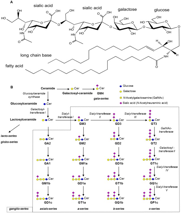
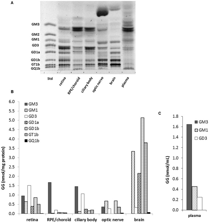
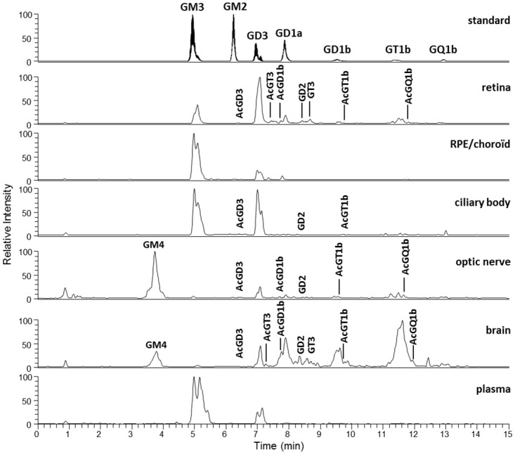
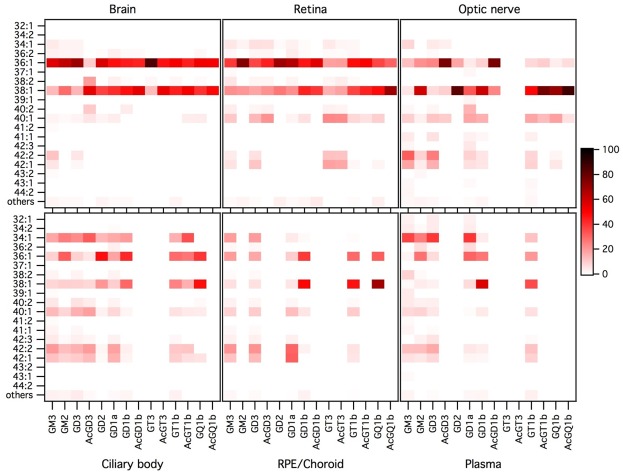
Gangliosides make a wide family of glycosphingolipids, highly heterogeneous in both the ceramide moiety and the oligosaccharide chain. While ubiquitously expressed in mammalian tissues, they are particularly abundant in the brain and the peripheral nervous system. Gangliosides are known to play a crucial role in the development, maintenance and functional integrity of the nervous system. However, the expression and roles of gangliosides in the retina, although often considered as a window on the brain, has been far less studied. We performed an in-depth analysis of gangliosides of the human retina, especially using powerful LC/MS methods. We compared the pattern of ganglioside classes and ceramide molecular species of this tissue with other ocular structures and with brain and plasma in elderly human individuals. About a hundred of ganglioside molecular species among 15 distinct classes were detected illustrating the huge structural diversity of these compounds. The retina exhibited a very diverse ganglioside profile and shared several common features with the brain (prominence of tetraosylgangliosides, abundance of d20:1 long chain base and 18:0 fatty acid…). However, the retina stood out with the specific expression of GD3, GT3 and AcGT3, which further presented a peculiar molecular species distribution. The unique ganglioside pattern we observed in the human retina suggests that these ganglioside species play a specific role in the structure and function of this tissue. This lipidomic study, by highlighting retina specific ganglioside species, opens up novel research directions for a better understanding of the biological role of gangliosides in the retina.


Conflict of interest statement

The authors have declared that no competing interests exist.

Figures

Fig 1
Fig 1. Structure of gangliosides and general scheme of biosynthesis (after Masson et al. [1]).
A. GGs are made of a ceramide moiety composed of a long chain base (LCB) and a fatty acid (FA) whose alkyl chains may vary, and on which an oligosaccharide chain is branched. GD3 34:1 (based on LIPID MAPS, systematic name: NeuAcα2-8NeuAcα2-3Galβ1-4Glcβ-Cer) is given as an example. The ceramide is composed of sphingosine (d18:1) as LCB and stearic acid (18:0) as FA. The oligosaccharide chain is made of one glucose, one galactose and two sialic acid residues. B. The formation of GGs is catalyzed by the sequential action of glycosyltransferases (italic). The main GG classes of the ganglio-series are represented with their common names, according to Svennerholm [2].
Fig 2
Fig 2. Ganglioside profile of the retina and other ocular tissues, brain and plasma.
A. Resorcinol-stained HPTLC plate of GGs extracted from retina, retinal pigment epithelium (RPE)/choroid, ciliary body, optic nerve, brain and plasma. GG aliquots (4–7 independent samples) were pooled for each tissue type to represent a total of 10 nmol GG-sialic acid spotted per lane. A standard mixture of ganglio-series GGs (Std, 5 nmol sialic acid) was also spotted. The plate was developed in CHCl3/CH3OH/0.2% CaCl2 (55:45:10, v/v/v) and revealed with resorcinol reagent. B. Quantitative distribution of GG classes calculated from a standard curve of each GG class. Results are expressed in nmol GG/mg protein or nmol GG/mL plasma.
Fig 3
Fig 3. Precursor ion chromatograms of the standard mixture, retina and other ocular tissues, brain and plasma.
A representative sample of each tissue was obtained by pooling an aliquot of every GG extract samples (4–7) for each tissue type. The QqQ mass spectrometer was operated in the precursor ion scanning of the characteristic fragment of GG, N-Acetylneuraminic acid, at m/z 290, in the negative ionization mode.
Fig 4
Fig 4. Molecular species profile of the ganglioside classes of the retina and other ocular tissues, brain and plasma.
Data (peak areas) were obtained by operating the QqQ mass spectrometer in negative SRM mode. The proportion of each ceramide species was expressed, as percentage, relatively to the sum of all detected species in its specific GG class, every GG class being considered separately. Molecular species accounting for less than 1% were grouped under the category “others”. Results were expressed as mean of 4 to 7 independent samples for each tissue, injected three times. The color intensity of a spot on the graph is proportional to the mean percentage of the ceramide species considered within a GG class.
Fig 5
Fig 5. Ceramide proportions in retinal GG classes (A, C, E) and in retinal and brain GT3 and AcGT3 (B, D and F).
Means of 4–7 independent samples and 95% confidence ellipses are represented on ternary diagrams for various groups of ceramides: (36:1 + 38:1), (40:1 + 42:2 + 42:1) ceramides and other molecular species (A and B); ceramides with 1, 2 and 3 unsaturations (C and D); ceramides with 34 carbons or less, 42 carbons or more and other molecular species (E and F). Axes from 0 to 1 for the three variables are represented on B and the proportions for retinal GT3 are shown as an example.

Similar articles

Cited by

References

    1. Masson EA, Sibille E, Martine L, Chaux-Picquet F, Bretillon L, Berdeaux O. Apprehending ganglioside diversity: a comprehensive methodological approach. J Lipid Res. 2015;56(9):1821–35. 10.1194/jlr.D060764 - DOI - PMC - PubMed
    1. Svennerholm L. Ganglioside designation. Adv Exp Med Biol. 1980;125:11 - PubMed
    1. Degroote S, Wolthoorn J, van Meer G. The cell biology of glycosphingolipids. Semin Cell Dev Biol. 2004;15(4):375–87. 10.1016/j.semcdb.2004.03.007 - DOI - PubMed
    1. Yu RK, Yanagisawa M., Ariga T.. Glycosphingolipid Structures In: Kamerling JP, editor. Comprehensive Glycoscience. 1: Elsevier; 2007. p. 73–120.
    1. Simons K, Ikonen E. Functional rafts in cell membranes. Nature. 1997;387(6633):569–72. 10.1038/42408 - DOI - PubMed

LinkOut - more resources