

Autophagy: an adaptive physiological countermeasure to cellular senescence and ischaemia/reperfusion-associated cardiac arrhythmias
- PMID: 27997746
- PMCID: PMC5431132
- DOI: 10.1111/jcmm.13053
Autophagy: an adaptive physiological countermeasure to cellular senescence and ischaemia/reperfusion-associated cardiac arrhythmias
Abstract
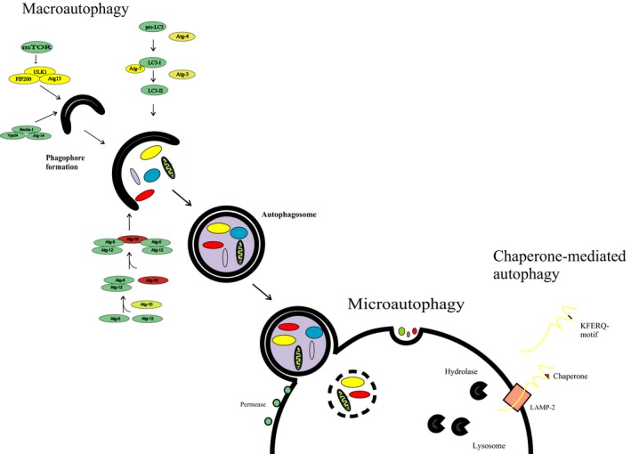
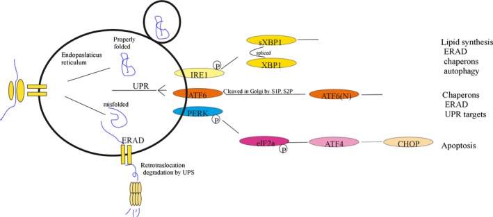
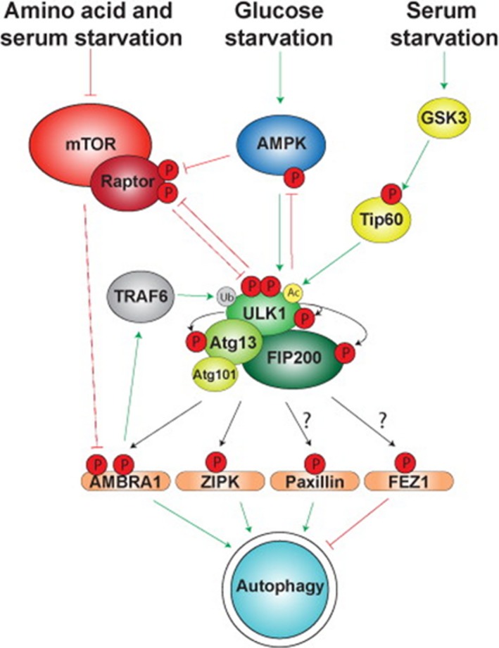
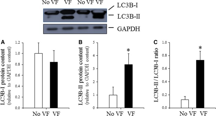
Oxidative stress placed on tissues that involved in pathogenesis of a disease activates compensatory metabolic changes, such as DNA damage repair that in turn causes intracellular accumulation of detritus and 'proteotoxic stress', leading to emergence of 'senescent' cellular phenotypes, which express high levels of inflammatory mediators, resulting in degradation of tissue function. Proteotoxic stress resulting from hyperactive inflammation following reperfusion of ischaemic tissue causes accumulation of proteinaceous debris in cells of the heart in ways that cause potentially fatal arrhythmias, in particular ventricular fibrillation (VF). An adaptive response to VF is occurrence of autophagy, an intracellular bulk degradation of damaged macromolecules and organelles that may restore cellular and tissue homoeostasis, improving chances for recovery. Nevertheless, depending on the type and intensity of stressors and inflammatory responses, autophagy may become pathological, resulting in excessive cell death. The present review examines the multilayered defences that cells have evolved to reduce proteotoxic stress by degradation of potentially toxic material beginning with endoplasmic reticulum-associated degradation, and the unfolded protein response, which are mechanisms for removal from the endoplasmic reticulum of misfolded proteins, and then progressing through the stages of autophagy, including descriptions of autophagosomes and related vesicular structures which process material for degradation and autophagy-associated proteins including Beclin-1 and regulatory complexes. The physiological roles of each mode of proteotoxic defence will be examined along with consideration of how emerging understanding of autophagy, along with a newly discovered regulatory cell type called telocytes, may be used to augment existing strategies for the prevention and management of cardiovascular disease.
Keywords: autophagy; cardiovascular system; proteotoxic stress.
© 2016 The Authors. Journal of Cellular and Molecular Medicine published by John Wiley & Sons Ltd and Foundation for Cellular and Molecular Medicine.
Figures
References
-
- Haines DD, Bak I, Ferdinandy P, et al Cardioprotective effects of the calcineurin inhibitor FK506 and the PAF receptor antagonist and free radical scavenger, EGb 761, in isolated ischemic/reperfused rat hearts. J Cardiovasc Pharmacol. 2000; 35: 37–44. - PubMed
-
- Hearse DJ, Tosaki A. Free radicals and calcium: simultaneous interacting triggers as determinants of vulnerability to reperfusion‐induced arrhythmias in the rat heart. J Mol Cell Cardiol. 1988; 20: 213–23. - PubMed
-
- Guarini G, Huqi A, Capozza P, et al Therapy against ischemic injury. Curr Pharm Des. 2013; 19: 4597–621. - PubMed
-
- Hearse DJ, Tosaki A. Free radicals and reperfusion‐induced arrhythmias: protection by spin trap agent PBN in the rat heart. Circ Res. 1987; 60: 375–83. - PubMed
-
- Yildirim F, Iskesen I, Kurdal AT, et al Is “Attenuation of Oxidative Stress” helpful to understand the mechanism of remote ischemic preconditioning in cardiac surgery? J Cardiothorac Vasc Anesth. 2016; 30: 134–40. - PubMed
Publication types
MeSH terms
Substances
LinkOut - more resources
Full Text Sources
Other Literature Sources
Medical
