. 2017 Apr;56(4):521-531.
doi: 10.1165/rcmb.2016-0225OC.

Glucose Transporter 1-Dependent Glycolysis Is Increased during Aging-Related Lung Fibrosis, and Phloretin Inhibits Lung Fibrosis

Affiliations

Glucose Transporter 1-Dependent Glycolysis Is Increased during Aging-Related Lung Fibrosis, and Phloretin Inhibits Lung Fibrosis

Soo Jung Cho et al. Am J Respir Cell Mol Biol. 2017 Apr.

Abstract

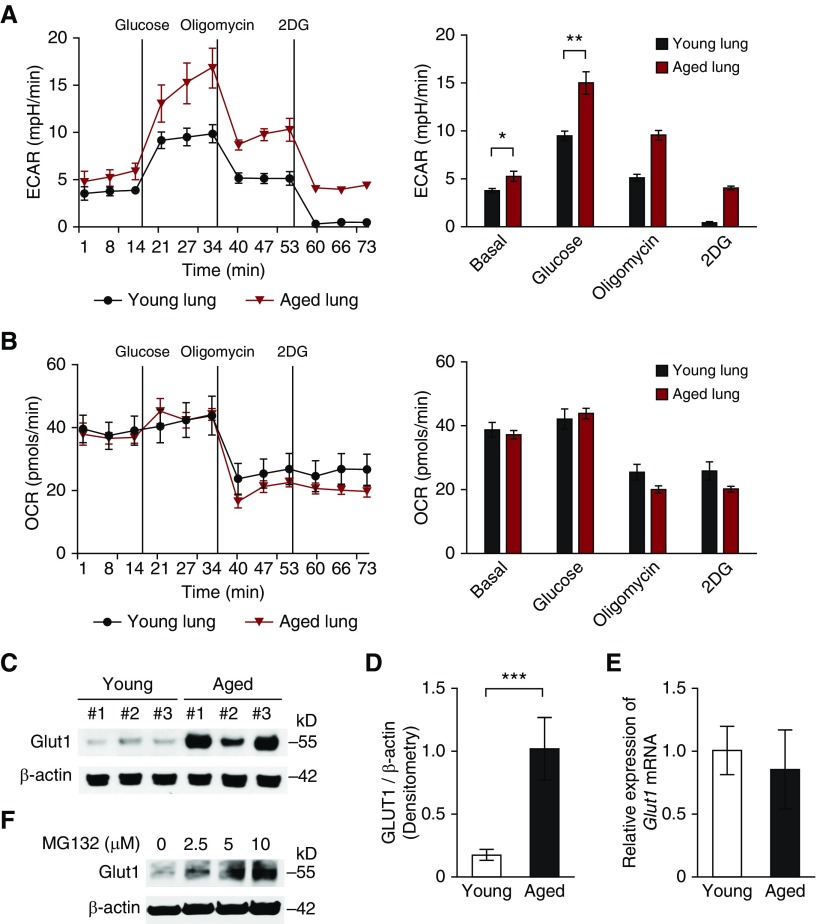
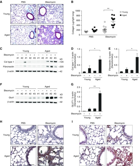
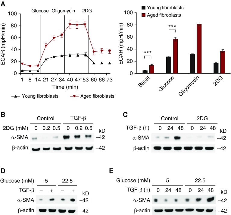
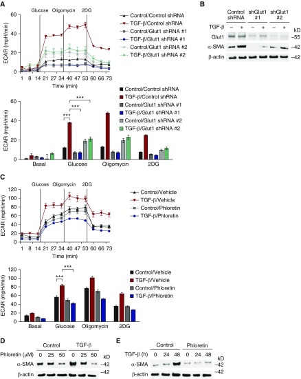
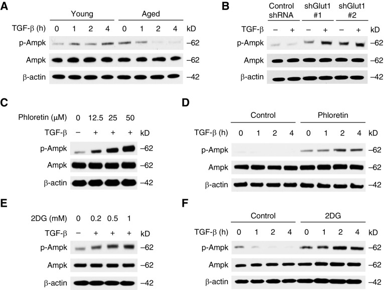
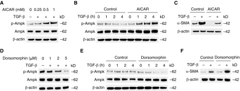
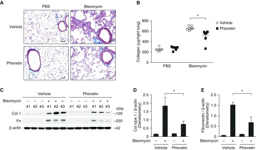
Aging is associated with metabolic diseases such as type 2 diabetes mellitus, cardiovascular disease, cancer, and neurodegeneration. Aging contributes to common processes including metabolic dysfunction, DNA damage, and reactive oxygen species generation. Although glycolysis has been linked to cell growth and proliferation, the mechanisms by which the activation of glycolysis by aging regulates fibrogenesis in the lung remain unclear. The objective of this study was to determine if glucose transporter 1 (GLUT1)-induced glycolysis regulates age-dependent fibrogenesis of the lung. Mouse and human lung tissues were analyzed for GLUT1 and glycolytic markers using immunoblotting. Glycolytic function was measured using a Seahorse apparatus. To study the effect of GLUT1, genetic inhibition of GLUT1 was performed by short hairpin RNA transduction, and phloretin was used for pharmacologic inhibition of GLUT1. GLUT1-dependent glycolysis is activated in aged lung. Genetic and pharmacologic inhibition of GLUT1 suppressed the protein expression of α-smooth muscle actin, a key cytoskeletal component of activated fibroblasts, in mouse primary lung fibroblast cells. Moreover, we demonstrated that the activation of AMP-activated protein kinase, which is regulated by GLUT1-dependent glycolysis, represents a critical metabolic pathway for fibroblast activation. Furthermore, we demonstrated that phloretin, a potent inhibitor of GLUT1, significantly inhibited bleomycin-induced lung fibrosis in vivo. These results suggest that GLUT1-dependent glycolysis regulates fibrogenesis in aged lung and that inhibition of GLUT1 provides a potential target of therapy of age-related lung fibrosis.

Keywords: bleomycin; glucose metabolism; idiopathic pulmonary fibrosis.


Figures

Figure 1.
Figure 1.
Glucose transporter 1 (GLUT1)–associated glycolysis is activated in aged lung. (A) The extracellular acidification rate (ECAR) was measured in young (2-month-old) and aged (18-month-old) cells isolated from whole lung (5 × 104 cells/well). Data are presented as mean ± SEM. *P < 0.05, **P < 0.01 by ANOVA. (B) The oxygen consumption rate (OCR) was measured in young and aged cells isolated from whole lung (5 × 104 cells/well). (C) Immunoblot assay of GLUT1 for lung lysates from young and aged mice. β-actin served as the standard. (D) Densitometry of immunoblot assay for GLUT1 in young and aged lung cells. Data are presented as mean ± SEM. ***P < 0.001 by ANOVA. (E) Analysis of GLUT1 mRNA expression in young and aged lung cells. Data are presented as mean ± SEM. Results are representative of three or more independent experiments. (F) Immunoblot assay for GLUT1 in aged lung fibroblasts treated with MG132 (0, 2.5, 5, 10 μM) for 1 hour. β-actin served as the standard. 2DG, 2-deoxyglucose.
Figure 2.
Figure 2.
Age-dependent lung fibrosis is associated with the expression of GLUT1 in vivo. Young and aged mice were instilled with bleomycin (0.01 mg/mouse via oropharyngeal aspiration) for 14 days. (A) Representative lung sections of young and aged mice stained with Masson trichrome staining. Scale bars, 200 μm. (B) Total lung collagen (Col) was quantified by Sircol assay (young/PBS, n = 11; aged/PBS, n = 9; young/bleomycin, n = 14, aged/bleomycin, n = 14). Data are presented as mean ± SEM. **P < 0.01 by ANOVA. (C) Immunoblot assay for Col type 1 and fibronectin in young and aged lung tissue lysates. β-actin served as the standard. (D) Densitometry of immunoblot assay for Col type 1 in young and aged lung tissue lysates. Densitometry of immunoblot assay for Col type 1 in young and aged lung tissue lysates. Data are presented as mean ± SEM. *P < 0.05 by ANOVA. (E) Densitometry of immunoblot assay for fibronectin in young and aged lung tissue lysates. Densitometry of immunoblot assay for fibronectin in young and aged lung tissue lysates. Data are presented as mean ± SEM. *P < 0.05 by ANOVA. (F) Immunoblot assay for GLUT1 in young and aged lung tissue lysates. β-actin served as the standard. (G) Densitometry of immunoblot assay for GLUT1 in young and aged lung tissue lysates. Densitometry of immunoblot assay for GLUT1 in young and aged lung tissue lysates. Data are presented as mean ± SEM. **P < 0.01 by ANOVA. Immunohistochemical staining of (H) GLUT1 and (I) α-smooth muscle actin in aged lung reveals enhanced signal in fibrotic foci (open arrow), epithelial cells (gray arrow), and inflammatory cells (solid arrow). Scale bars, 200 μm. Results are representative of three or more independent experiments.
Figure 3.
Figure 3.
Glycolysis is a critical metabolic pathway for the activation of lung fibroblasts. (A) ECAR was measured in young and aged lung fibroblasts (5 × 104 cells/well). Data are presented as mean ± SEM. ***P < 0.001 by ANOVA. (B) Immunoblot assay for α-smooth muscle actin (α-SMA) in aged lung fibroblasts pretreated with 2DG (0.2, 0.5 mM) for 1 hour before transforming growth factor-β (TGF-β) (20 ng/ml, 48 h) stimulation. (C) Immunoblot assay for α-SMA in aged lung fibroblasts pretreated with 2DG (0.5 mM) for 1 hour before TGF-β (20 ng/ml, 0, 24, 48 h) stimulation. (D) Immunoblot assay for α-SMA in cell lysates from aged lung fibroblasts pretreated with different concentrations of glucose (5.0 and 22.5 mM) before stimulation with TGF-β (20 ng/ml, 48 h). (E) Immunoblot assay for α-SMA in cell lysates from aged lung fibroblasts pretreated with different concentrations of glucose (5.0, and 22.5 mM) before stimulation with TGF-β (20 ng/ml, 0, 24, 48 h). β-actin served as the standard. Results are representative of three or more independent experiments.
Figure 4.
Figure 4.
GLUT1-dependent glycolysis is critical for the activation of lung fibroblasts and lung fibrogenesis. (A) ECAR was measured in aged lung fibroblasts (4 × 104 cells/well) transduced with lentiviruses expressing nontarget short hairpin RNA (shRNA) (control shRNA) or two independent shRNAs for GLUT1 before TGF-β (20 ng/ml, 48 h) stimulation. Data are presented as mean ± SEM. ***P < 0.001 by ANOVA. (B) α-SMA in cell lysates from aged lung fibroblasts transduced with lentiviruses expressing nontarget shRNA (control shRNA) or two independent shRNA for GLUT1 before TGF-β (20 ng/ml, 4 h) stimulation. β-actin served as the standard. (C) ECAR was measured in cell lysates from aged lung fibroblasts (4 × 104 cells/well) pretreated with phloretin (0, 25, 50 nM) 1 hour before TGF-β (20 ng/ml, 48 h) stimulation. Data are presented as mean ± SEM. ***P < 0.001 by ANOVA. (D) Immunoblot assay for α-SMA in cell lysates from aged lung fibroblasts pretreated with phloretin (0, 25, 50 nM) 1 hour before TGF-β (20 ng/ml, 4 h) stimulation. (E) Immunoblot assay for α-SMA in cell lysates from aged lung fibroblasts pretreated with phloretin (25 nM) 1 hour before TGF-β (20 ng/ml, 0, 24, 48 h) stimulation. β-actin served as the standard. Results are representative of three or more independent experiments. shGlut1, GLUT1 specific short hairpin RNA.
Figure 5.
Figure 5.
Inhibition of GLUT1-dependent glycolysis induces AMP-activated protein kinase (AMPK) activation. (A) Immunoblot analysis for phosphorylated AMPK (p-AMPK) and AMPK in cell lysates from young and aged lung fibroblasts treated with TGF-β (5 ng/ml) for 1–4 hours. (B) Immunoblot analysis for p-AMPK and AMPK in cell lysates from aged lung fibroblasts transduced with lentiviruses expressing nontarget shRNA (control shRNA) or two independent shRNA for GLUT1 before TGF-β (5 ng/ml, 2 h) stimulation. (C) Immunoblot analysis for p-AMPK and AMPK in cell lysates from aged lung fibroblasts pretreated with phloretin (0, 12.5, 25, 50 nM) 1 hour before TGF-β (5 ng/ml, 2 h) stimulation. (D) Immunoblot analysis for p-AMPK and AMPK in cell lysates from aged lung fibroblasts pretreated with phloretin (25 nM) 1 hour before TGF-β (5 ng/ml, 0, 1, 2, 4 h) stimulation. (E) Immunoblot analysis for p-AMPK and AMPK in cell lysates from aged lung fibroblasts pretreated with 2DG (0.2, 0.5, 1 mM) for 1 hour before TGF-β (5 ng/ml, 2 h) stimulation. (F) Immunoblot analysis for p-AMPK and AMPK in cell lysates from aged lung fibroblasts pretreated with 2DG (0.5 mM) for 1 hour before TGF-β (5 ng/ml, 0, 1, 2, 4 h) stimulation. Results are representative of three or more independent experiments.
Figure 6.
Figure 6.
The phosphorylation of AMPK attenuates the activation of lung fibroblasts. (A) Immunoblot analysis for p-AMPK and AMPK in cell lysates from aged lung fibroblasts pretreated with 5-aminoimidazole-4-carboxamide 1-β-D-ribofuranoside (AICAR) (0, 0.25, 0.5, 1 mM) for 1 hour before TGF-β (5 ng/ml, 2 h) stimulation. (B) Immunoblot analysis for p-AMPK and AMPK in cell lysates from aged lung fibroblasts pretreated with AICAR (0.5 mM) for 1 hour before TGF-β (5 ng/ml, 0, 1, 2, 4 h) stimulation. (C) Immunoblot assay for α-SMA in cell lysates from aged lung fibroblasts pretreated with AICAR (0.5 mM) for 1 hour before TGF-β (20 ng/ml, 48 h) stimulation. (D) Immunoblot analysis for p-AMPK and AMPK in cell lysates from aged lung fibroblasts pretreated with dorsomorphin (0, 1, 2, 5 μM) for 1 hour before TGF-β (5 ng/ml, 2 h) stimulation. (E) Immunoblot analysis for p-AMPK and AMPK in cell lysates from aged lung fibroblasts pretreated with dorsomorphin (2 μM) for 1 hour before TGF-β (5 ng/ml, 0, 1, 2, 4 h) stimulation. (F) Immunoblot assay for α-SMA in cell lysates from aged lung fibroblasts pretreated with dorsomorphin (2 μM) for 1 hour before TGF-β (20 ng/ml, 48 h) stimulation. β-actin served as the standard. Results are representative of three or more independent experiments.
Figure 7.
Figure 7.
Pharmacologic inhibition of GLUT1 attenuates lung fibrogenesis in vivo. Aged mice were treated with intraperitoneal injection of phloretin (10 mg/kg, every other day) after oropharyngeal bleomycin instillation (0.01 mg/mouse). (A) Representative lung sections of aged mice stained with Masson trichrome staining. Scale bars, 200 μm. (B) Total lung collagen was quantified by Sircol assay (PBS/vehicle, n = 5; PBS/phloretin, n = 6; bleomycin/vehicle, n = 6; bleomycin/phloretin, n = 8). Data are presented as mean ± SEM. *P < 0.05 by ANOVA. (C) Immunoblot assay for collagen type 1, fibronectin (Fn) in tissue lysates from aged lung at baseline and also after bleomycin challenge. β-actin served as the standard. (D) Densitometry of immunoblot assay for collagen type 1 in aged lung at baseline and also after bleomycin challenge. Data are presented as mean ± SEM. *P < 0.05 by ANOVA. (E) Densitometry of immunoblot assay for Fn in young and aged lung tissue lysates. Data are presented as mean ± SEM. *P < 0.05 by ANOVA.

References

    1. Raghu G, Weycker D, Edelsberg J, Bradford WZ, Oster G. Incidence and prevalence of idiopathic pulmonary fibrosis. Am J Respir Crit Care Med. 2006;174:810–816. - PubMed
    1. Fell CD, Martinez FJ, Liu LX, Murray S, Han MK, Kazerooni EA, Gross BH, Myers J, Travis WD, Colby TV, et al. Clinical predictors of a diagnosis of idiopathic pulmonary fibrosis. Am J Respir Crit Care Med. 2010;181:832–837. - PMC - PubMed
    1. Raghu G, Collard HR, Egan JJ, Martinez FJ, Behr J, Brown KK, Colby TV, Cordier JF, Flaherty KR, Lasky JA, et al. ATS/ERS/JRS/ALAT Committee on Idiopathic Pulmonary Fibrosis. An official ATS/ERS/JRS/ALAT statement: idiopathic pulmonary fibrosis: evidence-based guidelines for diagnosis and management. Am J Respir Crit Care Med. 2011;183:788–824. - PMC - PubMed
    1. Wynn TA, Ramalingam TR. Mechanisms of fibrosis: therapeutic translation for fibrotic disease. Nat Med. 2012;18:1028–1040. - PMC - PubMed
    1. Stout-Delgado HW, Cho SJ, Chu SG, Mitzel DN, Villalba J, El-Chemaly S, Ryter SW, Choi AM, Rosas IO. Age-dependent susceptibility to pulmonary fibrosis is associated with NLRP3 inflammasome activation. Am J Respir Cell Mol Biol. 2016;55:252–263. - PMC - PubMed

Publication types