

Rare presentation of subcapsular hepatic steatosis in a woman with uncontrolled diabetes without peritoneal dialysis: a case report
- PMID: 27998312
- PMCID: PMC5175298
- DOI: 10.1186/s13256-016-1152-8
Rare presentation of subcapsular hepatic steatosis in a woman with uncontrolled diabetes without peritoneal dialysis: a case report
Abstract
Background: Subcapsular hepatic steatosis is a rare atypical pattern of fatty deposition of the liver reported in patients with diabetic nephropathy receiving peritoneal dialysis with intraperitoneal insulin. To date, there has been only one pediatric and zero adult cases of subcapsular hepatic steatosis with no history of continuous ambulatory peritoneal dialysis. We report the first published case of subcapsular hepatic steatosis in an adult diabetic patient without any history of peritoneal dialysis or evidence of chronic renal disease.
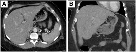
Case presentation: A 46-year-old Caucasian woman with type 2 diabetes mellitus without renal disease presented to our emergency department with vague abdominal symptoms and vomiting. Her blood glucose levels were poorly controlled with a range of 400 to 500 mg/dL. She was diagnosed as having subcapsular hepatic steatosis based on magnetic resonance imaging. Of note, after improved glucose control her subcapsular hepatic steatosis had nearly resolved.
Conclusions: Subcapsular hepatic steatosis has been exclusively described in patients with continuous ambulatory peritoneal dialysis and those on intraperitoneal insulin, except for one pediatric case, which was probably due to incorrect insulin administration. Our case demonstrates that a diagnosis of subcapsular hepatic diagnosis should not be restricted to those getting continuous ambulatory peritoneal dialysis, but rather expanded to all patients with uncontrolled blood glucose levels.
Keywords: CT; Case report; Diabetes; Hepatic steatosis; In and out-of-phase; MR; Subcapsular.
Figures


References
-
- Wanless IR, Bargman JM, Oreopoulos DG, Vas SI. Subcapsular steatonecrosis in response to peritoneal insulin delivery: a clue to the pathogenesis of steatonecrosis in obesity. Mod Pathol. 1989;2(2):69–74. - PubMed
-
- Baron RL, Gore RM. Textbook of Gastrointestinal Radiology. 2. Philadelphia: WB Saunders Company; 2000. pp. 1591–4.
Publication types
MeSH terms
Substances
LinkOut - more resources
Full Text Sources
Other Literature Sources
Medical
