. 2017 Feb 3;292(5):1573-1590.
doi: 10.1074/jbc.M116.769430. Epub 2016 Dec 20.

The Proprotein Convertase Subtilisin/Kexin Type 9-resistant R410S Low Density Lipoprotein Receptor Mutation: A NOVEL MECHANISM CAUSING FAMILIAL HYPERCHOLESTEROLEMIA

Affiliations

The Proprotein Convertase Subtilisin/Kexin Type 9-resistant R410S Low Density Lipoprotein Receptor Mutation: A NOVEL MECHANISM CAUSING FAMILIAL HYPERCHOLESTEROLEMIA

Delia Susan-Resiga et al. J Biol Chem. .

Abstract

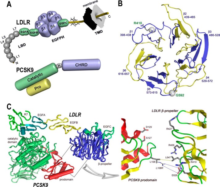
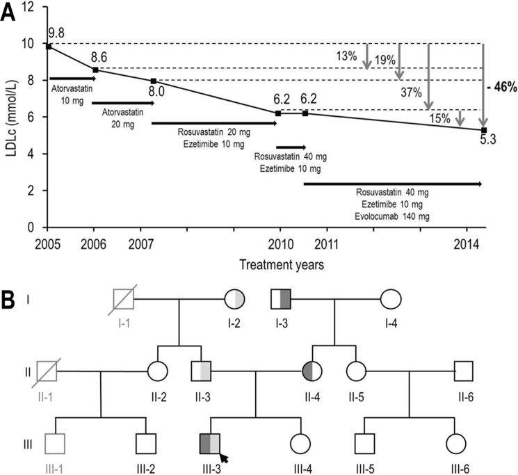
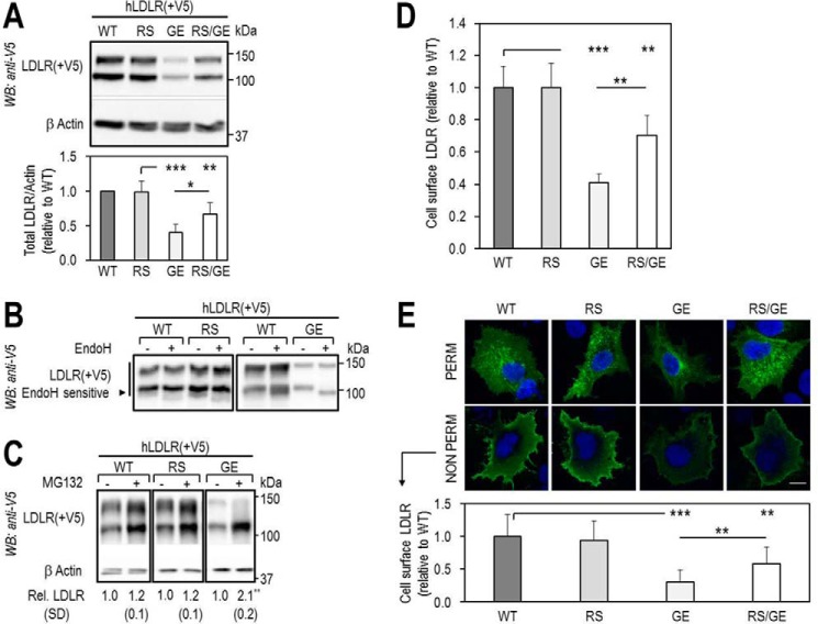
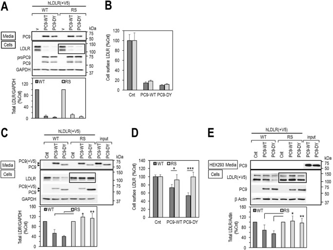
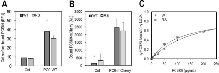
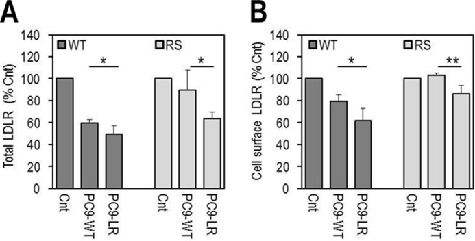
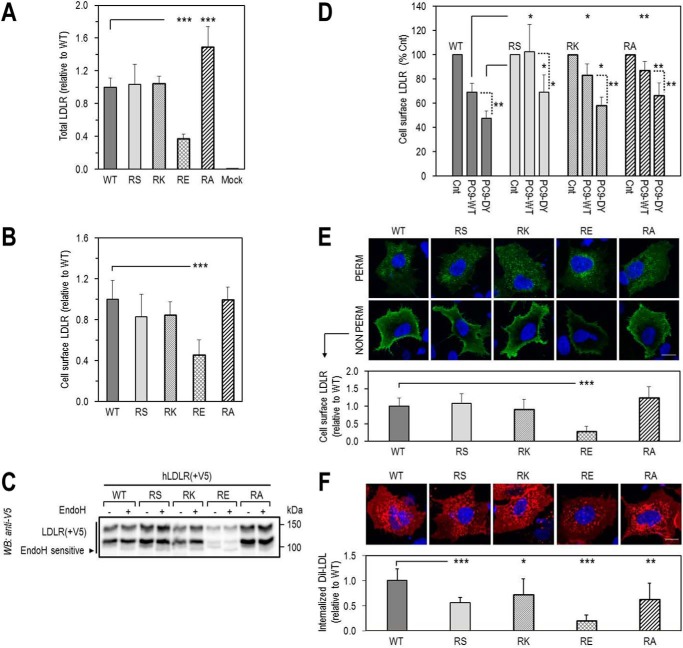
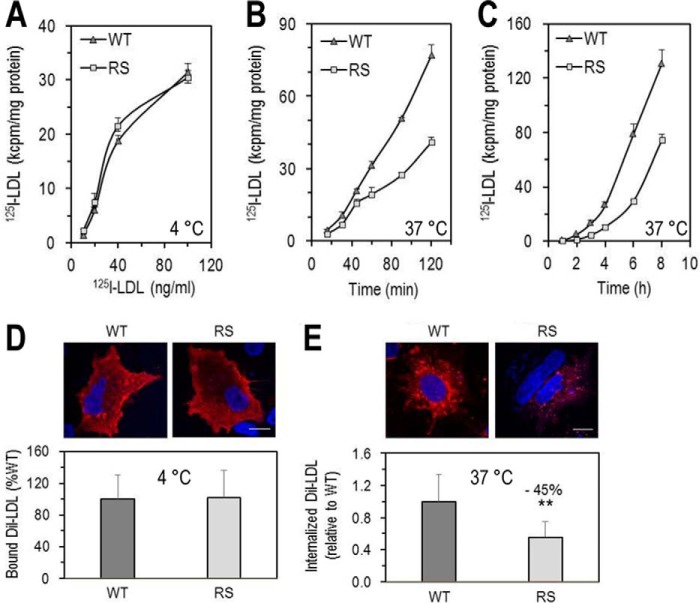
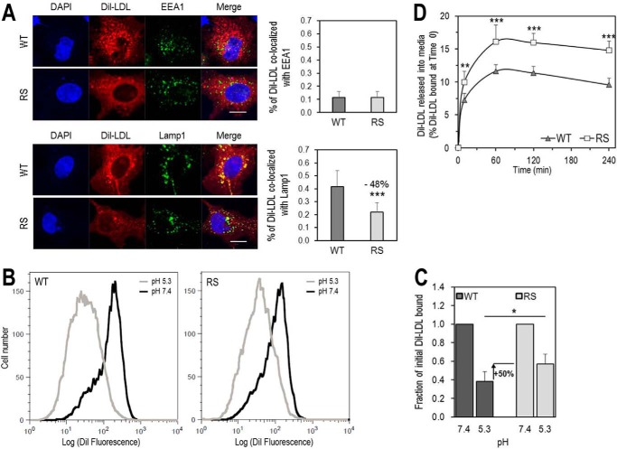
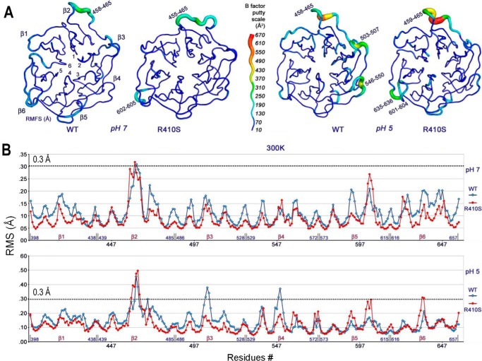
Familial hypercholesterolemia (FH) is characterized by severely elevated low density lipoprotein (LDL) cholesterol. Herein, we identified an FH patient presenting novel compound heterozygote mutations R410S and G592E of the LDL receptor (LDLR). The patient responded modestly to maximum rosuvastatin plus ezetimibe therapy, even in combination with a PCSK9 monoclonal antibody injection. Using cell biology and molecular dynamics simulations, we aimed to define the underlying mechanism(s) by which these LDLR mutations affect LDL metabolism and lead to hypercholesterolemia. Our data showed that the LDLR-G592E is a class 2b mutant, because it mostly failed to exit the endoplasmic reticulum and was degraded. Even though LDLR-R410S and LDLR-WT were similar in levels of cell surface and total receptor and bound equally well to LDL or extracellular PCSK9, the LDLR-R410S was resistant to exogenous PCSK9-mediated degradation in endosomes/lysosomes and showed reduced LDL internalization and degradation relative to LDLR-WT. Evidence is provided for a tighter association of LDL with LDLR-R410S at acidic pH, a reduced LDL delivery to late endosomes/lysosomes, and an increased release in the medium of the bound/internalized LDL, as compared with LDLR-WT. These data suggested that LDLR-R410S recycles loaded with its LDL-cargo. Our findings demonstrate that LDLR-R410S represents an LDLR loss-of-function through a novel class 8 FH-causing mechanism, thereby rationalizing the observed phenotype.

Keywords: LDL receptor; cell biology; cholesterol metabolism; familial hypercholesterolemia; human natural mutations; loss-of-function; low density lipoprotein (LDL); lysosome; proprotein convertase subtilisin/kexin type 9 (PCSK9); subcellular localization.


Conflict of interest statement

The authors declare that they have no conflicts of interest with the contents of this article

Figures

FIGURE 1.
FIGURE 1.
Domain organizations of LDLR and PCSK9 and structures of LDLR β-propeller domain and of PCSK9·LDLR complex. A, domain organizations of LDLR (top) and PCSK9 (bottom). LDLR is composed of the ligand binding domain (LBD), containing seven repeats (L1–L7) of ∼40 residues, the EGF precursor homology domain (EGFPH), consisting of EGFA, EGFB, six-bladed (1–6) β-propeller, and EGFC domains, the O-linked sugar regions, and the transmembrane domain (TMD). Mature PCSK9 is formed after auto-cleavage between the prodomain (Pro) and the catalytic domain (Catalytic). The hinge domain (H) and the C-terminal histidine-rich domain (CHRD) are indicated. B, LDLR β-propeller domain with its six blades and their corresponding residues. The positions of Arg410 and G592 in the β-propeller domain of the LDLR are displayed. C, overall structure of the PCSK9·LDLR complex (PDB code 3M0C) shows two interfaces of interaction between the two proteins as follows: a major interface between catalytic domain of PCSK9 (green) and the EGFA domain of LDLR (turquoise) and a minor interface (boxed) between the prodomain of PCSK9 (red) and the LDLR β-propeller domain (blue). Box, details of the prodomain/β-propeller interface; yellow dotted line shows the van der Waals interaction between Leu108 (PCSK9) and Leu647 (LDLR-WT), whereas the blue dotted line depicts the “putative” ionic interaction between the GOF mutation L108R (PCSK9) and Glu626 (LDLR-WT).
FIGURE 2.
FIGURE 2.
Evolution of LDLc concentration of patient III-3 following different therapies and family pedigree. A, LDLc concentrations following several lipid-lowering treatments. B, family pedigree. The prepositus is marked with an arrow. Round symbols, females. Square symbols, males. Black outlined symbols, family members for which data were available. Gray outlined symbols, family members for which no data were available. Symbols crossed through: deceased individuals. LDLR-R410S allele, dark gray shading. LDLR-G592E allele, light gray shading.
FIGURE 3.
FIGURE 3.
Total and cell surface expression of wild-type LDLR and its two mutants, R410S and G592E. HEK293 cells were transfected with V5-tagged LDLR-WT (WT), LDLR-R410S (RS), LDLR-G592E (GE), or both mutants (RS/GE). A, WB analysis of LDLR expression. B, LDLR sensitivity to endoglycosidase H (Endo H). C, LDLR levels after 18 h in absence (−) or presence (+) of 2.5 μmol/liter MG132, a proteasome inhibitor. D, FACS analyses of cell surface LDLR expression. E, immunofluorescence microscopy of total (PERM) and cell surface (NON PERM) LDLR in HepG2 cells overexpressing WT, RS, GE, or RS/GE. Quantification of cell surface LDLR is shown, using 12 transfected cells (EGFP-positive)/condition/experiment. Non-transfected cells expressed ∼100-fold less LDLR than transfected ones. Scale bar, 15 μm. Data are representative of at least two independent experiments. Quantifications are averages ± S.D. *, p < 0.05; **, p < 0.01; ***, p < 0.001 (t test).
FIGURE 4.
FIGURE 4.
Effects of PCSK9 on LDLR degradation. A and B, HEK293 cells were co-transfected with V5-tagged LDLR-WT (WT) or LDLR-R410S (RS) and PCSK9-WT (PC9-WT), PCSK9-DY (PC9-DY) or empty vector (v), as control, and analyzed by WB using anti-human LDLR (A), or by FACS for cell surface LDLR expression (B). C and D, HEK293 cells overexpressing V5-tagged WT or RS were incubated for 7 h with conditioned media from HEK293 cells (input): no PCSK9 control media (Cnt) or PCSK9 media (∼1.8 μg/ml), PC9-WT or GOF PC9-DY, and analyzed by WB with anti-human LDLR (C) or by FACS for cell surface LDLR expression (D). E, mouse primary hepatocytes lacking both PCSK9 and LDLR were transfected with V5-tagged WT or RS and incubated for 18 h with conditioned media from HEK293 as described in C. Cell lysates were immunoblotted with anti-V5-HRP for LDLR. Data are representative of at least three independent experiments. Quantifications are averages ± S.D. *, p < 0.05; **, p < 0.01; ***, p < 0.001 (t test).
FIGURE 5.
FIGURE 5.
PCSK9 displays similar binding to the cell surface LDLR-WT and LDLR-RS. A and B, HEK293 cells transfected with LDLR-WT (WT) or LDLR-R410S (RS) were incubated for 4 h at 4 °C with conditioned media from HEK293 cells: no PCSK9 control media (Cnt) or media containing PCSK9-WT. PCSK9 was non-tagged (PC9-WT) ∼1.3 μg/ml (A), or mCherry-tagged (PC9-mCherry), ∼1 μg/ml (B). PCSK9 binding to cell surface receptor was quantified by FACS (A) or fluorescence spectroscopy (B). Bars represent averages ± S.D. from three independent experiments. C, HEK293 cells overexpressing LDLR-WT (WT) or LDLR-RS (RS) were incubated with purified recombinant human PCSK9 for 4 h at 4 °C. The curve through the data is the fit to a rectangular hyperbola and is representative of two independent experiments. The average PCSK9 binding constants determined from the two experiments are 1.8 ± 0.8 μm (WT) and 1.3 ± 0.1 μm (RS).
FIGURE 6.
FIGURE 6.
GOF PCSK9-L108R in the prodomain rescues PCSK9-mediated degradation of LDLR-R410S via the extracellular pathway. HEK293 cells overexpressing LDLR-WT (WT) or LDLR-R410S (RS) were incubated for 18 h with conditioned media from HEK293 cells: no PCSK9 control-media (Cnt) or PCSK9-media (∼1.1 μg/ml), PCSK9-WT (PC9-WT), or GOF PCSK9-L108R (PC9-LR). A, total LDLR quantification by ELISA. B, FACS analyses of cell surface LDLR. Data are representative of four independent experiments. Bars are averages ± S.D. *, p < 0.05; **, p < 0.01 (t test).
FIGURE 7.
FIGURE 7.
Effect of loss of Arg410 on LDLR expression, functionality, and sensitivity to extracellular PCSK9-mediated degradation. A–D, HEK293 cells transfected with LDLR-WT (WT) or LDLR-R410 mutants (RS, RK, RE, RA) were analyzed for total LDLR expression by ELISA (A), cell surface LDLR expression by FACS (B), sensitivity of LDLR to endoglycosidase H, by WB (C), and cell surface LDLR expression, by FACS, following an 18-h incubation with conditioned media from HEK293 cells: no PCSK9 control media (Cnt), or PCSK9 media (∼1.1 μg/ml), PCSK9-WT (PC9-WT), or GOF PCSK9-DY (PC9-DY) (D). E and F, immunofluorescence microscopy in HepG2 cells transfected with WT or RS, RK, RE, RA mutants. E, total LDLR, under permeabilized conditions (PERM), and cell surface LDLR, under non-permeabilized conditions (NON PERM). F, DiI-LDL internalization after 4-h incubation with 5 μg/ml DiI-LDL at 37 °C. Quantifications in E and F were derived from analyses of 12 transfected cells (EGFP-positive)/condition/experiment. Scale bar, 15 μm. Bars in A, B, and F are averages ± S.D. of five independent experiments, and bars in C and D are averages ± S.D. of two independent experiments. *, p < 0.05; **, p < 0.01; ***, p < 0.001 (t test).
FIGURE 8.
FIGURE 8.
Functionality of LDLR-WT and LDLR-R410S. A–C, HEK293 cells transfected with LDLR-WT (WT) or LDLR-R410S (RS) were evaluated. A, 125I-LDL binding to LDLR after a 1-h incubation at 4 °C with increasing concentrations of 125I-LDL (200 cpm/pg). B, time-dependent 125I-LDL internalization after continuous incubation with 2 ng of 125I-LDL at 37 °C. C, time-dependent 125I-LDL degradation after continuous incubation with 2 ng of 125I-LDL (4 ng/ml) at 37 °C. At each time point, the medium was precipitated with ice-cold TCA, and the TCA-soluble material was used as a measure of the degraded 125I-LDL. Data are representative of three independent experiments each performed in four biological replicates. Points are the averages ± S.D. within one experiment. D and E, HepG2 cells overexpressing WT or RS were incubated with DiI-LDL (5 μg/ml) and analyzed by confocal microscopy as follows: DiI-LDL binding to the cell surface after 4-h incubation at 4 °C (D) or DiI-LDL internalization after 4 h at 37 °C (E). Quantifications in D and E were derived from analyses of 12 transfected cells (EGFP-positive)/condition/experiment. Scale bar, 15 μm. Data are representative of at least two independent experiments. Bars are averages ± S.D. **, p < 0.01 (t test).
FIGURE 9.
FIGURE 9.
Residue Arg410 of the LDLR is important for LDL release in acidic early endosomes and for its delivery to late endosomes. A, HepG2 cells overexpressing LDLR-WT (WT) or LDLR-R410S (RS) were incubated with DiI-LDL (5 μg/ml) for 4 h at 37 °C and analyzed by immunofluorescence under permeabilized conditions. Upper panels, co-localization of DiI-LDL with markers for early endosomal compartment (EEA1) and quantification. Lower panels, co-localization of DiI-LDL with markers for late endosomal/lysosomal compartment (Lamp1) and quantification. Quantifications were derived from analyses of 12 transfected cells (EGFP-positive)/condition/experiment. Scale bar, 15 μm. B and C, HEK293 cells overexpressing WT or RS were incubated for 1 h at 4 °C with 15 μg/ml DiI-LDL at pH 7.4, washed, and switched to pH 5.3 for an additional hour at 4 °C. At each pH, the amount of fluorescence from DiI-LDL bound to each receptor was measured by FACS. B, flow cytometry plots for WT (left) and RS (right). C, quantification of DiI-LDL bound at each pH relative to pH 7.4. D, HEK293 cells overexpressing WT or RS were incubated with DiI-LDL (6 μg/ml) for 1 h at 4 °C, washed, and then switched to fresh serum-free DMEM at 37 °C for the indicated times. DiI-LDL released into media was measured and reported relative to DiI-LDL bound at time 0. Data are averages ± S.D. of at least three independent experiments. *, p < 0.05; **, p < 0.01; ***, p < 0.001 (t test).
FIGURE 10.
FIGURE 10.
At pH 5, the β-propeller mobility of LDLR-R410S is reduced compared with LDLR-WT, and for both receptors the structures at pH 5 are more mobile than the ones at pH 7. A, β-propeller configurations of LDLR-WT (WT) and LDLR-R410S (RS) at pH 7 (left) and pH 5 (right) were generated by molecular dynamics simulations starting with the LDLR-WT structure from PDB code 1N7D. The six blades (β1–β6) and flexible amino acid regions are indicated. The B-factor putty scale is shown as a measure of region flexibility (blue-to-red translated into rigid-to-flexible). B, plots of root mean squared fluctuations values (Å) for each β-propeller residue of LDLR-WT (blue trace) and LDLR-R410S (red trace) at pH 7 (top) and pH 5 (bottom) that were used to calculate the B-factors and generate the configurations in A.

Similar articles

Cited by

References

    1. LaRosa J. C., Grundy S. M., Waters D. D., Shear C., Barter P., Fruchart J. C., Gotto A. M., Greten H., Kastelein J. J., Shepherd J., Wenger N. K., and Treating to New Targets (TNT) Investigators (2005) Intensive lipid lowering with atorvastatin in patients with stable coronary disease. N. Engl. J. Med. 352, 1425–1435 - PubMed
    1. Cannon C. P., Blazing M. A., Giugliano R. P., McCagg A., White J. A., Theroux P., Darius H., Lewis B. S., Ophuis T. O., Jukema J. W., De Ferrari G. M., Ruzyllo W., De Lucca P., Im K., Bohula E. A., et al. (2015) Ezetimibe added to statin therapy after acute coronary syndromes. N. Engl. J. Med. 372, 2387–2397 - PubMed
    1. Brown M. S., and Goldstein J. L. (1986) A receptor-mediated pathway for cholesterol homeostasis. Science 232, 34–47 - PubMed
    1. Goldstein J. L., Hobbs H. H., and Brown M. S. (2001) in The Metabolic and Molecular Bases of Inherited Disease (Valle D., Scrive C. R., Beaudet A. L., Sly W. S., Childs B., and Volgestein B., eds) pp. 2863–2913, McGraw-Hill, New York
    1. Fokkema I. F., Taschner P. E., Schaafsma G. C., Celli J., Laros J. F., and den Dunnen J. T. (2011) LOVD Version 2.0: the next generation in gene variant databases. Hum. Mutat. 32, 557–563 - PubMed

Publication types

Grants and funding

LinkOut - more resources