. 2017 Mar 10;292(10):4176-4184.
doi: 10.1074/jbc.M116.767954. Epub 2016 Dec 20.

The Human RecQ4 Helicase Contains a Functional RecQ C-terminal Region (RQC) That Is Essential for Activity

Affiliations

The Human RecQ4 Helicase Contains a Functional RecQ C-terminal Region (RQC) That Is Essential for Activity

Aditya Mojumdar et al. J Biol Chem. .

Abstract

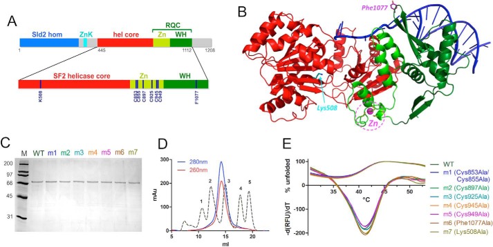
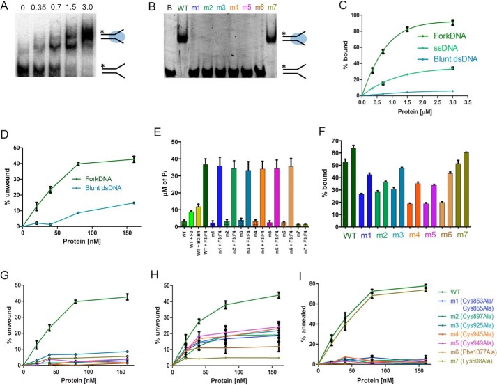
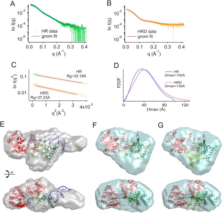
RecQ helicases are essential in the maintenance of genome stability. Five paralogues (RecQ1, Bloom, Werner, RecQ4, and RecQ5) are found in human cells, with distinct but overlapping roles. Mutations in human RecQ4 give rise to three distinct genetic disorders (Rothmund-Thomson, RAPADILINO, and Baller-Gerold syndromes), characterized by genetic instability, growth deficiency, and predisposition to cancer. Previous studies suggested that RecQ4 was unique because it did not seem to contain a RecQ C-terminal region (RQC) found in the other RecQ paralogues; such a region consists of a zinc domain and a winged helix domain and plays an important role in enzyme activity. However, our recent bioinformatic analysis identified in RecQ4 a putative RQC. To experimentally confirm this hypothesis, we report the purification and characterization of the catalytic core of human RecQ4. Inductively coupled plasma-atomic emission spectrometry detected the unusual presence of two zinc clusters within the zinc domain, consistent with the bioinformatic prediction. Analysis of site-directed mutants, targeting key RQC residues (putative zinc ligands and the aromatic residue predicted to be at the tip of the winged helix β-hairpin), showed a decrease in DNA binding, unwinding, and annealing, as expected for a functional RQC domain. Low resolution structural information obtained by small angle X-ray scattering data suggests that RecQ4 interacts with DNA in a manner similar to RecQ1, whereas the winged helix domain may assume alternative conformations, as seen in the bacterial enzymes. These combined results experimentally confirm the presence of a functional RQC domain in human RecQ4.

Keywords: DNA helicase; DNA repair; mutagenesis in vitro; protein purification; protein-DNA interaction; recombinant protein expression; small angle X-ray scattering (SAXS); structural model.


Conflict of interest statement

The authors declare that they have no conflicts of interest with the contents of this article

Figures

FIGURE 1.
FIGURE 1.
Expression and purification of human RecQ4. A, domain organization of human RecQ4 showing the Sld2 homologous region at the N terminus (blue) followed by a zinc knuckle (cyan), a helicase core (red), and RQC domain (consisting of a zinc-binding domain (bright green) and a winged helix domain (dark green)). Enlarged at the bottom of the panel is the catalytic core of RecQ4 (helicase and RQC domain) with the positions of the residues mutated in this work. B, the crystal structure of RecQ1 (PDB code 2WWY) with the same color code; critical residues are highlighted. C, SDS-PAGE gel of purified proteins. M, molecular markers; m1, C853A/C855A; m2, C897A; m3, C925A; m4, Cs945A; m5, C949A; m6, F1077A; m7, K508A. D, size exclusion chromatography indicates that the recombinant protein is a monomer. The calibration peaks correspond to protein markers: peak 1, ferritin, 440 kDa; peak 2, aldolase, 158 kDa; peak 3, ovalbumin, 44 kDa; peak 4, ribonuclease A, 13.7 kDa; peak 5, aprotinin, 6.5 kDa. E, heat denaturation profiles of RecQ4 variants. The curves depicting the percentage of unfolded proteins (positive y quadrant, 0–100) and the −(dRFU)/dT curves are plotted together (mostly in the negative y quadrant). The experiment suggests that all the mutants are folded and stable.
FIGURE 2.
FIGURE 2.
Biochemical activities of the purified recombinant WT and mutant proteins. A, EMSA gel showing the binding activity of increasing amounts (0–3 μm) of WT RecQ4 with 10 nm forked DNA (F1:F4). B, EMSA gel showing the DNA binding activity of the mutants at the highest concentration (3 μm), with 10 nm DNA substrate. Lane B, fork DNA (F1:F4); m1, C853A/C855A; m2, C897A; m3, C925A; m4, C945A; m5, C949A; m6, F1077A; m7, K508A. All the RQC mutants have no affinity in limiting substrate concentration. C, comparison of DNA binding activity of the WT protein on three substrates: blunt dsDNA (B1:B4), ssDNA (F1), and fork DNA (F1:F4) (10 nm). The protein has a preference for forked substrates. The one-site total function of GraphPad Prism was used to fit the data points. D, comparison of helicase activity of the WT protein on two substrates (10 nm): blunt dsDNA (B1:B2) and fork DNA (F1:F2). Forked substrates are preferred, although there is some activity on blunt substrates at higher protein concentrations. E, ATPase assay of WT and mutant proteins in the presence of 500 nm nucleic acid. ATP hydrolysis by WT protein is highly stimulated by fork DNA (F3:F4) compared with other substrates (ssDNA F3 and blunt dsDNA B3:B4). Only the Walker A mutant affects the hydrolysis. F, DNA binding activity of 0.5 and 1 μm of proteins (WT and mutants) at high fork DNA (F3:F4) concentration (100 nm). The bar graph shows the percentage of DNA bound by the proteins at respective concentrations. At this DNA concentration, the RQC mutants show a partial binding activity. G, comparison of helicase activity of mutants and WT at 10 nm DNA substrate (F1:F2). All the mutations abolish the DNA helicase activity. H, comparison of helicase activity of mutants and WT at 100 nm DNA substrate (F1:F2). Partial activity can be observed for the RQC mutants, consistent with the impairment in DNA binding observed in F, suggesting that the effect of the mutants is due to impairment in DNA binding. I, comparison of annealing activity of WT and mutants at 10 nm DNA. All the RQC mutants show a loss of DNA annealing activity. Each experiment was repeated at least three times.
FIGURE 3.
FIGURE 3.
SAXS analysis of human RecQ4. A, experimental SAXS profile (green) compared with the theoretical scattering curve calculated from the ab initio model (blue line) for the catalytic core of human RecQ4 (helicase-RQC (HR)). B, experimental SAXS profile (orange) compared with the theoretical scattering curve calculated from the ab initio model (magenta line) for the catalytic core of human RecQ4-DNA complex (HRD). C, Guinier's fits with experimental Rg values for HR and HRD in the allowed region (q < 1.3 Rg). D, pair distance distribution function (PDDF) for HR and HRD calculated in the allowed region (q > Π/Dmax). E, final model reconstructed from the scattering curve for the protein-DNA complex (HRD). The crystal structure of the RecQ1-DNA complex (PDB code 2WWY) was fitted onto the SAXS envelope. A long protrusion roughly corresponds to the location of DNA in the model. Some unaccounted density could correspond to two long insertions (90 residues in total) within the zinc domain. F, final model reconstructed from the scattering curve for the protein alone. In the absence of DNA, the envelope is more globular and lacks details, so that it is less easy to fit the RecQ1 model. G, to check for the possibility that the WH domain assumes an alternative conformation, the orientation of the WH seen in RecQ1 (dark green) has been overlapped with an alternative conformation (light green) based on the E. coli structure (PDB code 1OYY), suggesting that the protein in solution is a mixture of multiple states.

References

    1. Croteau D. L., Popuri V., Opresko P. L., and Bohr V. A. (2014) Human RecQ helicases in DNA repair, recombination, and replication. Annu. Rev. Biochem. 83, 519–552 - PMC - PubMed
    1. Keijzers G., Maynard S., Shamanna R. A., Rasmussen L. J., Croteau D. L., and Bohr V. A. (2014) The role of RecQ helicases in non-homologous end-joining. Crit. Rev. Biochem. Mol. Biol. 49, 463–472 - PMC - PubMed
    1. Larsen N. B., and Hickson I. D. (2013) RecQ helicases: conserved guardians of genomic integrity. Adv. Exp. Med. Biol. 767, 161–184 - PubMed
    1. Oshima J., Sidorova J. M., and Monnat R. J. Jr. (2016) Werner syndrome: clinical features, pathogenesis and potential therapeutic interventions. Ageing Res. Rev. pii, S1568–S1637 - PMC - PubMed
    1. de Renty C., and Ellis N. A. (2017) Bloom's syndrome: why not premature aging?: a comparison of the BLM and WRN helicases. Ageing Res. Rev. 33, 36–51 - PMC - PubMed

Publication types

LinkOut - more resources