. 2017 Feb 7;8(6):9216-9229.
doi: 10.18632/oncotarget.13985.

MutY-Homolog (MYH) inhibition reduces pancreatic cancer cell growth and increases chemosensitivity

Affiliations

MutY-Homolog (MYH) inhibition reduces pancreatic cancer cell growth and increases chemosensitivity

George Sharbeen et al. Oncotarget. .

Abstract

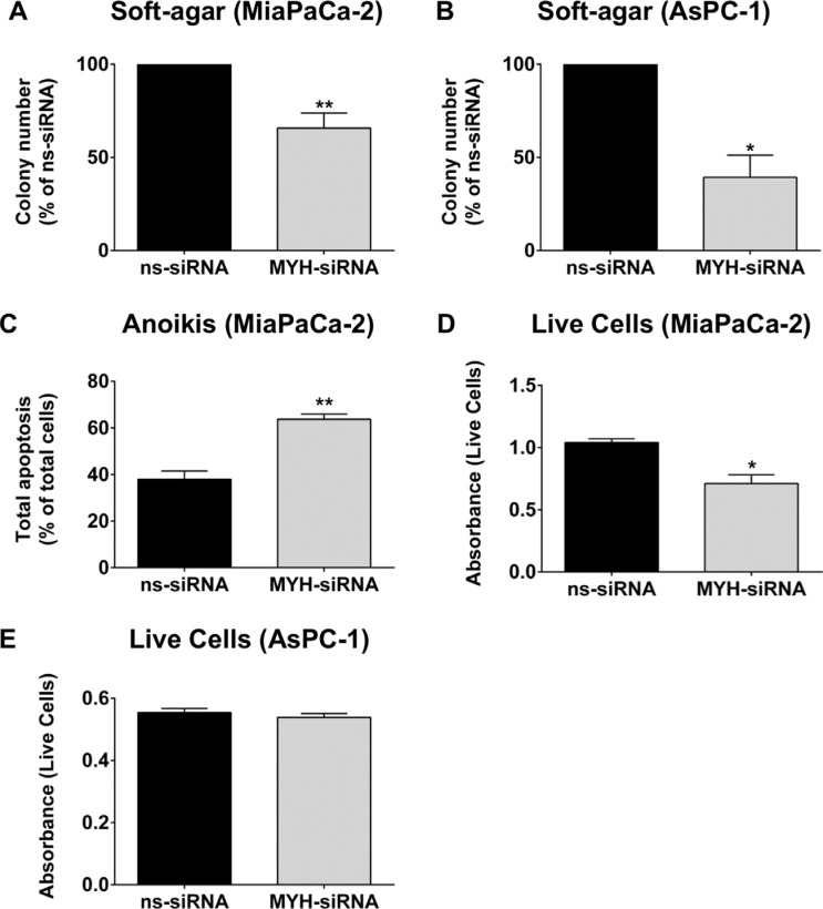
Patients with pancreatic ductal adenocarcinoma (PC) have a poor prognosis due to metastases and chemoresistance. PC is characterized by extensive fibrosis, which creates a hypoxic microenvironment, and leads to increased chemoresistance and intracellular oxidative stress. Thus, proteins that protect against oxidative stress are potential therapeutic targets for PC. A key protein that maintains genomic integrity against oxidative damage is MutY-Homolog (MYH). No prior studies have investigated the function of MYH in PC cells. Using siRNA, we showed that knockdown of MYH in PC cells 1) reduced PC cell proliferation and increased apoptosis; 2) further decreased PC cell growth in the presence of oxidative stress and chemotherapy agents (gemcitabine, paclitaxel and vincristine); 3) reduced PC cell metastatic potential; and 4) decreased PC tumor growth in a subcutaneous mouse model in vivo. The results from this study suggest MYH may be a novel therapeutic target for PC that could potentially improve patient outcome by reducing PC cell survival, increasing the efficacy of existing drugs and reducing metastatic spread.

Keywords: DNA repair; chemoresistance; mutY-homolog (MYH); oxidative stress; pancreatic cancer.


Conflict of interest statement

CONFLICTS OF INTEREST

No potential conflicts of interest to disclose.

Figures

Figure 1
Figure 1. Expression of MYH in human pancreatic adenocarcinoma tissue and cell lines
(A) Panels show control tissue (normal pancreas or benign pancreatic conditions) or pancreatic cancer tissue stained with MYH antibody (brown). Closer views of normal acinar cells (top panels) or pancreatic cancer cells (bottom panels) are shown in insets. (B) Magnified field of view showing human pancreatic cancer tissue stained with either isotype control antibody (i) or MYH antibody (ii-iii). The isotype control was negative and tumor elements had strong immunoreactivity for MYH. Labels demonstrate cytoplasmic+nuclear staining (*) versus cytoplasmic only staining (#). Panel (iii) demonstrates low MYH staining in normal acinar cells. (C) Western blot analysis of MYH in protein extracts from normal human pancreatic ductal epithelial cells (HPDE cells) and pancreatic cancer cell lines. GAPDH was used as a loading control.
Figure 2
Figure 2. Knockdown of MYH in pancreatic cancer cells
RNA and protein was extracted from cells 96 h after transfection with control siRNA (ns-siRNA) or MYH-siRNA. (A–B) qPCR analysis of MYH knockdown in RNA extracts from (A) MiaPaCa-2 and (B) AsPC-1. Samples were standardised to 18S RNA. (CD) Western blot analysis of MYH silencing in protein extracts from (C) MiaPaCa-2 and (D) AsPC-1 cells. GAPDH was used as a loading control. Graphs show densitometry of Western blots for MYH (representative Western blots shown in top panel). Asterisks indicate significance (**p ≤ 0.01, ***p ≤ 0.001; n = 3).
Figure 3
Figure 3. The effect of MYH knockdown on pancreatic cancer cell proliferation and sensitivity to oxidative stress
(AB) Proliferation assay of (A) MiaPaCa-2 cells and (B) AsPC-1 cells transfected with control siRNA (ns-siRNA) or MYH-siRNA. 48 h post-transfection, cells were cultured ± t-butyl hydroperxoide (t-BHP) for a further 48 h. Cells were then lifted and live cells counted on an automated BioRAD cell counter (trypan blue staining). Bars represent the total live cell count as a fraction of ns-siRNA control (± s.e.m.). (CD) MitoSOXTM assay of oxidative stress in (C) MiaPaCa-2 and (D) AsPC-1 cells after stimulation with 37 μM or 148 μM t-BHP, respectively. Asterisks indicate significance relative to ns-siRNA controls (*p ≤ 0.05, **p ≤ 0.01, ***p ≤ 0.001; n ≥ 3).
Figure 4
Figure 4. MYH knockdown alters cell cycle and induces apoptosis in pancreatic cancer cells
(AB) Cell cycle: (A) MiaPaCa-2 cells or (B) AsPC-1 cells were transfected with ns-siRNA or MYH-siRNA. 48 h post-transfection, cells were cultured in t-butyl hydroperoxide (t-BHP). 48 h later, cells were stained with PI and DNA content measured by flow cytometry. (CD) Apoptosis: (C) MiaPaCa-2 or (D) AsPC-1 cells were transfected with ns-siRNA or MYH-siRNA. Apoptosis was determined 96 h post-transfection, by AnnexinV + 7AAD staining and flow cytometry. Bars represent the fraction of total MiaPaCa-2 or AsPC-1 cells that were apoptotic (± s.e.m.). Asterisks indicate significance relative to ns-siRNA controls (*p < 0.05, **p < 0.01, ***p < 0.001, ****p < 0.0001; n ≥ 3).
Figure 5
Figure 5. MYH knockdown reduces pancreatic cancer cell clonogenicity and increases chemosensitivity
Bars represent the number of MiaPaCa-2 colonies (mean+s.e.m. as a % of control siRNA (ns-siRNA) 0 nM drug) that formed from low density seeding following transfection with ns-siRNA or MYH-siRNA and 72 h culture in gemcitabine (A), paclitaxel (B) or vincristine (C). (DF) as per A–C, except experiments were carried out with AsPC-1 cells. Asterisks indicate significance relative to the 0nM control for the same siRNA (*p ≤ 0.05, ***p ≤ 0.001, ****p ≤ 0.0001; n ≥ 4). Hashes indicate significance relative to the ns-siRNA control of the same drug dose (#p ≤ 0.05, ##p ≤ 0.01, ###p ≤ 0.001, ####p ≤ 0.0001; n ≥ 4).
Figure 6
Figure 6. MYH knockdown reduces pancreatic cancer cell metastatic potential
(A) MiaPaCa-2 or (B) AsPC-1 transfected with control siRNA (ns-siRNA) or MYH-siRNA were embedded in soft-agarose at 48 h post-transfection and allowed to form colonies. Bars represent the number of colonies that formed (mean+s.e.m. as a % of ns-siRNA). Asterisks indicate significance (*p ≤ 0.05, **p ≤ 0.01; n ≥ 3). (C) MiaPaCa-2 cells were transfected with ns-siRNA or MYH-siRNA and 24 h post-transfection cells were cultured under anchorage independent conditions for a further 48 h (wells coated with Poly-HEMA). Bars represent the apoptotic fraction determined by AnnexinV + 7AAD staining and flow cytometry (mean + s.e.m.). Asterisks indicate significance (**p ≤ 0.01; n = 3). (DE) MiaPaCa-2 or AsPC-1 cells were cultured as in C), before the number of live cells was measured using cell counting kit 8 (cck8; colorimetric assay whose absorbance directly correlates with live cell number). Bars represent the absorbance for each siRNA treatment (mean + s.e.m). Asterisks indicate significance (*p ≤ 0.05; n = 3).
Figure 7
Figure 7. Silencing MYH in pancreatic cancer cells reduces subcutaneous tumor growth
(A) Immunohistochemistry for MYH in subcutaneous pancreatic (MiaPaCa-2) tumor sections treated with control siRNA or MYH-siRNA. (B) Representative photomicrographs of subcutaneous tumors treated with control siRNA or MYH-siRNA. (C) Growth curves showing tumor volume, over the course of JetPEI® + control siRNA or MYH-siRNA treatments (mean ± s.e.m.). (D) Tumor volumes, as a fraction of starting volume, calculated from ex vivo measurements at endpoint (excised tumors). Asterisks indicate significance (*p ≤ 0.05; n = 4 for control siRNA and n = 6 for MYH-siRNA).

References

    1. Miller KD, Siegel RL, Lin CC, Mariotto AB, Kramer JL, Rowland JH, Stein KD, Alteri R, Jemal A. Cancer treatment and survivorship statistics, 2016. CA-A Cancer J Clin. 2016;66:271–289. - PubMed
    1. Siegel RL, Miller KD, Jemal A. Cancer statistics, 2016. CA-A Cancer J Clin. 2016;66:7–30. - PubMed
    1. Hidalgo M. Pancreatic cancer. New Engl J Med. 2010;362:1605–1617. - PubMed
    1. Vincent A, Herman J, Schulick R, Hruban RH, Goggins M. Pancreatic cancer. Lancet. 2011;378:607–620. - PMC - PubMed
    1. Wolfgang CL, Herman JM, Laheru DA, Klein AP, Erdek MA, Fishman EK, Hruban RH. Recent progress in pancreatic cancer. CA-A Cancer J Clin. 2013;63:318–348. - PMC - PubMed

MeSH terms