Review
. 2016 Dec 12:10:27.
doi: 10.1186/s13037-016-0115-8. eCollection 2016.

Intraoperative radiation safety in orthopaedics: a review of the ALARA (As low as reasonably achievable) principle

Affiliations
Review

Intraoperative radiation safety in orthopaedics: a review of the ALARA (As low as reasonably achievable) principle

Daniel J Kaplan et al. Patient Saf Surg. .

Abstract

The use of fluoroscopy has become commonplace in many orthopaedic surgery procedures. The benefits of fluoroscopy are not without risk of radiation to patient, surgeon, and operating room staff. There is a paucity of knowledge by the average orthopaedic resident in terms proper usage and safety. Personal protective equipment, proper positioning, effective communication with the radiology technician are just of few of the ways outlined in this article to decrease the amount of radiation exposure in the operating room. This knowledge ensures that the amount of radiation exposure is as low as reasonably achievable. Currently, in the United States, guidelines for teaching radiation safety in orthopaedic surgery residency training is non-existent. In Europe, studies have also exhibited a lack of standardized teaching on the basics of radiation safety in the operating room. This review article will outline the basics of fluoroscopy and educate the reader on how to safe fluoroscopic image utilization.

Keywords: Fluoroscopy; Operating room safety; Orthopaedic surgery; Radiation; Radiation exposure; Radiation safety; Surgical training; c arm.


Figures
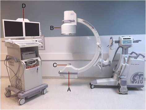
Fig. 1
Fig. 1
Basic c-arm unit. a. X-ray Tube; b. Image Intensifier; c. Collimator; d. Display Monitor
Fig. 2
Fig. 2
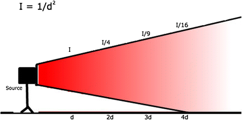
Inverse-square law: I = 1/d2, where I = magnitude of scatter and d = distance from the source. By doubling the distance from the x-ray tube, you receive only one fourth of the exposure from scatter
Fig. 3
Fig. 3
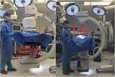
a shows a setup with the x-ray tube on the bottom. The red arrows represent radiation beams that scatter after they deflect off of the object being imaged. With the x-ray tube on the bottom most of the scattered (deflected) radiation is towards the legs and feet of the surgeon. b shows a setup with the x-ray tube on the top. Here the scattered radiation is towards the head and neck region of the surgeon
Fig. 4
Fig. 4
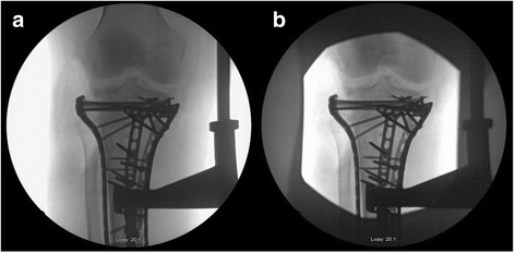
a shows a standard x-ray taken without collimation. b is an x-ray taken with collimation; collimation helps reduce exposure and may also help produce sharper images

References

    1. Nugent M, Carmody O, Dudeney S. Radiation safety knowledge and practices among Irish orthopaedic trainees. Ir J Med Sci. 2014;184(2):369–373. doi: 10.1007/s11845-014-1121-4. - DOI - PubMed
    1. Khan FR, Ul-Abadin Z, Rauf S, et al. Awareness and attitudes amongst basic surgical trainees regarding radiation in orthopaedic trauma surgery. Biomed Imaging Interv J. 2010;6(3):1–4. doi: 10.2349/biij.6.3.e25. - DOI - PMC - PubMed
    1. Bushberg JT, Seibert JA, Leidholdt EM, et al. The essential physics of medical imaging, third edition. Med Phys. 2013;40(7):077301. doi: 10.1118/1.4811156. - DOI - PubMed
    1. Dewey P, George S, Gray A. (i) Ionising radiation and orthopaedics. Curr Orthop. 2005;19(1):1–12. doi: 10.1016/j.cuor.2005.01.002. - DOI
    1. Hafez MA, Smith RM, Matthews SJ, et al. Radiation exposure to the hands of orthopaedic surgeons: are we underestimating the risk? Arch Orthop Trauma Surg. 2005;125(5):330–335. doi: 10.1007/s00402-005-0807-5. - DOI - PubMed