

POST OPERATIVE REHABILITATION OF GRADE III MEDIAL COLLATERAL LIGAMENT INJURIES: EVIDENCE BASED REHABILITATION AND RETURN TO PLAY
- PMID: 27999730
- PMCID: PMC5159640
POST OPERATIVE REHABILITATION OF GRADE III MEDIAL COLLATERAL LIGAMENT INJURIES: EVIDENCE BASED REHABILITATION AND RETURN TO PLAY
Abstract
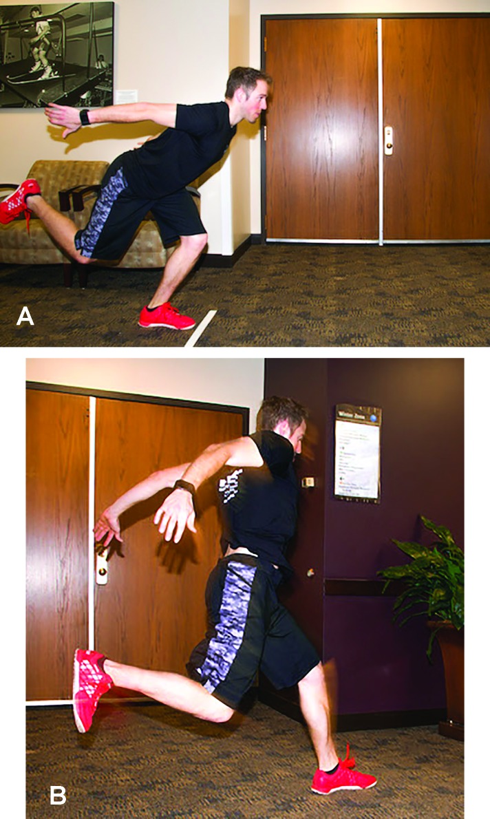
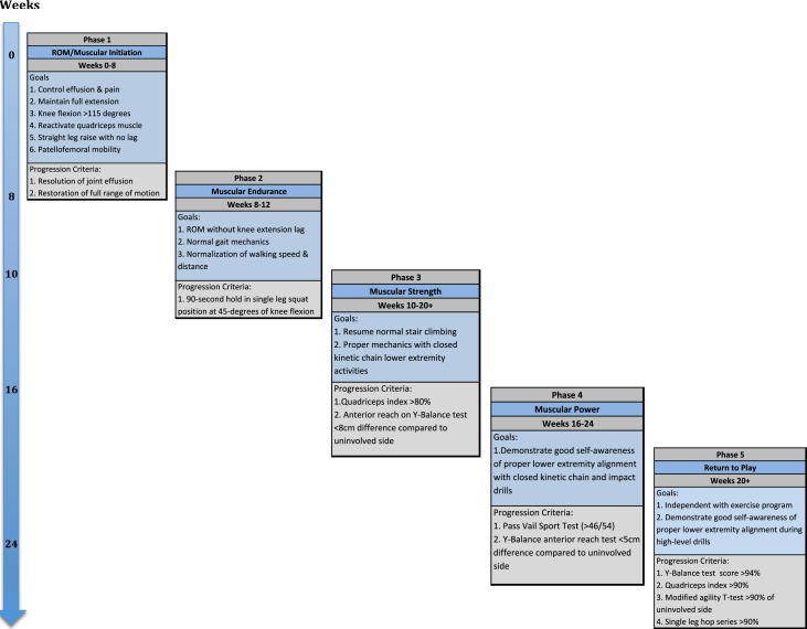
The medial collateral ligament is the most commonly injured ligament of the knee, with injury generally sustained in the athletic population as a result of valgus contact with or without tibial external rotation. The capacity of the medial collateral ligament to heal has been demonstrated in both laboratory and clinical studies; however, complete ruptures heal less consistently and may result in persistent instability. When operative intervention is deemed necessary, anatomical medial knee reconstruction is recommended. Post-operative rehabilitation focuses on early motion and the return of normal neuromuscular firing patterns with progression based on attainment of specific phase criteria and goals. The purpose of this clinical commentary is to discuss the determinants of phase progression and the importance of objectively assessing readiness for advancement that is consistent with post-operative healing. Additional tests and validated measures to assess readiness for sport are also presented.
Level of evidence: 5.
Keywords: Medial knee; medial collateral ligament; periodization; reconstruction; rehabilitation; return to sport.
Figures
References
-
- Miyamoto RG, Bosco JA, Sherman OH. Treatment of medial collateral ligament injuries. J Am Acad Orthop Surg. 2009;17(3):152-161. - PubMed
-
- Giannotti BF, Rudy T, Graziano J. The non-surgical management of isolated medial collateral ligament injuries of the knee. Sports Med Arthrosc. 2006;14(2):74-77. - PubMed
-
- Indelicato P. Isolated Medial Collateral Ligament Injuries in the Knee. J Am Acad Orthop Surg. 1995;3(1):9-14. - PubMed
-
- Roach CJ, Haley CA, Cameron KL, Pallis M, Svoboda SJ, Owens BD. The epidemiology of medial collateral ligament sprains in young athletes. Am J Sports Med. 2014;42(5):1103-1109. - PubMed
LinkOut - more resources
Full Text Sources
Research Materials