

PD-L1, PD-L2 and PD-1 expression in metastatic melanoma: Correlation with tumor-infiltrating immune cells and clinical outcome
- PMID: 27999753
- PMCID: PMC5139635
- DOI: 10.1080/2162402X.2016.1235107
PD-L1, PD-L2 and PD-1 expression in metastatic melanoma: Correlation with tumor-infiltrating immune cells and clinical outcome
Abstract
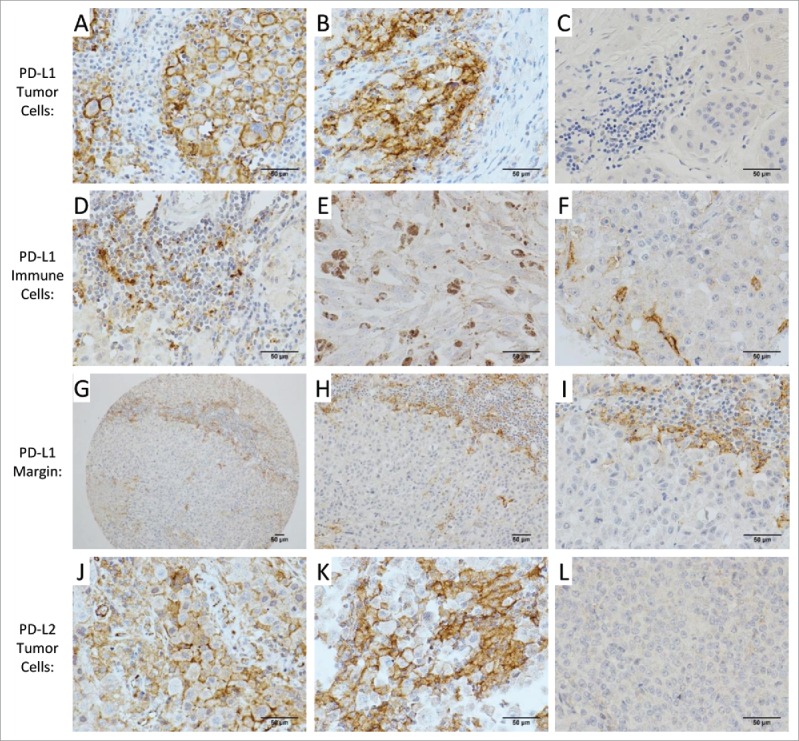
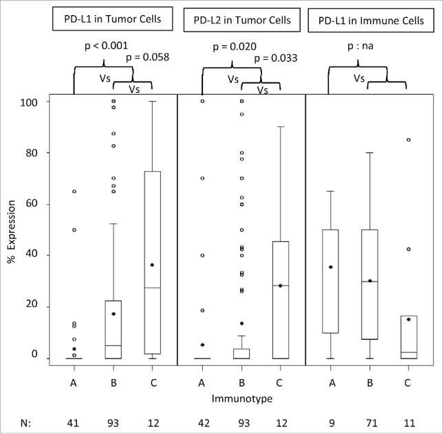
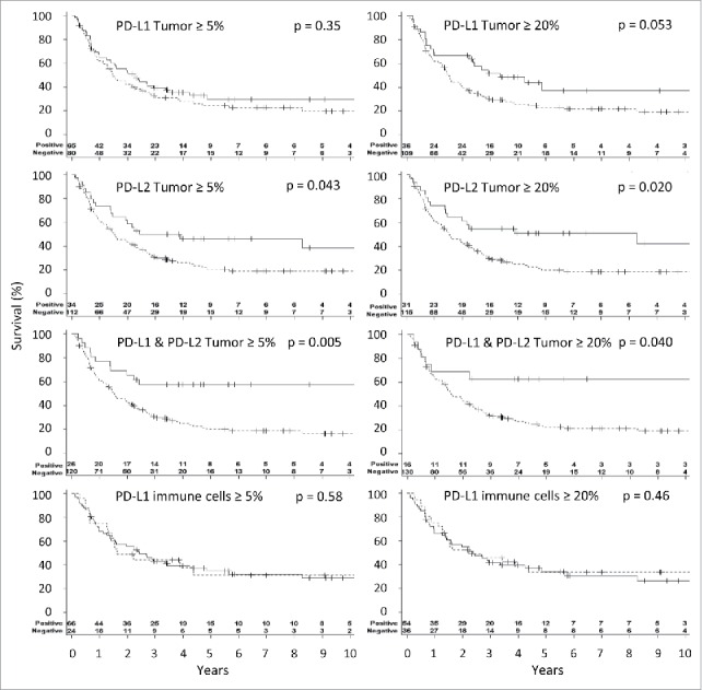
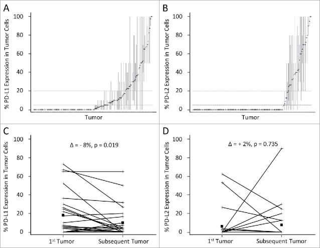
Therapeutic blockade of PD-1/PD-L1 can have dramatic therapeutic benefit in some patients; however, the prognostic associations of PD-1 and its ligands, in the absence of therapeutic blockade have not been definitively addressed. In particular, associations of PD-L2 with immune infiltrates and with outcome have yet to be explored. We hypothesized that surface expression of both PD-L1 and PD-L2 by melanoma cells would be associated with immune cell infiltration and with overall patient survival, independent of checkpoint blockade therapy. We also characterized the heterogeneity of their distribution within a tumor and within tumors of the same patient. Tissue microarrays of metastatic melanoma samples from 147 patients were quantified for CD8+, CD45, CD4+, CD3, CD163, CD20, CD138, FoxP3, PD-1, PD-L1 and PD-L2 markers by immunohistochemistry. Relationships between the proportions of PD-L1 and PD-L2 expressing tumor cells with the immune cell count, distribution (immunotype) and patient survival were studied. Expressions of both PD-L1 and PD-L2 correlated significantly with increasing densities of immune cells in the tumor specimens and with immunotype. Positive PD-L2 expression was associated with improved overall survival and the simultaneous positive expression of both PD-1 ligands showed a higher association with survival. Significant heterogeneity of PD-L1 and PD-L2 expressions within tumors were observed, however, they were less pronounced with PD-L2. In conclusion, both are markers of immune infiltration and PD-L2, alone or in combination with PD-L1, is a marker for prognosis in metastatic melanoma patients. Larger tumor samples yield more reliable assessments of PD-L1/L2 expression.
Keywords: Immune checkpoint; PD-1; PD-L1; PD-L2; immune infiltrates; metastatic melanoma; patient outcomes; tumor-infiltrating lymphocytes.
Figures
References
-
- Rosenberg SA, Dudley ME. Adoptive cell therapy for the treatment of patients with metastatic melanoma. Curr Opin Immunol 2009; 21:233-40; PMID:19304471; http://dx.doi.org/10.1016/j.coi.2009.03.002 - DOI - PMC - PubMed
-
- Topalian SL, Sznol M, McDermott DF, Kluger HM, Carvajal RD, Sharfman WH, Brahmer JR, Lawrence DP, Atkins MB, Powderly JD et al.. Survival, durable tumor remission, and long-term safety in patients with advanced melanoma receiving nivolumab. J Clin Oncol 2014; 32:1020-30; PMID:24590637; http://dx.doi.org/10.1200/JCO.2013.53.0105 - DOI - PMC - PubMed
-
- Homet Moreno B, Parisi G, Robert L, Ribas A. Anti-PD-1 therapy in melanoma. Seminars Oncol 2015; 42:466-73; PMID:25965365; http://dx.doi.org/10.1053/j.seminoncol.2015.02.008 - DOI - PubMed
-
- Zou W, Wolchok JD, Chen L. PD-L1 (B7-H1) and PD-1 pathway blockade for cancer therapy: Mechanisms, response biomarkers, and combinations. Sci Transl Med 2016; 8:328rv4; PMID:26936508; http://dx.doi.org/2106119710.1126/scitranslmed.aad7118 - DOI - PMC - PubMed
-
- Jin HT, Ahmed R, Okazaki T. Role of PD-1 in regulating T-cell immunity. Curr Topics Microbiol Immunol 2011; 350:17-37; PMID:21061197; http://dx.doi.org/10.1007/82_2010_116 - DOI - PubMed
Publication types
Grants and funding
LinkOut - more resources
Full Text Sources
Other Literature Sources
Medical
Research Materials
Miscellaneous