Review
. 2017 Mar;67(2):271-281.
doi: 10.1007/s12576-016-0514-8. Epub 2016 Dec 20.

Adaptation to microgravity, deconditioning, and countermeasures

Affiliations
Review

Adaptation to microgravity, deconditioning, and countermeasures

Kunihiko Tanaka et al. J Physiol Sci. 2017 Mar.

Abstract

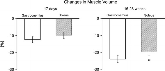
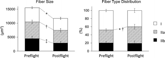
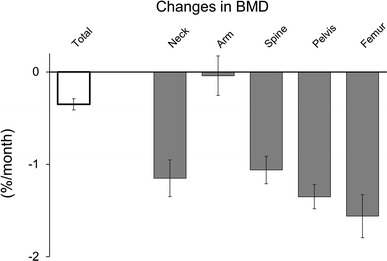
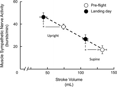
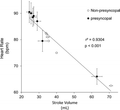
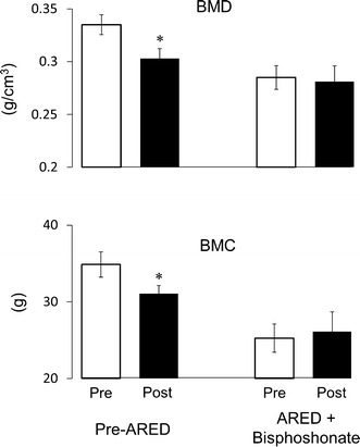
Humans are generally in standing or sitting positions on Earth during the day. The musculoskeletal system supports these positions and also allows motion. Gravity acting in the longitudinal direction of the body generates a hydrostatic pressure difference and induces footward fluid shift. The vestibular system senses the gravity of the body and reflexively controls the organs. During spaceflight or exposure to microgravity, the load on the musculoskeletal system and hydrostatic pressure difference is diminished. Thus, the skeletal muscle, particularly in the lower limbs, is atrophied, and bone minerals are lost via urinary excretion. In addition, the heart is atrophied, and the plasma volume is decreased, which may induce orthostatic intolerance. Vestibular-related control also declines; in particular, the otolith organs are more susceptible to exposure to microgravity than the semicircular canals. Using an advanced resistive exercise device with administration of bisphosphonate is an effective countermeasure against bone deconditioning. However, atrophy of skeletal muscle and the heart has not been completely prevented. Further ingenuity is needed in designing countermeasures for muscular, cardiovascular, and vestibular dysfunctions.

Keywords: Atrophy; Bisphosphonate; Bone mineral density; Gravity; Hydrostatic pressure; Orthostatic intolerance; Spaceflight.


Conflict of interest statement

The authors declare that they have no conflict of interest.

Figures

Fig. 1
Fig. 1
Changes in muscle volume of triceps surae in astronauts after short (17 days) and long (16–28 weeks) spaceflights (drawn from data in Ref. [27]). *p < 0.05 vs gastrocnemius
Fig. 2
Fig. 2
Changes in muscle fiber size (left panel) and fiber type distribution (right panel) of the vastus lateralis in astronauts after 11 days of spaceflight (drawn from data in Ref. [28]). *p < 0.05 vs type I of preflight, p < 0.05 vs type IIa of preflight, p < 0.05 vs type IIb of preflight
Fig. 3
Fig. 3
Changes in bone mineral density (BMD) of various portions of astronauts after 4–14.4 months of spaceflight (drawn from data in Ref. [44])
Fig. 4
Fig. 4
Muscle sympathetic nerve activity plotted as a function of left ventricular stroke volume before and on landing day after 16 days of spaceflight (mean ± SE). With a change in position from supine to upright, stroke volume decreased, and sympathetic nerve activity increased. Stroke volume significantly decreased, and sympathetic nerve activity increased after spaceflight in both supine and upright positions. However, a relationship was maintained similar to that before spaceflight. *p < 0.05 for comparison between pre- and postflight muscle sympathetic nerve activity and stroke volumes (redrawn from data in Ref. [71])
Fig. 5
Fig. 5
Heart rate plotted as a function of left ventricular stroke volume during tilt test before and after 4–215 days of spaceflight in astronauts who completed 10 min of tilt test (non-presyncopal) and those who could not stand for the entire 10 min (mean ± SE). Values are plotted regardless of flight duration, and values during 6 min after the onset of tilt are plotted (drawn from data in Ref. [72]). The relationship between stroke volume and HR in presyncopal subjects is maintained, similar to that in non-presyncopal subjects after spaceflight
Fig. 6
Fig. 6
Bone mineral density (BMD) and bone mineral content (BMC) in the total hip of before (pre) and after (post) 4.5–6.2 months of spaceflight (mean ± SE). Values are from astronauts who flew before the advanced resistance exercise device (ARED) was employed as a countermeasure (pre-ARED) and those who used combined ARED and medicated bisphosphonate (ARED + bisphosphonate). Before the use of ARED and bisphosphonates, total hip BMD and BMC decreased after spaceflight. However, a countermeasure using both ARED and bisphosphonates maintained the BMD and BMC during 4.5–6.2 months of spaceflight. *p < 0.05 vs preflight (drawn from data in Ref. [121])

References

    1. Joseph J. Locomotor system. In: Hamilton WJ, editor. Textbook of human anatomy. London: The macmillan press; 1976.
    1. Kumakura H, Inokuchi S. Lay-out of the human triceps surae muscle: with special concern for the origin of the human bipedal posture. Showa Univ J Med Sci. 1991;3:79–89. doi: 10.15369/sujms1989.3.79. - DOI
    1. Suzuki A, Hayama S. Individual variation in myofiber type composition in the triceps surae and flexor digitorum superficialis. Anthropol Sci. 1994;102(Suppl):127–138. doi: 10.1537/ase.102.Supplement_127. - DOI
    1. St George RJ, Fitzpatrick RC. The sense of self-motion, orientation and balance explored by vestibular stimulation. J Physiol. 2011;589:807–813. doi: 10.1113/jphysiol.2010.197665. - DOI - PMC - PubMed
    1. Watenpaugh DE, Hargens AR. The cardiovascular system in microgravity. In: Fregly MJ, Blatteis CM, editors. Handbook of physiology, the gravitational environment. Maryland: American Physiological Society; 1996.

LinkOut - more resources