

Role of contrast-enhanced harmonic endoscopic ultrasound in submucosal tumors
- PMID: 28000626
- PMCID: PMC5206823
- DOI: 10.4103/2303-9027.190928
Role of contrast-enhanced harmonic endoscopic ultrasound in submucosal tumors
Abstract
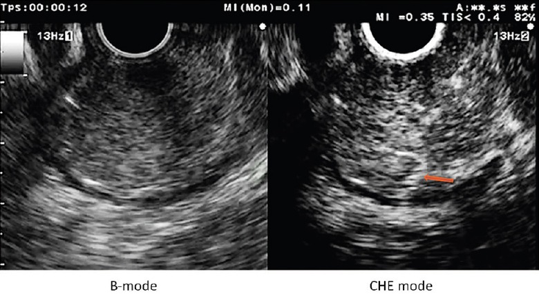
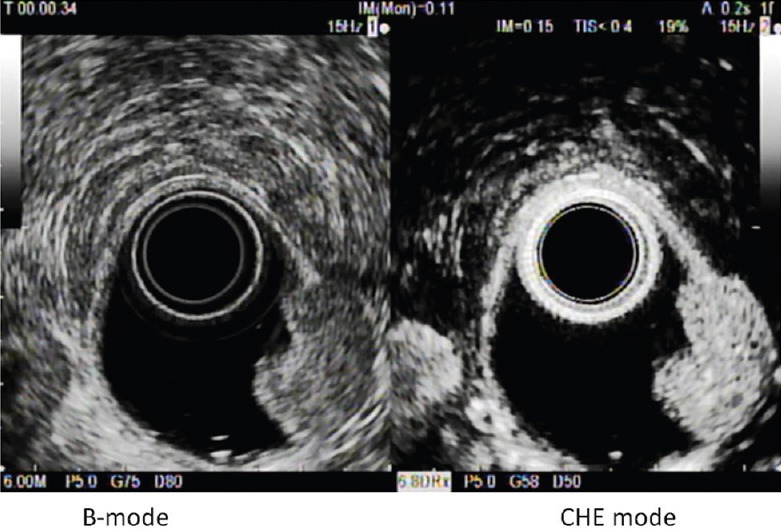
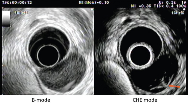
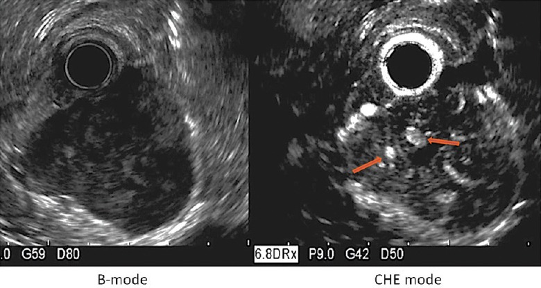
With the widespread use of endoscopy, gastrointestinal submucosal lesions are now more commonly discovered. Although endoscopic ultrasound (EUS) is superior to all other imaging techniques for the diagnosis of submucosal tumors (SMTs), it is still suboptimal for differentiating hypoechoic lesions arising from the fourth sonographic gastrointestinal wall layer, which encompass tumors with very different prognosis. EUS tissue acquisition has provided with the unique opportunity to obtain histological confirmation, but it is not accurate enough to evaluate the malignant potential of gastrointestinal stromal tumors (GISTs). In the last years, contrast-enhanced harmonic EUS (CH-EUS) emerged as a powerful imaging modality to assess the microperfusion patterns of pancreatic tumors. Based on the distinct microvascularity of malignant SMTs, it was hypothesized that CH-EUS might also assist in the differential diagnosis of SMTs. Preliminary experience in this field is now available and suggests CH-EUS as a performant modality to distinguish between benign SMTs and GISTs and to evaluate the malignant potential of GISTs. High expectations are also relied on CH-EUS for the monitoring of antiangiogenic treatments of GISTs and the evaluation of gastrointestinal neuroendocrine tumors (NETs).
Conflict of interest statement
There are no conflicts of interest.
Figures
References
-
- Hedenbro JL, Ekelund M, Wetterberg P. Endoscopic diagnosis of submucosal gastric lesions. The results after routine endoscopy. Surg Endosc. 1991;5:20–3. - PubMed
-
- Nishida T, Kawai N, Yamaguchi S, et al. Submucosal tumors: Comprehensive guide for the diagnosis and therapy of gastrointestinal submucosal tumors. Dig Endosc. 2013;25:479–89. - PubMed
-
- Hwang JH, Saunders MD, Rulyak SJ, et al. A prospective study comparing endoscopy and EUS in the evaluation of GI subepithelial masses. Gastrointest Endosc. 2005;62:202–8. - PubMed
-
- Mekky MA, Yamao K, Sawaki A, et al. Diagnostic utility of EUS-guided FNA in patients with gastric submucosal tumors. Gastrointest Endosc. 2010;71:913–9. - PubMed
Publication types
LinkOut - more resources
Full Text Sources
Other Literature Sources
