. 2016 Dec 21:6:36411.
doi: 10.1038/srep36411.

Hematopoietic Stem Cells in Neural-crest Derived Bone Marrow

Affiliations

Hematopoietic Stem Cells in Neural-crest Derived Bone Marrow

Nan Jiang et al. Sci Rep. .

Abstract

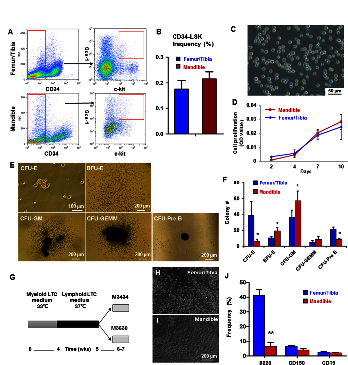
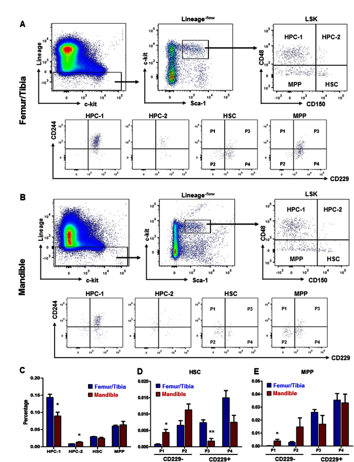
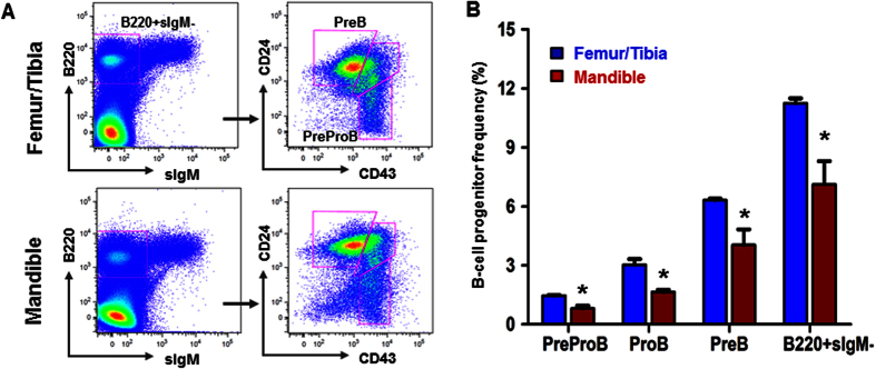
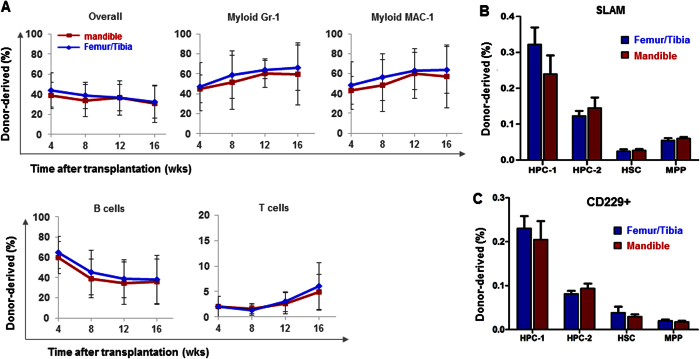
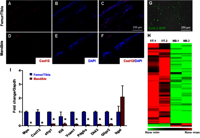
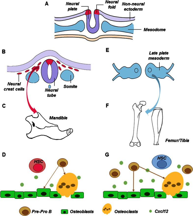
Hematopoietic stem cells (HSCs) in the endosteum of mesoderm-derived appendicular bones have been extensively studied. Neural crest-derived bones differ from appendicular bones in developmental origin, mode of bone formation and pathological bone resorption. Whether neural crest-derived bones harbor HSCs is elusive. Here, we discovered HSC-like cells in postnatal murine mandible, and benchmarked them with donor-matched, mesoderm-derived femur/tibia HSCs, including clonogenic assay and long-term culture. Mandibular CD34 negative, LSK cells proliferated similarly to appendicular HSCs, and differentiated into all hematopoietic lineages. Mandibular HSCs showed a consistent deficiency in lymphoid differentiation, including significantly fewer CD229 + fractions, PreProB, ProB, PreB and B220 + slgM cells. Remarkably, mandibular HSCs reconstituted irradiated hematopoietic bone marrow in vivo, just as appendicular HSCs. Genomic profiling of osteoblasts from mandibular and femur/tibia bone marrow revealed deficiencies in several HSC niche regulators among mandibular osteoblasts including Cxcl12. Neural crest derived bone harbors HSCs that function similarly to appendicular HSCs but are deficient in the lymphoid lineage. Thus, lymphoid deficiency of mandibular HSCs may be accounted by putative niche regulating genes. HSCs in craniofacial bones have functional implications in homeostasis, osteoclastogenesis, immune functions, tumor metastasis and infections such as osteonecrosis of the jaw.


Conflict of interest statement

The authors declare no competing financial interests.

Figures

Figure 1
Figure 1. Characterization of HSC-like cells in neural-crest derived mandible.
(A) Flow cytometry profiles of CD34ˉLSK cells. Lineage-depleted cells with CD34 negative and c-Kit and Sca-1 double positive selection. (B) Quantitative counts of CD34ˉLSK cells from femur/tibia and mandible at 0.176 ± 0.034 and 0.216 ± 0.027, respectively (mean ± s.d., n = 5). (C) Sorted mandibular CD34ˉLSK cells cultured for 10 days. (D) Proliferation rates of CD34ˉLSK cells from mandible and femur/tibia (mean ± s.d., n = 3). (E) Representative images of colony-forming mandibular CD34ˉLSK cells, including colony-forming unit erythroid (CFU-E), mature burst-forming unit-erythroid (BFU-E), colony-forming unit granulocyte/macrophage (CFU-GM), CFU-granulocyte/ erythroid/ megakaryocyte/ macrophage (CFU-GEMM) and pre-B lymphoid (CFU-Pre B). (F) Colony numbers of mandibular CD34ˉLSK cells (mean ± s.d., n = 3, *p < 0.05). (G) Diagram of LTC-IC assay. (H,I) HSCs cultured on a feeder layer in the myeloid medium for 4 wks and lymphoid medium for 1 wk. (J) Flow cytometry of differentiated long-term culture-initiating cells in methylcellulose culture medium (mean ± s.d., n = 3, **p < 0.01).
Figure 2
Figure 2. SLAM family markers by LSK HSC stem/progenitors.
(A,B) Gating for femur/tibia and mandibular LSK cells, CD150 and CD48. The CD150CD48+ LSK was labeled as HPC-1, CD150+ CD48+ LSK were labeled as HPC-2, CD150+ CD48−/low LSK were labeled as HSC and CD150CD48−/low LSK were labeled as MPP. Doublets, red blood cells, and dead cells were excluded prior to analysis. HSC and MPP were gated by CD229 and CD244. CD229CD244+. CD229CD244, CD229+ CD244+ and CD229+ CD244 HSC/MPP were labeled as P1, P2, P3 and P4, respectively. (C) Frequency subdivided by SLAM markers (mean ± s.d., n = 3, *p < 0.05). (D,E) Frequency of stem and progenitor fractions subdivided by CD229 and CD244 of HSC and MPP populations. (mean ± s.d., n = 3, *p < 0.05, **p < 0.01).
Figure 3
Figure 3. Flow cytometry for isolation of lymphoid progenitor cells.
(A) Representative gates to isolate lymphoid progenitor cells. B220 + sIgM-CD43 + CD24- labeled as Pre-Pro B cells; B220 + sIgM-CD43 + CD24 + labeled as Pro B cells; B220 + sIgM-CD43- labeled as Pre B cells. (B) Frequency of lymphoid progenitor cells. (mean ± s.d., n = 3, *p < 0.05).
Figure 4
Figure 4. In vivo rescue of irradiated bone marrow.
(A) A total of 3 × 105 donor HSC-like cells from the mandible and from femur/tibia both reconstituted marrow hematopoiesis following sublethal irradiation, with similarly restored levels of myeloid cell, B-cell and T-cell (mean ± s.d., three experiments with a total of 12–15 recipient mice per group). (B) Frequency of stem and progenitor cells subpopulated by SLAM family markers (mean ± s.d., n = 5). (C) Frequency of CD229 positive cells of stem and progenitor cells (mean ± s.d., n = 5).
Figure 5
Figure 5. Profiling of HSC niche signals.
(A–C) Cxcl12 immunofluorescence of the femur/tibia and (D–F) mandible. (G) Representative images of GFP positive, osteoblasts isolated from the mandible. (H) Heat map of global gene expression with significant change (p < 0.05). Up-regulated genes are indicated as red color and down-regulated genes are indicated as green color. (I) Selected genes differentially expressed in femur/tibia and mandible by using qRT-PCR (mean ± s.d., n = 3, *p < 0.05).
Figure 6
Figure 6. Schematics of neural-crest derived vs. mesoderm derived hematopoietic stem cells.
Neural tube derives from neural epithelium early in development, with adjacent paraxial mesoderm (A). Neural crest cells split from neural tube and begin to migrate towards the presumptive face and form the first branchial arch (B), from which the mandible is derived (C). Here, we demonstrated that neural-crest derived bone (mandible) harbors HSCs that benchmark with mesoderm derived femur/tibia HSCs, and yet with a severe deficiency in lymphoid differentiation (D). Signals in HSC niche in the form of bound or secreted molecules, including osteoblast derived Cxcl12, regulate HSC functions (D). Contrastingly, HSCs derive from paraxial mesoderm reside in appendicular bones such as femur and tibia (E,F) and have been well characterized, including progenitors of osteoclasts and Cxcl12 as a pivotal signal that regulates HSC niche (G).

References

    1. Spangrude G. J., Heimfeld S. & Weissman I. L. Purification and characterization of mouse hematopoietic stem cells. Science 241, 58–62 (1988). - PubMed
    1. Warr M. R., Pietras E. M. & Passegue E. Mechanisms controlling hematopoietic stem cell functions during normal hematopoiesis and hematological malignancies. Wiley interdisciplinary reviews. Systems biology and medicine 3, 681–701 (2011). - PubMed
    1. Lo Celso C. & Scadden D. T. The haematopoietic stem cell niche at a glance. J Cell Sci 124, 3529–3535 (2011). - PMC - PubMed
    1. Morrison S. J. & Scadden D. T. The bone marrow niche for haematopoietic stem cells. Nature 505, 327–334 (2014). - PMC - PubMed
    1. Doan P. L. et al.. Tie2(+) bone marrow endothelial cells regulate hematopoietic stem cell regeneration following radiation injury. Stem cells 31, 327–337 (2013). - PMC - PubMed

Publication types