. 2017 Sep;10(5):1248-1258.
doi: 10.1038/mi.2016.115. Epub 2016 Dec 21.

Mycobacterium tuberculosis cell wall released fragments by the action of the human lung mucosa modulate macrophages to control infection in an IL-10-dependent manner

Affiliations

Mycobacterium tuberculosis cell wall released fragments by the action of the human lung mucosa modulate macrophages to control infection in an IL-10-dependent manner

J Arcos et al. Mucosal Immunol. 2017 Sep.

Abstract

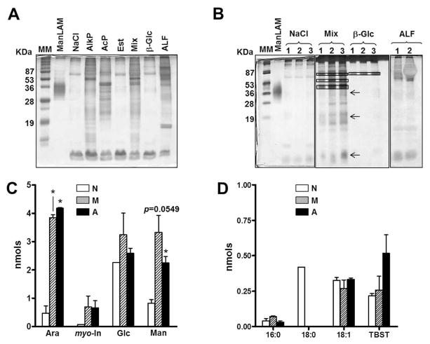
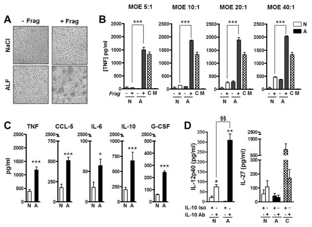
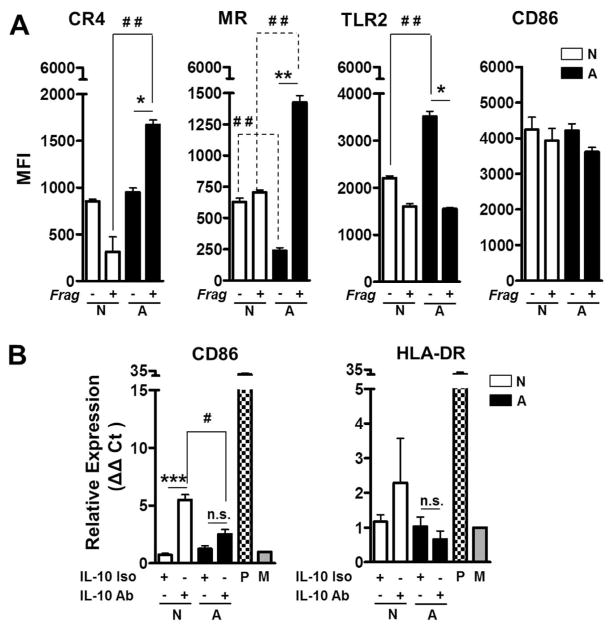
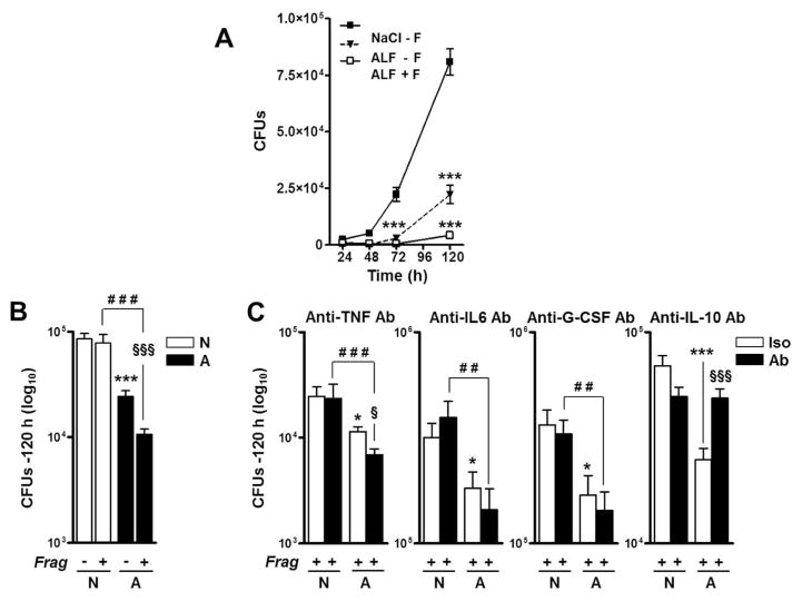
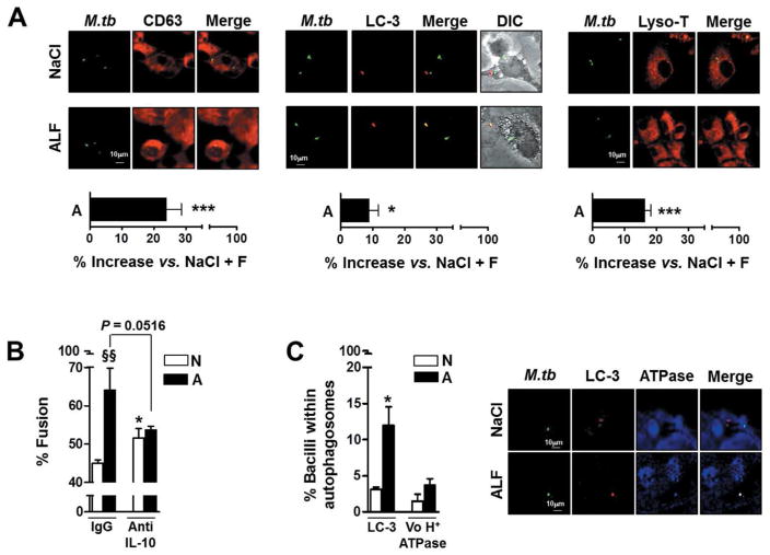
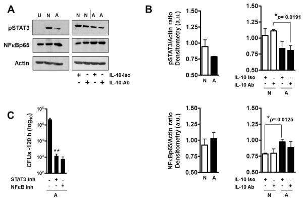
Mycobacterium tuberculosis (M.tb), the causative agent of tuberculosis, is a major public health challenge facing the world. During infection, M.tb is deposited in the lung alveolar space where it comes in contact with the lung mucosa, known as alveolar lining fluid (ALF), an environment that M.tb encounters at different stages of the infection and disease. ALF is abundant in homeostatic and antimicrobial hydrolytic enzymes, also known as hydrolases. Here we demonstrate that ALF hydrolases, at their physiological concentrations and upon contact with M.tb, release M.tb cell envelope fragments into the milieu. These released fragments are bioactive, but non-cytotoxic, regulate the function of macrophages, and thus are capable of modulating the immune response contributing to the control of M.tb infection by human macrophages. Specifically, macrophages exposed to fragments derived from the exposure of M.tb to ALF were able to control the infection primarily by increasing phagosome-lysosome fusion and acidification events. This enhanced control was found to be dependent on fragment-induced interleukin-10 (IL-10) production but also involves the STAT3 signaling pathway in an IL-10-independent manner. Collectively our data indicate that M.tb fragments released upon contact with lung mucosa hydrolases participate in the host immune response to M.tb infection through innate immune modulation.


Conflict of interest statement

Disclosure/Conflict of Interest

The authors declared no conflict of interest.

Figures

Fig. 1
Fig. 1. Analysis of ALF exposed-M.tb released fragments
Single cell suspensions of M.tb were exposed to 0.9% NaCl (control), human ALF, single hydrolases or to a mixture (Mix) of the most active hydrolases [AlkP + AcP + Est] for 12 h, 37°C, 5% CO2. Supernatants containing released fragments were analyzed. (A, B) A representative SDS-PAGE analysis of released fragments followed by a periodic acid silver stain. (A) M.tb released fragments from 5×10⁶ M.tb exposed to different ALF hydrolases and human ALF. AlkP and AcP: alkaline and acid phosphatases; Est: non-specific esterase; β-Glc (β-glucosidase). (B) Released fragments from 3 independent experiments (–3) showing reproducibility. Arrows indicate the locations of ManLAM, LM and PIMs on the gel, from high to low molecular mass respectively, and boxes indicate the location of the hydrolase. (C, D) Released fragments (normalized by bacterial number and volume) were chemically hydrolyzed and their sugar and fatty acid composition determined by mass spectrometry. (C) Neutral sugar analyses (n=3, in duplicate) showing that fragments are composed of myo-inositol (myo-In), mannose (Man), glucose (Glc), and arabinose (Ara). (D) Fatty acid analyses of methyl ester derivatives show fragments containing fatty acids (n=2, duplicate). TBST: Tuberculostearic acid. Student’s t test, *P<0.05. N: Fragments generated by M.tb exposure to NaCl; M: Fragments generated by M.tb exposure to the mixture of hydrolases; A: Fragments generated by M.tb exposure to ALF. For each ‘n’ value, ALFs were obtained from different human donors.
Fig. 2
Fig. 2. Cytokine and chemokine production induced by macrophages exposed to M.tb cell wall fragments
(A) Macrophages exposed to M.tb cell wall fragments trigger cell aggregation. Human macrophage monolayers were exposed to fragments (+F) or controls (−F) for 24 h at 37 °C, 5%CO2. Monolayers exposed to human ALF developed cell aggregations. Aggregations were observed as early as 12 h (data not shown) and increased in number and size through 240 h until the monolayer lost integrity. Fragments obtained upon M.tb exposure to 0.9% NaCl or exposure to β-Glc (not shown) served as controls. (B) TNF production by human macrophages exposed to fragments at different MOE. Human macrophages were exposed to fragments (+F) or controls (−F) at different MOE. A representative (M±SD) experiment in triplicate of n=3 is shown. Student t test was used to compare fragments generated by M.tb exposure to ALF with fragments generated by M.tb exposure to NaCl, ***P<0.0005. C: LPS (positive control), M: Media (background). (C) Human macrophage monolayers were exposed to M.tb cell wall fragments at a MOE of 20:1 for 12 h, 37°C, 5% CO2, and cytokine and chemokine secretion was measured from cell supernatants by ELISA; between n=4 and 9 experiments were performed depending of the measured immune component in triplicate. (D) IL-12p40 and IL-27 secretion in the absence [(−) isotype control] or presence (+) of anti-IL-10 neutralizing antibody (n=3). In (C/D), shown M±SEM, Student’s t test was used to compare the immune response induced by fragments generated by M.tb exposure to ALF with fragments generated by M.tb exposure to NaCl in the absence (*P<0.05; **P<0.005; ***P<0.0005) or in the presence of anti-IL-10 Ab (§§P<0.005). NaCl-Frag [N]; ALF-Frag [A]. For each ‘n’ value, both ALF and macrophages were obtained from different human donors.
Fig. 3
Fig. 3. Effects of ALF fragments on macrophage receptors and activation
(A) Macrophages exposed to ALF exposed M.tb [A] or 0.9% NaCl exposed M.tb [N] released cell wall fragments induce the surface expression of the MR and CR4, but not TLR2. Differences in the macrophage activation marker CD86 on the macrophage surface were not observed. Experiments were performed n=3 and cells analyzed by flow cytometry. Shown M±SEM, Student’s t test was used to compare the receptor/marker surface expression induced by fragments generated by M.tb exposure to ALF with fragments generated by M.tb exposure to NaCl (*P<0.05; **P<0.005). (B) Expression of CD86 and HLA-DR as measured by RT-PCR (n=3). P: Positive Control; M: Medium. Student’s t test was used to compare the mRNA levels induced by fragments generated by M.tb exposure to ALF with fragments generated by M.tb exposure to NaCl in the presence (#P<0.05; ***P<0.0005) or in the absence of anti-IL-10 Ab. For each ‘n’ value, both ALF and macrophages were obtained from different human donors.
Fig. 4
Fig. 4. Effects of ALF on M.tb intracellular survival in human macrophages
M.tb bacilli were exposed to 0.9% NaCl (control), Mix, or ALF at relevant in vivo concentrations. Macrophages were infected at a MOI 1:1 with exposed M.tb in the absence or presence of fragments and in the absence or presence of anti-TNF, anti-IL-10, anti-IL-6 or anti-G-CSF neutralizing Abs. Intact macrophage monolayers were verified by inverted phase microscopy throughout the infection period. M.tb survival was determined by CFUs at the indicated intervals. (A) A representative experiment in triplicate is shown (M±SD), Student t test was used to compare control with vs. without fragments (±F), ***p<0.0005. (B) Overall data at 120 h from n=3 each performed in triplicate (M±SEM). (C) Effects of neutralizing TNF, IL-10, IL-6 or G-CSF on macrophages in the presence of fragments during exposed M.tb infection (n=3 in triplicate, M±SEM). Notice that y-axes are different among the graphs shown. One-way ANOVA, Tukey-Posttest was used to compare the effects on the M.tb cell wall (*, white bars), ± F (§, white vs. black bars), and M.tb exposed to different conditions in the presence of fragments (#, black bars), *P<0.05; **P<0.005; ***P<0.0005; §P<0.05; §§§P<0.0005; ##P<0.005; ###P<0.0005. NaCl-M.tb + Frag [N]; ALF-M.tb + Frag Frag [A]. For each ‘n’ value, both ALF and macrophages were obtained from different human donors.
Fig. 5
Fig. 5. Effects of human ALF on phagolysosome (P-L) fusion in macrophages
Macrophage monolayers on coverslips were incubated with NaCl- or ALF-exposed-GFP-M.tb (MOI 10:1) in the presence (MOE 10:1) or absence of their respective released fragments (± F) for 2 h. Cells were washed, fixed, permeabilized, and stained with anti-human CD63, LC-3, V0H+ATPAse, their respective IgG controls, or with Lyso-Tracker (Lyso-T) (A) Shown are merged images where CD63 (left panel), LC-3 (middle panel) or Lyso-T (right panel) positive compartments are red, GFP-exposed-M.tb bacilli in unfused vesicles are green, and those co-localized are yellow. Phagolysosome (CD63/M.tb), autophagosome (LC-3/M.tb) fusion events and acidification (Lyso-T/M.tb) were examined and enumerated via confocal microscopy, n=3–4 counting >150 events per coverslip, in triplicate (original magnification X600). Overall percent increase from n=3–4 in triplicate (M±SEM); Student t test was used to compare ALF exposed M.tb with NaCl exposed M.tb (control) in the presence of fragments, *P<0.05; **P<0.005; ***P<0.0005. (B) Shown are merged images where LC-3 positive compartments are red, V0H+ATPAse compartments are blue, and those co-localized are yellow. Student t test was used to compare Mix or ALF exposed M.tb with NaCl-exposed M.tb (control) in the presence of fragments, *P<0.05. (C) P-L fusion was quantified in the presence of anti-IL-10 neutralizing Ab. Student t test was used to compare ALF exposed M.tb with NaCl-exposed M.tb (control) in the presence of fragments, *P<0.05 (between white bars), §§P<0.005 (between white and black bars under the same condition). For each ‘n’ value, both ALF and macrophages were obtained from different human donors.
Fig. 6
Fig. 6. Effects of ALF on macrophage STAT3 and NFκB activation
Following infections as described in Fig.4, lysates from ALF-M.tb [A] or NaCl-M.tb [N] infected macrophages in the presence of their respective fragments were obtained. (A) Representative Western blots show that ALF-exposed M.tb in the presence of fragments decreased the activation of STAT3 signaling pathway and increased NFκB activation. This was not an IL-10 dependent mechanism. U: Uninfected; N and A: NaCl- or –ALF-exposed M.tb in the presence of fragments, respectively. (B) Densitometry analysis of n=3 showing phosphorylated STAT3 and NFκB vs. Actin ratios. Student t test was used to compare ALF exposed M.tb with NaCl-exposed M.tb (control) in the presence of fragments, §§P<0.005 (between white and black bars under the same condition). (C) Inhibition of STAT3 and NFκB both allowed macrophages a further enhanced control of ALF-M.tb infection in the presence of fragments. Student t test was used to compare ALF exposed M.tb infection in the presence of fragments in the absence or presence of inhibitors, **P<0.005. For each ‘n’ value, both ALF and macrophages were obtained from different human donors.

References

    1. WHO. WHO Tuberculosis Fact Sheet 2015. World Health Organization; 15 A.D. Mar 20, Available from: http://www.who.int/mediacentre/factsheets/fs104/en/
    1. Torrelles JB. Broadening our view about the role of Mycobacterium tuberculosis cell envelope components during infection: A battle for survival. In: Cardona PJ, editor. Understanding Tuberculosis - Analyzing the Origin of Mycobacterium tuberculosis Pathogenicity. Rijeka, Croatia: Intech; 2012. pp. 77–122.
    1. Arcos J, Sasindran SJ, Fujiwara N, Turner J, Schlesinger LS, Torrelles JB. Human lung hydrolases delineate Mycobacterium tuberculosis-macrophage interactions and the capacity to control infection. J Immunol. 2011;187:372–381. - PMC - PubMed
    1. Schlesinger LS. Phagocytosis and Toll-like Receptors in Tuberculosis. In: Rom WN, Garay SM, editors. Tuberculosis. 2. Lippincott, Williams, & Wilkins; 2004. pp. 203–214.
    1. Sasindran J, Torrelles JB. Mycobacterium tuberculosis infection and inflammation: What is beneficial for the host and for the bacterium? Front Microbiol. 2011;2:1–16. - PMC - PubMed

Publication types