. 2016 Dec 21:6:39420.
doi: 10.1038/srep39420.

The matrix protein of rabies virus binds to RelAp43 to modulate NF-κB-dependent gene expression related to innate immunity

Affiliations

The matrix protein of rabies virus binds to RelAp43 to modulate NF-κB-dependent gene expression related to innate immunity

Youcef Ben Khalifa et al. Sci Rep. .

Abstract

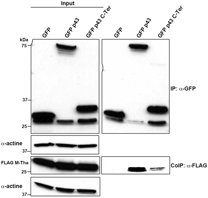
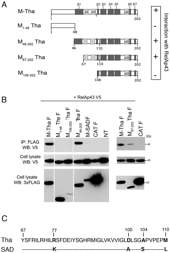
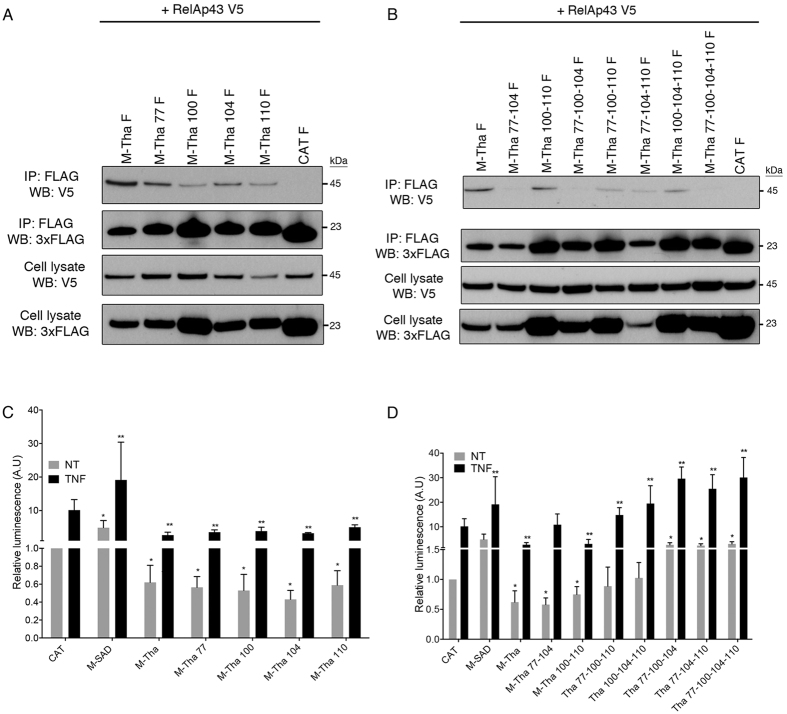
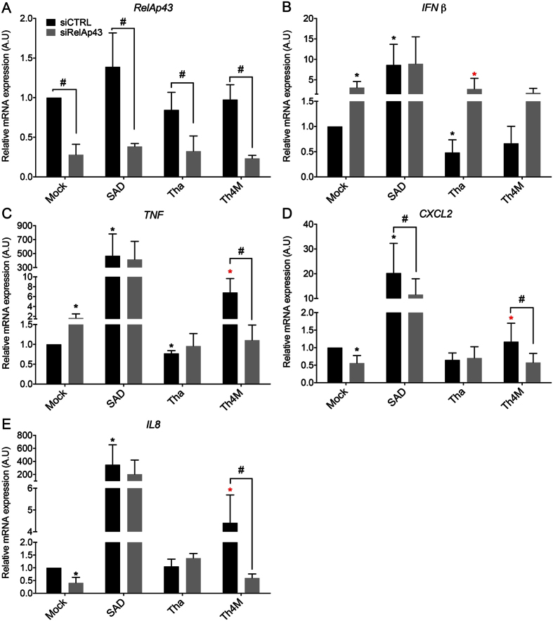
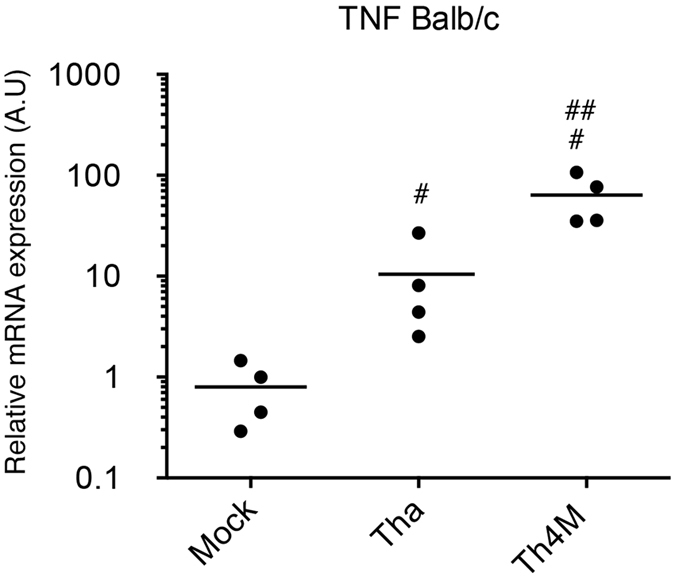
The matrix (M) protein of wild isolates of rabies virus such as Tha (M-Tha) was previously shown to be able to interact with RelAp43, a protein of the NF-κB family, and to efficiently suppress NF-κB-dependent reporter gene expression, in contrast with the vaccine strain SAD. Here, we analyze the mechanisms involved in RelAp43-M protein interaction. We demonstrate that the central part of M-Tha, and the specific C-terminal region of RelAp43 are required for this interaction. Four differences in the corresponding amino acid sequences of the M-Tha and M-SAD are shown to be crucial for RelAp43 interaction and subsequent modulation of innate immune response. Furthermore, the capacity of M-Tha to interact with RelAp43 was shown to be crucial for the control of the expression of four genes (IFN, TNF, IL8 and CXCL2) during viral infection. These findings reveal that RelAp43 is a potent regulator of transcription of genes involved in innate immune response during rabies virus infection and that the M protein of wild isolates of rabies virus is a viral immune-modulatory factor playing an important role in this RelAp43-mediated host innate immunity response in contrast to M protein of vaccine strains, which have lost this property.


Conflict of interest statement

The authors declare no competing financial interests.

Figures

Figure 1
Figure 1. M-Tha interacts with the specific C-terminal region of RelAp43.
HeLa cells were co-transfected with FLAG-tagged M-Tha and GFP as a negative control, GFP p43 as a positive control or GFP p43 C-Ter. Co-IP were performed using GFP-Trap and the presence of GFP or FLAG-tagged protein was analyzed by western blot using specific antibodies in cell lysates either before (Input, left panel) or after IP (right panel). The western blots depicted here are the results of a representative experiment (three repeats).
Figure 2
Figure 2. Amino acids 67 to 110 of M-Tha are involved in the interaction with RelAp43.
(A) Schematic representation of the truncated mutants of M-Tha used in this study. Their capacity of interaction with RelAp43 is summarized on the right part of the Figure (+: interaction with RelAp43; - : absence of interaction). (B) HeLa cells were used to co-express V5-tagged RelAp43 in combination with the FLAG-tagged CAT as a negative control or the indicated FLAG-tagged M construct. After 24 h, cells were lysed, protein expression in cell lysates was controlled (cell lysate: WB V5; cell lysate: WB 3xFLAG) and co-IP experiments were performed using anti-FLAG M2 beads. Interaction of RelAp43 with the different M constructs or CAT was visualized by western blot (IP: 3xFLAG; WB: V5). The western blots depicted here are the results of a representative experiment (three repeats). (C) Alignment of the 67 to 110 amino acids sequences of M-Tha and M-SAD using the ClustalX software. Four differences appeared in the sequences (in bold), respectively at position 77 (R in M-Tha, K in M-SAD), 100 (D in M-Tha, A in M-SAD), 104 (A in M-Tha, S in M-SAD) and 110 (M in M-Tha, L in M-SAD).
Figure 3
Figure 3. Residues at positions 77 and 104 are necessary for the interaction of M-Tha with RelAp43 and should be coupled with mutations D100A and/or M110L to restore a level of activation of the NF-κB reporter vector comparable to M-SAD.
(A and B) HeLa cells were used to co-express V5-tagged RelAp43 in combination with the indicated FLAG-tagged single (A) or multiple (B) mutants of M-Tha. After 24 h, cells were lysed, protein expression in cell lysates was controlled (cell lysate: WB V5; cell lysate: WB 3xFLAG on the Figure) and co-IP experiments were performed using anti-FLAG M2 beads. Interaction of RelAp43 with the different M constructs or CAT was visualized by western blot (IP: 3xFLAG; WB: V5 on the picture). The western blots depicted here are the results of a representative experiment (three repeats). (C) Modulation of NF-κB activation in 293T cells in the presence of single or double mutants of M-Tha compared to M-Tha, M-SAD or CAT. The NF-κB pathway was exogenously activated using 10 ng/mL TNF-α during 5 h (black bars) or left untreated (grey bars). The CAT control without the TNF treatment was used as the reference condition. (D) Modulation of NF-κB activation in 293T cells in the presence of triple or quadruple mutants of M-Tha compared to M-Tha, M-SAD or CAT. The NF-κB pathway was exogenously activated using 10 ng/mL TNF-α during 5 h (black bars) or left untreated (grey bars). The CAT control without the TNF treatment was used as the reference condition. Significant results (p < 0.05) in comparison to untreated (*) and TNF treated (**) CAT transfected cells were annotated.
Figure 4
Figure 4. Mutation K77R or S104A in M-SAD is sufficient to induce its interaction with RelAp43 and should be coupled with mutations D100A and M110L to abolish the M-SAD activation of the NF-κB reporter vector.
(A) HeLa cells were used to co-express V5-tagged RelAp43 in combination with the FLAG-tagged CAT as a negative control, M-Tha as a positive control or the indicated FLAG-tagged single, double or quadruple mutants of M-SAD. After 24 h, cells were lysed, protein expression in cell lysates was controlled (Input: WB V5 and WB 3xFLAG) and co-IP experiments were performed using anti-FLAG M2 beads. Interaction of RelAp43 with the different M constructs or CAT was visualized by western blot (IP: 3xFLAG; WB: V5). The western blots depicted here are the results of a representative experiment (three repeats). (B) Modulation of NF-κB activation in 293T cells in the presence of single, double or quadruple mutants of M-SAD compared to M-Tha, M-SAD or CAT. The NF-κB pathway was exogenously activated using 10 ng/mL TNF-α during 5 h (black bars) or left untreated (grey bars). The M protein of vaccine strain SAD with the TNF treatment was arbitrary considered as a reference. Significant results (p < 0.05) in comparison to untreated (*) and TNF treated (**) CAT transfected cells were annotated.
Figure 5
Figure 5. M-Tha positions 77, 100, 104, 110 are important for viral escape.
HeLa cells were infected by Tha, Th4M or SAD. After 43 hours of infection, the NF-κB pathway was exogenously activated using 10 ng/mL TNF-α during 5 h (black bars) or left untreated (grey bars) and total RNA was extracted. The expression level of the following genes: IFNβ (A), TNF (B), IL8 (C), CXCL2 (D) was studied by RT-qPCR analysis. Significant results (p < 0.05) compared to the untreated (*) or TNF treated (**) mock and to the untreated (#) or TNF treated (##) Th4M infected cells are annoted. The levels of gene expression were normalized according to the level of GAPDH reporter gene in non-infected and untreated cells.
Figure 6
Figure 6. RelAp43 is involved in the modulation of genes related to innate immunity.
HeLa cells were infected by Tha, Th4M or SAD virus and treated with a siRNA control (siCTRL) or a siRNA directed against RelAp43 (siRelAp43). After 48 hours of infection, total RNA was extracted. The efficiency of siRNA treatment was evaluated by the measure of RelAp43 mRNA expression by RT-qPCR (A). The expression level of the following genes: IFNβ (B), TNF (C), IL8 (D), CXCL2 (E) was studied by RT-qPCR analysis. Significant results (p < 0.05) compared to the siCTRL of mock (*) and Tha () infected cells are annoted. (#) indicated significant results between the siCTRL treated sample and the siRelAp43 respective one (p < 0.05). The levels of gene expression were normalized according to the level of GAPDH reporter gene in non-infected and untreated cells.
Figure 7
Figure 7. Matrix protein of Tha virus modulates the transcription of TNF in vivo.
Relative quantification of TNF mRNA expression was performed in the brain of mice infected with Tha and Th4M viruses. Four six-weeks-old BALB/c were infected in intramuscular injection of 1000 FFU and monitored over 21 days. The mice were sacrified upon the apparition of late infection symptoms (humane endpoint) and RNA were extracted from the brain. The expression of the TNF mRNA was studied by RT-qPCR analysis. The levels of gene expression were normalized according to the level of GAPDH reporter gene in non-infected mice. Significant results (p < 0.05) compared to uninfected mice (#) and Tha (##) infected mice are annoted.
Figure 8
Figure 8. Location of residues of the M protein involved in the interaction with RelAp43.
The M protein structure presented is that of Lagos bat virus. Residues 77 and 104, the crucial residues for interaction with RelAp43 are colored in green. Positions 100 and 110 are in yellow. The residues from the N-terminal regions that interact with the globular domains are shown as sticks. The two views are rotated by 90 degrees.

Similar articles

Cited by

References

    1. Luco S. et al. RelAp43, a member of the NF-kappaB family involved in innate immune response against Lyssavirus infection. PLoS Pathog 8, e1003060, doi: 10.1371/journal.ppat.1003060 (2012). - DOI - PMC - PubMed
    1. Thanos D. & Maniatis T. Virus induction of human IFN beta gene expression requires the assembly of an enhanceosome. Cell 83, 1091–1100, doi: 0092-8674(95)90136-1 (1995). - PubMed
    1. Verma I. M., Stevenson J. K., Schwarz E. M., Van Antwerp D. & Miyamoto S. Rel/NF-kappa B/I kappa B family: intimate tales of association and dissociation. Genes Dev 9, 2723–2735 (1995). - PubMed
    1. Saccani S., Pantano S. & Natoli G. Modulation of NF-kappaB activity by exchange of dimers. Mol Cell 11, 1563–1574, doi: S1097276503002272 (2003). - PubMed
    1. Oeckinghaus A. & Ghosh S. The NF-kappaB family of transcription factors and its regulation. Cold Spring Harb Perspect Biol 1, a000034, doi: 10.1101/cshperspect.a000034 (2009). - DOI - PMC - PubMed

Publication types