. 2016 Dec 21:6:39616.
doi: 10.1038/srep39616.

In vivo label-free photoacoustic flow cytography and on-the-spot laser killing of single circulating melanoma cells

Affiliations

In vivo label-free photoacoustic flow cytography and on-the-spot laser killing of single circulating melanoma cells

Yun He et al. Sci Rep. .

Abstract

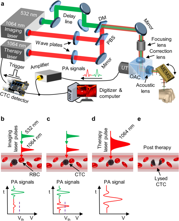
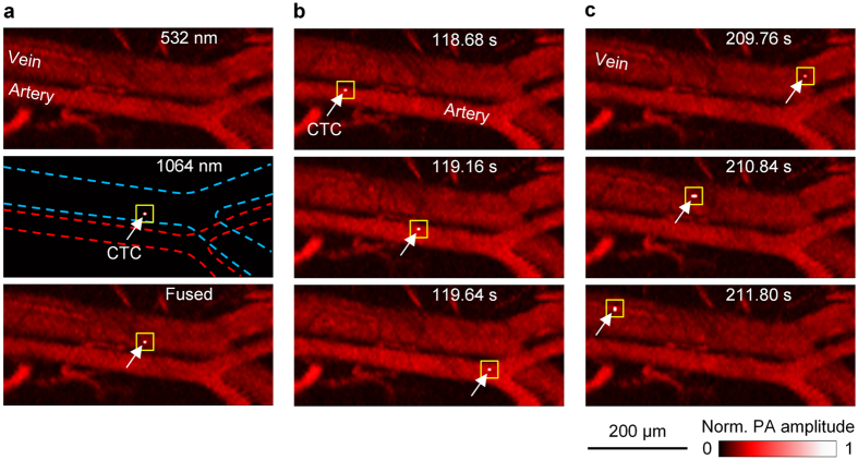
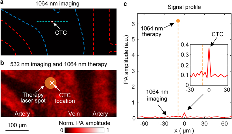
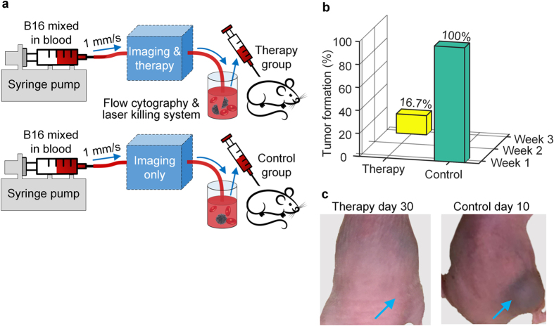
Metastasis causes as many as 90% of cancer-related deaths, especially for the deadliest skin cancer, melanoma. Since hematogenous dissemination of circulating tumor cells is the major route of metastasis, detection and destruction of circulating tumor cells are vital for impeding metastasis and improving patient prognosis. Exploiting the exquisite intrinsic optical absorption contrast of circulating melanoma cells, we developed dual-wavelength photoacoustic flow cytography coupled with a nanosecond-pulsed melanoma-specific laser therapy mechanism. We have successfully achieved in vivo label-free imaging of rare single circulating melanoma cells in both arteries and veins of mice. Further, the photoacoustic signal from a circulating melanoma cell immediately hardware-triggers a lethal pinpoint laser irradiation to kill it on the spot in a thermally confined manner without causing collateral damage. A pseudo-therapy study including both in vivo and in vitro experiments demonstrated the performance and the potential clinical value of our method, which can facilitate early treatment of metastasis by clearing circulating tumor cells from vasculature.


Conflict of interest statement

L.V.W. has a financial interest in Microphotoacoustics, Inc., which, however, did not support this work. The remaining authors declare no competing financial interest.

Figures

Figure 1
Figure 1. CTC imaging and destruction by dual-wavelength PA flow cytography combined with laser therapy.
(a) Schematic of selected components of the experimental system. DM, dichroic mirror; MEMS, micro-electro-mechanical-system scanning mirror; OAC, optical-acoustic combiner; PBS, polarizing beamsplitter; UT, ultrasonic transducer. The 1064 nm and 532 nm imaging lasers are employed to image CTCs and vasculature, respectively. (b–e) Scheme of real-time detection and laser killing of CTCs. The CTC detector compares the earlier 1064 nm laser-induced CTC-specific PA signal against an optimized threshold level (purple dashed line in (b) and (c)) above the Hb signal, and thus can reliably distinguish CTCs and trigger the therapy laser (c). Within ~10 μs, the therapy laser is fired and focused to the detected CTC location to photomechanically kill the CTC (e).
Figure 2
Figure 2. Snapshots showing single CTCs travelling in vasculature.
(a) 532 nm laser-induced (Top), 1064 nm laser-induced (Middle), and fused (Bottom) flow cytography images. In the 1064 nm laser-induced image, the white arrow and yellow square indicate the detected CTC; the red and blue dashed lines delineate the artery and vein boundaries, respectively. (b) Three fused snapshots spanning ~1 s, showing a single CTC traveling in the artery. (c) Three fused snapshots spanning ~2 s, showing a single CTC traveling in the vein. The times labeled in (b) and (c) are relative to CTC injection.
Figure 3
Figure 3. In vivo detection and expected photothermal killing of a CTC.
The CTC was first detected in the 1064 nm laser-induced flow cytography image (a) and then lethally irradiated by a therapy laser pulse, also at 1064 nm, with a 50-μm focal diameter (b). In (b), The much-higher-amplitude PA signal induced by the therapy laser pulse is illustrated by the filled yellow circle. (c) Profile of the PA signals from the region across the CTC, indicated by the dashed cyan line in (a). The x axis, parallel with the imaging laser’s scanning direction (from right to left), was centered at the CTC location. In the 1064 nm imaging laser-induced signals (red solid line), the CTC signal (shown in detail by the inset) had a contrast-to-noise-ratio of ~25. The therapy laser-induced PA signal’s peak location (dashed orange line) was only ~10 μm away from the detected CTC location, which indicated that the CTC position (white cross in (b)) was within the therapy laser’s focal spot (circle in (b)).
Figure 4
Figure 4
Scheme (a) and results (b,c) of the pseudo-therapy study. (a) The control experiment (bottom) was conducted after the therapy experiment (top) by simply switching off the therapy laser. The flow rate was set at ~80 μL/hr with ~20 CTCs flowing through the system every second. (b) Only 1 out of 6 therapy experiments had a tumor detected at week 3, compared to 100% tumor formation by week 1 in the control group. (c) Representative photos of the mice after experiments. (Left) No tumor was detected around the inoculation site (blue arrow) in 30 days following a therapy experiment. (Right) A raised tumor (blue arrow) was observed 10 days after a control experiment.

Similar articles

Cited by

References

    1. Chaffer C. L. & Weinberg R. A. A perspective on cancer cell metastasis. Science 331, 1559–1564 (2011). - PubMed
    1. Steeg P. S. Tumor metastasis: mechanistic insights and clinical challenges. Nature medicine 12, 895–904 (2006). - PubMed
    1. Plaks V., Koopman C. D. & Werb Z. Circulating tumor cells. Science 341, 1186–1188 (2013). - PMC - PubMed
    1. Kaiser J. Cancer’s circulation problem. Science 327, 1072–1074 (2010). - PubMed
    1. Chambers A. F., Groom A. C. & MacDonald I. C. Metastasis: dissemination and growth of cancer cells in metastatic sites. Nature Reviews Cancer 2, 563–572 (2002). - PubMed

Publication types