. 2017 Jan 12;541(7636):176-181.
doi: 10.1038/nature20793. Epub 2016 Dec 21.

Piezo2 senses airway stretch and mediates lung inflation-induced apnoea

Affiliations

Piezo2 senses airway stretch and mediates lung inflation-induced apnoea

Keiko Nonomura et al. Nature. .

Abstract

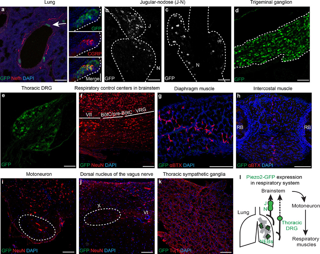
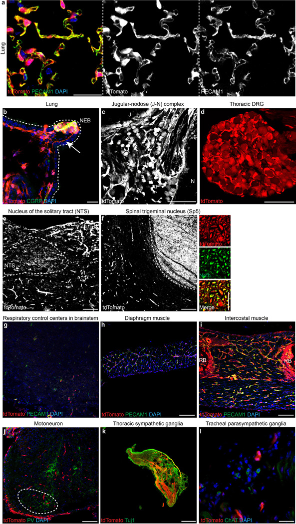
Respiratory dysfunction is a notorious cause of perinatal mortality in infants and sleep apnoea in adults, but the mechanisms of respiratory control are not clearly understood. Mechanical signals transduced by airway-innervating sensory neurons control respiration; however, the physiological significance and molecular mechanisms of these signals remain obscured. Here we show that global and sensory neuron-specific ablation of the mechanically activated ion channel Piezo2 causes respiratory distress and death in newborn mice. Optogenetic activation of Piezo2+ vagal sensory neurons causes apnoea in adult mice. Moreover, induced ablation of Piezo2 in sensory neurons of adult mice causes decreased neuronal responses to lung inflation, an impaired Hering-Breuer mechanoreflex, and increased tidal volume under normal conditions. These phenotypes are reproduced in mice lacking Piezo2 in the nodose ganglion. Our data suggest that Piezo2 is an airway stretch sensor and that Piezo2-mediated mechanotransduction within various airway-innervating sensory neurons is critical for establishing efficient respiration at birth and maintaining normal breathing in adults.


Figures

Extended Data Figure 1
Extended Data Figure 1. Characterization of lung development in Piezo2−/− mice
a, b, Whole-mount staining for αSMA in the left lobe of lungs from E16.5 (a) and E18.5 (b) wild-type and Piezo2−/− mice. L1–L6, lateral branches of the left lobe. E16.5: wild-type n = 4, Piezo2−/− n = 3; E18.5: wild-type n = 4, Piezo2−/− n = 5. A normal conducting airway and vessel pattern was observed in Piezo2−/− lungs at late embryonic stages. c, d, Whole-mount immunostaining for E-cadherin and Muc-1 (c) or E-cadherin, T1α, and SpC (d) in left lobes from E18.5 wild-type and Piezo2−/− mice. Arrows indicate proximal (P) to distal (D) direction. Left panels, lower magnification; right panels, higher magnification of the distal region. Solid circles in right panels indicate alveolar progenitors in the distal region. Solid hexagons in left panels indicate nascent type I pneumocytes. Yellow dashed circles in left panels indicate nascent type II pneumocytes. A normal epithelial morphology and no defects in alveolar epithelial patterning and differentiation were observed Piezo2−/− lungs. e, Representative ultrastructures of wild-type and Piezo2−/− newborn lungs. Black arrows mark type I pneumocyte extensions. White dotted circles mark type II pneumocytes. White arrowheads within white dotted circles mark lamellar bodies. RBC, red blood cells. Samples from four mice per genotype were analysed. Normal morphology and similar abundances of type I and II pneumocytes, endothelial cells, red blood cells and surfactant proteins were observed in Piezo2−/− lungs compared to wild-type lungs. f, Wet-to-dry lung ratio 6 h after delivery at E18.5 to assess clearance of fetal pulmonary fluid. NS, statistically not significant. Unpaired Student’s t-test. Bars represent mean ± s.d. g, Per cent weight of postnatal day (P)0 lung, heart, and liver normalized to whole body weight. Bars represent mean ± s.e.m. NS, statistically not significant. Kruskal–Wallis nonparametric test (lung and liver) or one-way ANOVA (heart). Scale bars, 500 µm (a, b); 33 µm (c, d, left); 22 µm (c, d, right); 5 µm (e).
Extended Data Figure 2
Extended Data Figure 2. Piezo2 expression in the respiratory system
a, Co-immunostaining for GFP and Nefh in P0 Piezo2GFP reporter lung. Arrow, NEB. Smaller panels on right show a magnified view of NEB stained for GFP and CGRP (a marker of NEBs). b, c, GFP immunostaining in the jugular–nodose complex from P0 (b) and adult (c) Piezo2GFP reporter mice. Dotted line demarcates the boundary of the jugular– nodose complex. d, e, GFP immunostaining in P0 trigeminal ganglion (d) or adult thoracic DRG (e) from Piezo2GFP reporter mice. Dotted line in (d) demarcates the boundary of the trigeminal ganglion. f, Co-immunostaining for GFP and NeuN in a sagittal section of P0 Piezo2GFP brainstem. g, h, GFP and αBTX (a marker of neuromuscular junction) co-staining in P0 Piezo2GFP diaphragm (g) and intercostal muscles (h). i, Co-immunostaining for GFP and NeuN in adult Piezo2GFP lumbar spinal cord. Dotted circle indicates motor neuron localization. j, Co-immunostaining for GFP and NeuN in adult Piezo2GFP brainstem. Dotted circle marks dorsal nucleus of the vagus nerve, X. k, Co-immunostaining for GFP and Tuj1 in thoracic sympathetic ganglia from adult Piezo2GFP reporter mice. l, Schematic summary of Piezo2–GFP expression in the respiratory system. VII, facial nucleus; BötC, the Bötzinger complex; VRG, ventral respiratory group; RB, rib bone; Vt, ventricle. Scale bars, 20 µm (smaller panels on right in a), 100 µm (all other panels).
Extended Data Figure 3
Extended Data Figure 3. Characterization of tdTomato expression in the respiratory system of Piezo2-GFP-IRES-Cre (Piezo2GFP);Ai9 reporter mice
a, b, Immunostaining for PECAM1 (a) and CGRP (b) with tdTomato epifluorescence in postnatal lungs. Arrow in (b) indicates tdTomato+ nerve fibre innervating NEB. Dotted line in (b) demarcates the lung epithelium. c, tdTomato epifluorescence in P0 jugular–nodose complex. Dotted line in (c) demarcates the boundary of the jugular– nodose complex. d, tdTomato epifluorescence in adult thoracic DRG. e, f, tdTomato epifluorescence in adult nucleus of the solitary tract (NTS) (e) and in adult spinal trigeminal nucleus (Sp5) (f), where axons of nodose and jugular/trigeminal sensory neurons project, respectively,. Smaller panels on right in f show a magnified view of Sp5 with Nefh staining. g–i, PECAM1 immunostaining with tdTomato epifluorescence in P0 brainstem (g), adult diaphragm (h), and P0 intercostal muscle (i). j, PV immunostaining with tdTomato epifluorescence in postnatal lumbar spinal cord. Dotted circle marks motor neuron localization. k, Tuj1 immunostaining with tdTomato epifluorescence in adult thoracic sympathetic ganglia. l, ChAT immunostaining and tdTomato epifluorescence in tracheal parasympathetic ganglia from adult reporter mice. Scale bars, 25 µm (a, l, smaller panels on right in f), 20 µm (b), 100 µm (c–k).
Extended Data Figure 4
Extended Data Figure 4. Characterization of tissue-specific Cre activities via Ai9 reporters
a, b, tdTomato epifluorescence in P0 Tie2Cre;Ai9 lung with PECAM1 staining (a) or CGRP staining (b). c, tdTomato epifluorescence in P0 Tie2Cre;Ai9 jugular–nodose complex with PECAM1 staining. Dotted line demarcates the boundary of the jugular–nodose complex. d, tdTomato epifluorescence in adult Tie2Cre;Ai9 thoracic DRG. Dotted line demarcates the boundary of the DRG. e, tdTomato epifluorescence in P0 Tie2Cre;Ai9 trigeminal ganglion with Advillin staining. TdTomato signal co-localizes with PECAM1+ endothelial cells in a and c. f–i, tdTomato epifluorescence in P0 Phox2bCre;Ai9 lung with CGRP staining (f), P0 jugular–nodose complex with Advillin staining (g), adult thoracic DRG (h), and P0 trigeminal ganglion with Advillin staining (i). TdTomato signal is present in nodose ganglia (g), but absent in lung cells and NEBs (f), jugular ganglia (g), DRG (h), and trigeminal ganglia (i). Arrows in f indicate tdTomato+ vagal nerve fibre innervating the lung epithelium. Dotted line in h demarcates the boundary of DRG. j–m, tdTomato epifluorescence in P0 Wnt1Cre;Ai9 lung with CGRP staining (j), P0 jugular–nodose complex (k), adult thoracic DRG (l), and adult jugular–nodose complex with Piezo2 staining (m). m′, m″, Higher magnification images of m. Arrows show tdTomato expression in both jugular neuronal cell bodies and satellite cells. Arrowheads show tdTomato expression only in satellite cells. TdTomato signal is present in neuronal cell bodies of jugular ganglia (k, m) and DRG (l), and satellite cells in the jugular–nodose complex and DRG (k–m), but absent in lung cells and NEBs (j) and neuronal cell bodies of nodose ganglia (k, m). TdTomato+ nerve fibres innervate the lung (j). Dotted lines indicate boundaries of the ganglia. VII + VIII, facial-acoustic complex. Scale bars, 50 µm (b), 12.5 µm (m′, m″), 100 µm (all other panels).
Extended Data Figure 5
Extended Data Figure 5. Characterization of Piezo2 knockdown in Piezo2 conditional knockout mice
a, Characterization of Piezo2 knockdown by qRT–PCR using FACS-sorted CD31+ (or PECAM1+) lung cells from adult wild-type and Tie2Cre;Piezo2cKO mice. b, Piezo2 in situ hybridization in the jugular–nodose complex of adult wild-type and Phox2bCre;Piezo2cKO mice. Dotted circles mark nodose ganglia (N). c, qRT–PCR using DRG isolated from P0 wild-type and Wnt1Cre;Piezo2cKO pups. * P < 0.05, *** P < 0.001, **** P < 0.0001, unpaired Welch’s t-test for a, c. Data are presented as mean ± s.e.m. Scale bar, 100 µm.
Extended Data Figure 6
Extended Data Figure 6. Optogenetic activation of Piezo2+ vagal sensory neurons in Piezo2GFP;lox-ChR2 mice
a, A compound action potential response following brief optogenetic stimulation (blue lightning sign) of vagus nerve in Piezo2GFP;lox-ChR2 mice. b, A and C currents classified on the basis of corresponding peak area in the compound action potential. Dashed line: A–C ratio of 1. Data are presented as mean ± s.e.m. c, GFP, Nefh and IB4 co-staining in adult Piezo2GFP jugular–nodose complex. Red arrows indicate GPF+ Nefh+ IB4 cells; blue arrows indicate GFP+ Nefh IB4+ cells. d, Percentage of Nefh or IB4 positive cells among GFP+ cells in adult Piezo2GFP jugular–nodose complex. e–g, State of respiratory trapping following optogenetic stimulation (50 Hz, 10 s) of vagus nerve in Piezo2GFP;lox-ChR2 mice. Representative trace showing changes in lung volume following optogenetic activation in Piezo2GFP;lox-ChR2 mice (e). Per cent change in total lung volume in Piezo2GFP;lox-ChR2 mice without and with light (f). The percentage of time in a high lung volume state (greater than mean volume during tidal breathing) in Piezo2GFP;lox-ChR2 mice without and with light (g). *** P < 0.001, **** P < 0.0001, paired t-test, mean ± s.e.m. h, Brainstem of Piezo2GFP mice with AAV-flex-tdTomato injection to the jugular–nodose complex. Sol, solitary tract; CC, central canal; AP, area postrema; L-NTS; ventral, lateral, ventrolateral, interstitial, and intermediate NTS subnuclei; M-NTS; dorsolateral, dorsomedial, medial, and commissural NTS subnuclei. Scale bars, 100 µm.
Extended Data Figure 7
Extended Data Figure 7. Characterization of AdvillinCreERT2 and PvalbCre activity via Ai9 reporter
a, tdTomato epifluorescence and DAPI staining in the lung from adult AdvillinCreERT2;Ai9 +Tam reporter mice. TdTomato is not expressed in lung cells. Instead, tdTomato+ nerve fibres innervate the lung. b, tdTomato epifluorescence, CGRP and DAPI staining in lungs from adult AdvillinCreERT2;Ai9 +Tam reporter mice. TdTomato is not expressed in NEBs. Dotted line demarcates the lung epithelium. Bronch, bronchioles. c, d, tdTomato epifluorescence in adult jugular–nodose complex (c) and tdTomato epifluorescence and DAPI staining in adult thoracic DRG (d) from AdvillinCreERT2;Ai9 +Tam reporter mice. TdTomato is expressed in both the jugular–nodose complex (c) and the DRG (d). e, f, tdTomato epifluorescence and DAPI staining in the lung (e) and jugular–nodose complex (f) of adult PvalbCre;Ai9 reporter mice. TdTomato is not expressed in lung cells and neuronal cell bodies in the jugular–nodose complex. Scale bars, 100 µm.
Extended Data Figure 8
Extended Data Figure 8. Respiratory properties of various Piezo2-deficient mouse lines
a, b, Average frequency (a) and average tidal volume (b) of adult wild-type and Phox2bCre;Piezo2cKO mice under anaesthesia. * P < 0.05, unpaired Student’s t-test, mean ± s.e.m. c, Respiration activity during lung inflation (0.3 ml air) normalized to baseline in adult wild-type and AdvillinCreERT2;Piezo2cKO mice. * P < 0.05, unpaired Welch’s t-test, mean ± s.e.m. d, e, Phenylbiguanide (PBG)-induced chemoreflex in adult AdvillinCreERT2;Piezo2cKO mice. Representative traces of respiratory air flow from wild-type and AdvillinCreERT2;Piezo2cKO mice with 2.0 µg PBG intravenous injection (d). Baseline and longest breath interval after PBG injection. Bars represent mean ± s.e.m. (e). f, g, Average frequency (f) and average tidal volume (g) of adult wild-type and PvalbCre;Piezo2cKO mice under anaesthesia. Unpaired Student’s t-test, mean ± s.e.m. h, Respiration activity during lung inflation (0.3 ml air) normalized to baseline in adult wild-type and PvalbCre;Piezo2cKO mice. Unpaired Welch’s t-test, mean ± s.e.m. NS, statistically not significant.
Extended Data Figure 9
Extended Data Figure 9. Roles of Piezo2 in respiratory system
a, Piezo2 in nodose sensory neurons. It has been widely reported that nodose sensory neurons contain low-threshold mechanosensors innervating the lower airway tract including the lungs, while jugular sensory neurons contain high-threshold mechanosensors that innervate the upper airway tract, such as the larynx and the trachea. In addition, nodose sensory neurons project to the NTS,, a synaptic station required for the Hering–Breuer reflex in the brainstem. Consistent with these findings, Piezo2 expression is detected in the jugular–nodose ganglia complex, and Piezo2+ nerve fibres project to the NTS. Moreover, adult mice lacking Piezo2 in the nodose ganglion show abolished vagal nerve responses to lung inflation, increased tidal volume, and an impaired Hering–Breuer inspiratory reflex. In addition to the nodose ganglia, Piezo2 is also expressed in the jugular, trigeminal, and dorsal root ganglia. We observed similar phenotypes in mice with Piezo2 depletion induced in virtually all sensory neurons in the adult. These data suggest that Piezo2 in nodose sensory neurons is the major stretch sensor required for lung volume regulation and the Hering–Breuer reflex response in adult mice. b, Piezo2 in jugular, trigeminal and/or spinal sensory neurons. In newborn mice, Piezo2 in sensory neurons of the neural crest origin is required for proper lung expansion and establishing efficient respiration as both global Piezo2 knockout and neural crest-derived sensory neuron-specific Piezo2 conditional knockout newborn mice showed hypoventilation, decreased inspiratory activity, altered expiratory pattern and unexpanded lungs. Our genetic studies also suggest that Piezo2 is not required in nodose ganglia for newborn lung expansion and respiration; however, this lack of requirement does not imply lack of involvement, and could be due to functional redundancy (that is, Piezo2 in jugular, trigeminal and/or dorsal root ganglia can compensate for Piezo2 deficiency in nodose ganglia). Although sensory neuronal control of respiration in newborn animals remains largely unknown, the newborn airway experiences a large pressure change in the course of lung expansion. Piezo2-mediated mechanosensory feedback of the airway might be crucial for subsequent motor output (for example, control of diaphragm discharge or prevention of upper airway narrowing) to establish proper breathing patterns in newborn animals. The data presented here do not identify the exact cause of lethality in Piezo2-deficient newborn animals; however, we speculate that lethality in pups might be due to a combined effect of hypoventilation and lack of nutrients owing to inability to suckle. c, Piezo2 in NEBs. In addition to airway-innervating sensory neurons, Piezo2 is also expressed in pulmonary NEB cells, which are likely to be innervated by Piezo2+ afferents (Extended Data Fig. 3b, arrow). NEBs are enigmatic pulmonary cells whose physiological function is unclear,,. Previous studies have suggested that the inflation-induced vagal nerve responses that are responsible for the Hering–Breuer reflex are slowly adapting,,,. While Piezo2 channels are generally rapidly adapting when assayed in cultured cells, Piezo2 channels in Merkel cell–neurite complexes in the skin give rise to slowly adapting firing responses, that are proposed to be caused by dual Piezo2 expression in both epidermal Merkel cells and associated afferents,. Therefore, it is possible that NEBs also contribute to sensing lung inflation in concert with Piezo2+ mechanosensory afferents. Future efforts will explore whether pulmonary NEBs function as mechanosensory cells, similar to Merkel cells.
Figure 1
Figure 1. Respiratory distress and lethality observed in Piezo2−/− newborn mice
a, Survival curve. WT, wild-type. b, A representative picture of newborn mice. Arrow indicates milk in the stomach. c, % SpO2 in wild-type and Piezo2−/− newborn mice. *** P < 0.001, Mann–Whitney test. Bars represent mean ± s.e.m. dg, Respiratory patterns. d, f, Representative traces of respiratory air flow (ml s−1). E, expiration; I, inspiration. e, Breaths per min; g, per cent of inspirations followed by expiratory peak; * P < 0.05, ** P < 0.01, unpaired Student’s t-test, mean ± s.e.m. h–j, Haematoxylin and eosin staining of sections of the left lung. Lungs were isolated immediately after birth (h), about 6 h after birth (i), or at embryonic day (E)18.5 (j). Scale bars, 100 µm. Number of animals shown in parentheses.
Figure 2
Figure 2. Characterization of tissue-specific Piezo2 conditional knockout (cKO) mice
a, b, Tie2Cre;Piezo2cKO newborn mice. c, d, Phox2bCre;Piezo2cKO newborn mice. e–j Wnt1Cre;Piezo2cKO newborn mice. a, c, e, % SpO2. ** P < 0.01, Mann–Whitney test. NS, statistically not significant. Bars represent mean ± s.e.m. b, d, f, Haematoxylin and eosin staining of sections of the left lung. Right panels in d and f show Piezo2-depleted tissues in red. J, jugular ganglion. N, no-dose ganglion. g, i, Representative traces of respiratory air flow (ml s−1). h, Breaths per minute. j, Per cent of inspirations followed by expiratory peak. * P < 0.05, unpaired Student’s (h) or Welch’s (j) t-test, mean ± s.e.m. All scale bars, 100 µm. Number of animals shown in parentheses.
Figure 3
Figure 3. Characterization of Piezo2+ vagal sensory neurons
a, b, GFP immunostaining in the jugular–nodose (J–N) ganglia complex (a) and thoracic DRG (b) of Piezo2GFP reporter mice injected with DiO retrograde tracer into the lower tracheal lumen. Yellow arrowheads mark GFP+ DiO+ cells. Samples from four mice were analysed. Scale bars, 100 µm. c, Respiratory responses to focal illumination (blue shading) of the vagus nerve in Piezo2GFP;lox-ChR2 mice. Respiratory rhythms (representative traces, bottom) were measured by recording tracheal pressure. d, Light-induced changes in breathing rate over 10-s trial. **** P < 0.0001, unpaired Student’s t-test, mean ± s.e.m.
Figure 4
Figure 4. Respiratory characteristics in adult
AdvillinCreERT2;Piezo2cKO mice. a, Left, whole vagus nerve recording during lung inflation. Repeated measures two-way ANOVA: genotype F(1,18) = 8.077, P = 0.0108; air flow F(3,54) = 28.62, P < 0.0001; interaction F(3,54) = 6.372, P = 0.0009. * P < 0.05, *** P < 0.001, Sidak’s post-hoc test; mean ± s.e.m. Right, Piezo2-depleted tissues shown in red. b–d, Whole-body plethysmograph recordings under anaesthesia. Average frequency (b) and average tidal volume (c); * P < 0.05, unpaired Welch’s (b) or Student’s (c) t-test, mean ± s.e.m. d, Representative traces of respiratory air flow. E, expiration; I, inspiration. Number of animals shown in parentheses.
Figure 5
Figure 5. Impaired detection of lung inflation in adult Phox2bCre;Piezo2cKO mice.
a, b, Whole vagus nerve recordings. a, Representative traces; b, Normalized nerve activity (mean ± s.e.m.). Repeated measures two-way ANOVA: genotype F(1,10) = 18.93, P = 0.0014; air flow F(3,30) = 10.75, P < 0.0001; interaction F(3,30) = 13.94, P < 0.0001. * P < 0.05, *** P < 0.001, **** P < 0.0001, Sidak’s post-hoc test. Piezo2-depleted tissues shown in red (b, right). c, d, Respiratory patterns during lung inflation. c, Representative traces of tracheal pressure. Steps indicate induced pressure increases and each deflection is a breath. d, Normalized respiration rate (mean ± s.e.m.). Repeated measures two-way ANOVA: genotype F(1,10) = 188.7, P < 0.0001; air flow F(3,30) = 97.48, P < 0.0001; interaction F(3,30) = 126.4, P < 0.0001, **** P < 0.0001, Sidak’s post-hoc test. Number of animals shown in parentheses.

Comment in

References

    1. Rhoades RA, Bell DR. Medical Physiology: Principles for Clinical Medicine. 3rd. Vol. 328. Lippincott Williams & Wilkins; 2009.
    1. Schelegle ES, Green JF. An overview of the anatomy and physiology of slowly adapting pulmonary stretch receptors. Respir. Physiol. 2001;125:17–31. - PubMed
    1. Lee LY, Yu J. Sensory nerves in lung and airways. Compr. Physiol. 2014;4:287–324. - PubMed
    1. Zhang JW, Walker JF, Guardiola J, Yu J. Pulmonary sensory and reflex responses in the mouse. J. Appl. Physiol. 2006;101:986–992. - PubMed
    1. Carr MJ, Undem BJ. Bronchopulmonary afferent nerves. Respirology. 2003;8:291–301. - PubMed

Publication types

MeSH terms