. 2016 Dec 21;12(12):e1006089.
doi: 10.1371/journal.ppat.1006089. eCollection 2016 Dec.

EhNPC1 and EhNPC2 Proteins Participate in Trafficking of Exogenous Cholesterol in Entamoeba histolytica Trophozoites: Relevance for Phagocytosis

Affiliations

EhNPC1 and EhNPC2 Proteins Participate in Trafficking of Exogenous Cholesterol in Entamoeba histolytica Trophozoites: Relevance for Phagocytosis

Jeni Bolaños et al. PLoS Pathog. .

Abstract

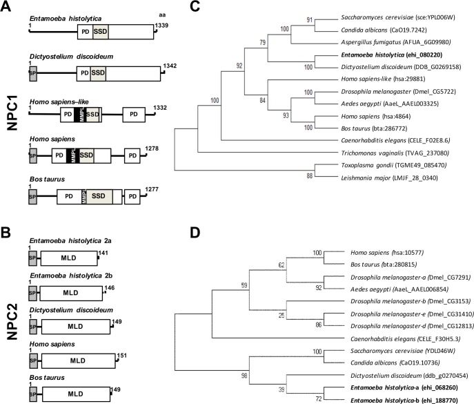
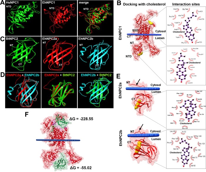
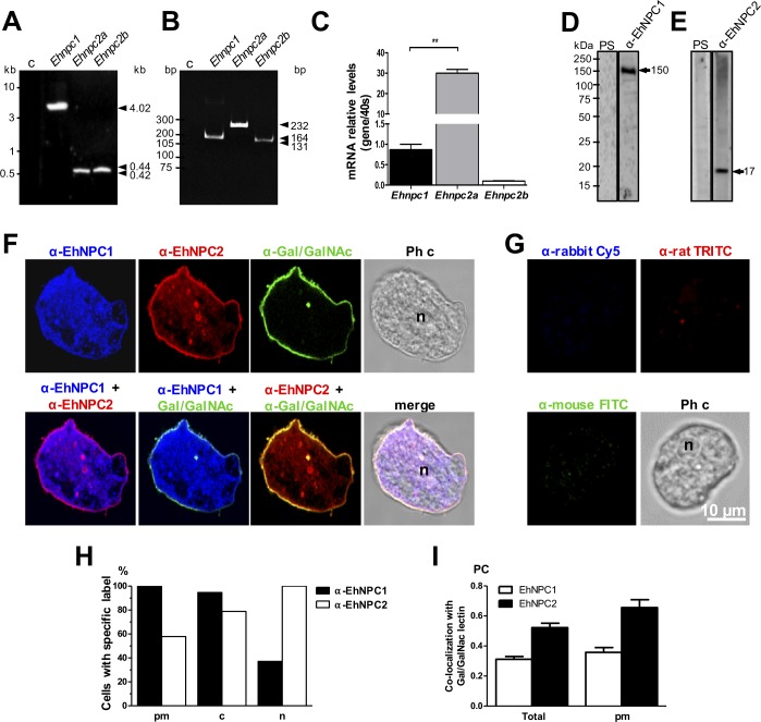
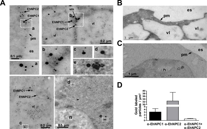
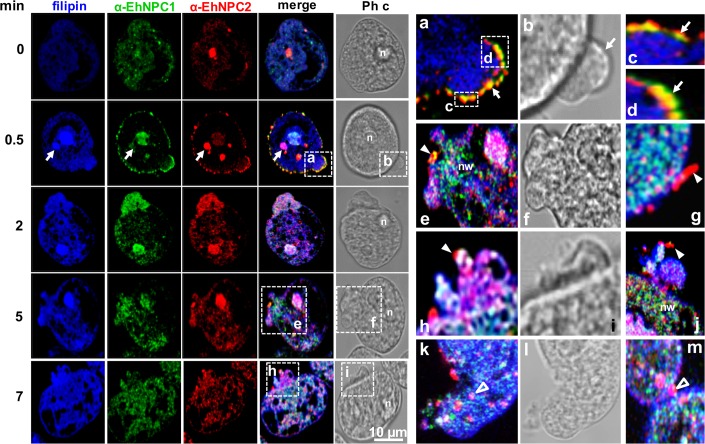
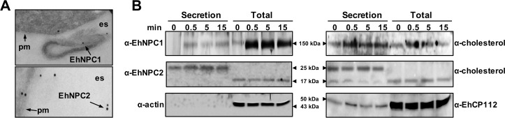
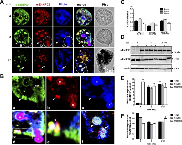
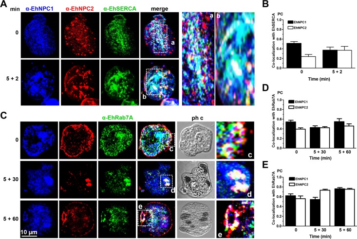
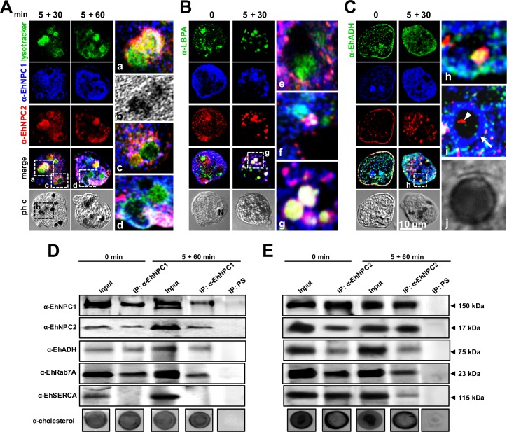
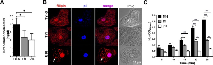
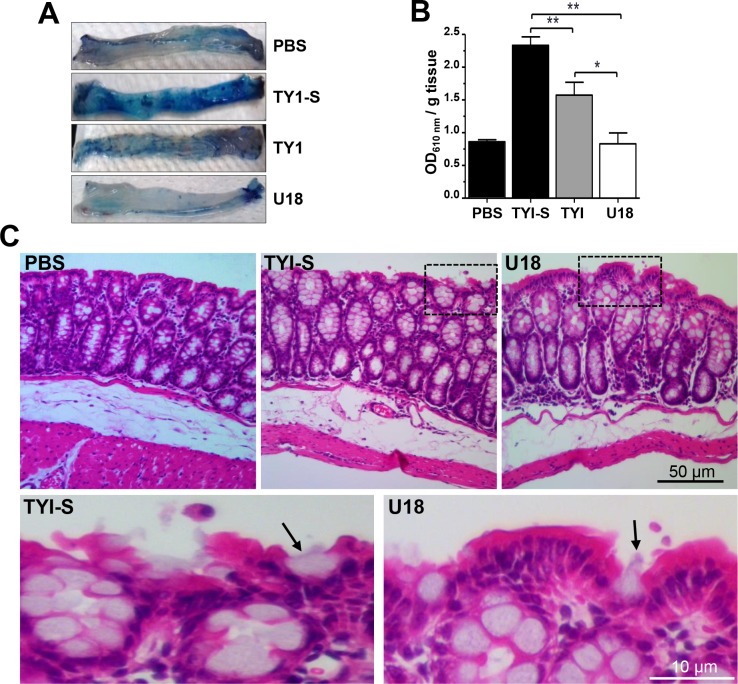
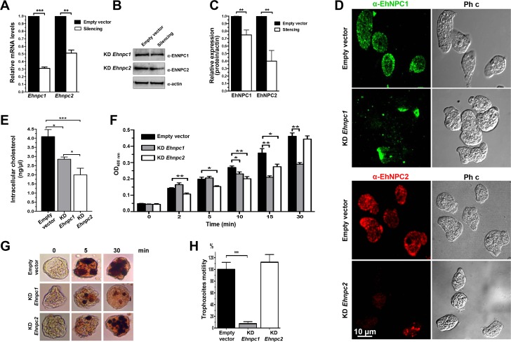
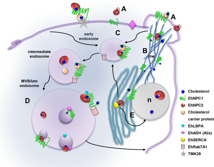
Entamoeba histolytica, the highly phagocytic protozoan causative of human amoebiasis lacks the machinery to synthesize cholesterol. Here, we investigated the presence of NPC1 and NPC2 proteins in this parasite, which are involved in cholesterol trafficking in mammals. Bioinformatics analysis revealed one Ehnpc1 and two Ehnpc2 genes. EhNPC1 appeared as a transmembrane protein and both EhNPC2 as peripheral membrane proteins. Molecular docking predicted that EhNPC1 and EhNPC2 bind cholesterol and interact with each other. Genes and proteins were identified in trophozoites. Serum pulse-chase and confocal microscopy assays unveiled that after trophozoites sensed the cholesterol source, EhNPC1 and EhNPC2 were organized around the plasma membrane in a punctuated pattern. Vesicles emerged and increased in number and size and some appeared full of cholesterol with EhNPC1 or EhNPC2 facing the extracellular space. Both proteins, but mostly EhNPC2, were found out of the cell associated with cholesterol. EhNPC1 and cholesterol formed networks from the plasma membrane to the nucleus. EhNPC2 appeared in erythrocytes that were being ingested by trophozoites, co-localizing with cholesterol of erythrocytes, whereas EhNPC1 surrounded the phagocytic cup. EhNPC1 and EhNPC2 co-localized with EhSERCA in the endoplasmic reticulum and with lysobisphosphatidic acid and EhADH (an Alix protein) in phagolysosomes. Immunoprecipitation assays confirmed the EhNPC1 and EhNPC2 association with cholesterol, EhRab7A and EhADH. Serum starved and blockage of cholesterol trafficking caused a low rate of phagocytosis and incapability of trophozoites to produce damage in the mouse colon. Ehnpc1 and Ehnpc2 knockdown provoked in trophozoites a lower intracellular cholesterol concentration and a diminished rate of phagocytosis; and Ehnpc1 silencing also produced a decrease of trophozoites movement. Trafficking of EhNPC1 and EhNPC2 during cholesterol uptake and phagocytosis as well as their association with molecules involved in endocytosis strongly suggest that these proteins play a key role in cholesterol uptake.


Conflict of interest statement

The authors have declared that no competing interests exist.

Figures

Fig 1
Fig 1. Structural domains and phylogenetic trees of EhNPC1, EhNPC2a and EhNPC2b.
(A, B) Schemes show the main structural characteristics of NPC1 (A) and NPC2 (B) from distinct organisms. PD: patched domain, SSD: sterol sensing domain, MMPL: putative integral membrane domain, SP: signal peptide, MLD: MD-2 related lipid recognition domain. Numbers at the right correspond to the amino acids forming the proteins. (C, D) Phylogenetic tree indicating the position of E. histolytica NPC1 (C), NPC2a and NPC2b (D) proteins among different species. Numbers on horizontal lines in the trees indicate the confidence percentages of the tree topology from bootstrap analysis of 1000 replicates.
Fig 2
Fig 2. 3D structures of NTD-EhNPC1, EhNPC2a and EhNPC2b and molecular dockings between them and with cholesterol.
(A) NTD-EhNPC1 model (1 to 250 amino acids) predicted by RaptorX server was compared with the crystal of human NTD-NPC1 protein (HsNPC1) (23 to 254 amino acids). (B) Docking simulation of EhNPC1 with cholesterol performed using the AutoDock Tools V1.5.6 program. SSD: sterol sensing domain. NTD: amino terminal domain. (C, D) The EhNPC2a and EhNPC2b 3D structures (full-length amino acid sequences) were compared between them and with the NPC2 B. taurus crystal (BtNPC2). (E) Docking simulation of EhNPC2a and EhNPC2b with cholesterol. Arrows: amino acids tail at the amino terminus that it is not present in the crystal of Btaurus NPC2. Squares at the right in B and E show the amino acids involved in the protein-cholesterol interaction. (F) Docking simulation of EhNPC1 and EhNPC2a binding. ΔG: binding energy. Blue lines in dockings: plasmatic membrane. NT: amino terminus. CT: carboxy terminus.
Fig 3
Fig 3. Expression and localization of EhNPC1 and EhNPC2 in trophozoites.
(A) Ehnpc1, Ehnpc2a and Ehnpc2b full-length genes were PCR amplified using specific primers and genomic DNA. (B) RT-PCR amplification of transcript fragments using specific primers and cDNA. c: Controls without gDNA or with mRNA as template. (C) The relative expression of the three genes was measured by RT-qPCR in trophozoites, using as a control the 40s ribosomal S2 protein gene. ** p<0.01. (D, E) Total extracts of E. histolytica were separated by 10% SDS-PAGE and analyzed by western blot assays using pre-immune serum (PS) or rabbit α-EhNPC1 (D) or rat α-EhNPC2 (E) antibodies. (F) Representative images of laser confocal microscopy of PFA-fixed trophozoites using rabbit α-EhNPC1 or rat α-EhNPC2 or mouse α-Gal/GalNAc lectin antibodies. (G) Controls using only secondary antibodies. Ph c: phase contrast images. (H) Protein localization of EhNPC1 and EhNPC2 in plasma membrane (pm), cytoplasm (c) or nucleus (n). Counts were performed in 50 cells. (I) Pearson coefficient (PC) correlation measured in at least 15 confocal images, indicating co-localization of EhNPC1 or EhNPC2 with Gal/GalNAc lectin in the entire cell and in the plasma membrane. Laser sections = 0.5 μm.
Fig 4
Fig 4. Localization of EhNPC1 and EhNPC2 in trophozoites analyzed by TEM.
(A) Thin sections of trophozoites were incubated with rabbit α-EhNPC1 and rat α-EhNPC2 antibodies, followed by incubation with gold labeled α-rabbit and α-rat secondary antibodies (20 and 10 nm gold particles, respectively). Squares indicate the magnified areas marked with the corresponding lower case letters. pm: plasma membrane, vl: vesicle lumen, vm: vesicle membrane, es: extracellular space, n: nucleus. (B, C) Controls using only secondary antibodies. (D) Graph showing number of EhNPC1 and EhNPC2 molecules recognized by the respective gold-labeled antibodies and their co-localization.
Fig 5
Fig 5. Localization of EhNPC1 and EhNPC2 in trophozoites after an ABS pulse.
Trophozoites were serum starved for 12 h by culturing in TYI medium on coverslips. Then, ABS was added as a cholesterol source at 37°C for 0.5 to 7 min. Cells were washed, fixed and incubated with filipin, rabbit α-EhNPC1 and rat α-EhNPC2 antibodies, secondary antibodies, and examined by laser confocal microscopy. Arrows: cytoplasmic dots. Squares marked with lower case letters are magnified at the right. (a-d) Membrane vacuoles containing EhNPC1 and EhNPC2. Arrows: show the plasma membrane with EhNPC2 or EhNPC1 facing the extracellular space. (e) Networks stained by α-EhNPC1 antibody. (e-j) rod and spherical structures (arrowheads) facing the extracellular space, stained mainly by α-EhNPC2 antibody. (k-m) Spherical structures of 1 to 2 μm (empty arrowheads) inside the cell. n: nucleus, Ph c: phase contrast images, nw: networks like structures.
Fig 6
Fig 6. Secretion of EhNPC1 and EhNPC2 after ABS stimulus.
(A) TEM of trophozoites under basal condition showing EhNPC1 and EhNPC2 in the extracellular space. pm: plasma membrane, es: extracellular space. (B) After the ABS stimulus, supernatants were collected and trophozoites were lysed at the indicated times. Secreted products and total extracts were analyzed by SDS-PAGE and western blot assays using rabbit α-EhNPC1, rat α-EhNPC2, rabbit α-cholesterol, rabbit α-EhCP112 and mouse α-actin antibodies and corresponding secondary antibodies. Actin was used as a control of cell integrity and EhCP112 as a secretion control.
Fig 7
Fig 7. Localization of EhNPC1, EhNPC2 and cholesterol during phagocytosis.
(A) Trophozoites were incubated with erythrocytes (1:25) at 37°C, for the indicated times. Then, samples were prepared for laser confocal microscopy and stained with filipin, rabbit α-EhNPC1 and rat α-EhNPC2 antibodies and secondary antibodies. Ph c: phase contrast images, e’: erythrocytes, n: nucleus. Arrow head: cytoplasmic vesicles. (B) (a-d) Dotted circles: Magnification of an erythrocyte that is being internalized. (e) Magnification of vesicles close to the plasma membrane. (f) Magnification of a huge phagosome (ph) containing digested erythrocytes (arrows). nw: networks. (C) PC of the co-localization between filipin and EhNPC1 or EhNPC2 or EhNPC1 with EhNPC2 at distinct phagocytosis time. (D) Trophozoites were incubated with erythrocytes at 37°C for 5 min, then adherent and non-ingested erythrocytes were removed by washing with a mixture of water-TYI medium. Later, trophozoites were incubated at 37°C again for 60 min with TYI to continue the process. Trophozoites were lysed and also cellular fractionation was carried out. Samples were processed for 12% SDS-PAGE and western blot assays. Representative blots of three experiments are shown. T: total extracts, S: soluble fraction, I: insoluble fraction. (E,F) Densitometry of the bands in (D) corresponding to EhNPC1 (E) and EhNPC2 (F) using actin as loading control.
Fig 8
Fig 8. Co-localization of EhNPC1 and EhNPC2 with EhSERCA and EhRab7A.
Trophozoites were incubated with erythrocytes at 37°C and treated as Fig 7. Samples were processed for confocal microscopy using α-EhNPC1, α-EhNPC2 and (A) α-EhSERCA or (C) α-EhRab7A antibodies. (a-e) Magnification of the white squares. (B, D, E) PC of the co-localization between EhNPC1 or EhNPC2 with EhSERCA (B), or with EhRab7A in the whole cell (D) or in cellular structures stained by the three antibodies (E).
Fig 9
Fig 9. Co-localization and association of EhNPC1 and EhNPC2 with cholesterol and endosomal molecules.
Trophozoites were incubated with erythrocytes as in Fig 7 and processed for confocal microscopy after incubation with α-EhNPC1 and α-EhNPC2 antibodies and (A) Lysotracker or (B) α-LBPA or (C) α-EhADH antibody and corresponding secondary antibodies. (a-d,g,h) Magnification of white squares. (e,f,i,j) Magnifications from other images. Ph c: phase contrast images, arrow in I: EhNPC1 outside endosomes, arrowhead: EhNPC2 inside endosomes. D, E) Trophozoites in basal condition or after 5+60 min of erythrophagocytosis were lysed and immunoprecipitation assays (IP) were performed using α-EhNPC1 (D) or α-EhNPC2 (E) antibodies or preimmune serum (PS). Immunoprecipitated proteins of trophozoites were analyzed by western blot and dot blot experiments, using α-EhNPC1, α-EhNPC2, α-EhADH, α-EhRab7A, α-EhSERCA and α-cholesterol antibodies.
Fig 10
Fig 10. Erythrophagocytosis of trophozoites cultured in TYI-S, TYI and TYI plus U18.
Trophozoites were incubated ON at 37°C in TYI-S, TYI and TYI plus U18. (A) Cholesterol concentration in the respective trophozoites was measured as described in material and methods. (B) Laser confocal microscopy showing the morphology and the cholesterol localization by filipin staining. Nuclei were counterstained with propidium iodide (pi). Images were false colored to obtain a better contrast. Ph c: phase contrast images, arrows: cytoplasmic dots. (C) Rate of erythrophagocytosis spectrophotometrically measured by hemoglobin (Hb) concentration inside trophozoites.
Fig 11
Fig 11. In vivo virulence of trophozoites cultured in TYI-S, TYI and TYI plus U18.
Capability of trophozoites to impair the mouse intestinal barrier. (A) Distal parts of the mouse colons after treatment with trophozoites or with PBS. Intestinal barrier impairment was measured as the ability of the Evans blue dye to permeate the mouse intestinal epithelium after contact with trophozoites. n = 5. (B) Data represent the mean ± standard error. PBS: mice undergoing surgery, but not inoculated with trophozoites. * p<0.05, ** p<0.01. (C) Hematoxylin-eosin staining of tissues. Squares were magnified in the corresponding lower panels. Arrows: trophozoites.
Fig 12
Fig 12. Silencing of Ehnpc1 and Ehnpc2 genes in E. histolytica.
(A) Trophozoites clone G3 were transfected with psAP-2, psAP-2Ehnpc1 or psAP-2Ehnpc2a plasmids and stable populations were selected with 4 μg/ml G-418. RT-qPCR assays were performed using mRNA from transfected trophozoites (empty vector, KD Ehnpc1 and KD Ehnpc2), using specific primers for Ehnpc1 and Ehnpc2a genes and as a housekeeping the 40s ribosomal S2 protein gene. (B) Western blot assays of transfected trophozoites extracts, using α-EhNPC1 and α-EhNPC2 antibodies and respectively secondary antibody. As a loading control, the same membrane was reblotted with α-actin antibodies. (C) Densitometry analysis of bands showed in (B) normalized again actin protein. (D) Confocal microscopy of transfected trophozoites in basal conditions using α-EhNPC1 or α-EhNPC2 antibodies. Ph c: phase contrast. (E) Cholesterol concentration in the transfected trophozoites was measured as described in material and methods. (F) Rate of erythrophagocytosis of transfected trophozoites. * p<0.05, ** p<0.01. (G) Diaminobenzidine-stained trophozoites that ingested erythrocytes for different times. (H) Motility assays of transfected trophozoites cultured in transwell inserts. *** p<0.001.
Fig 13
Fig 13. Working model of the EhNPC1 and EhNPC2 participation in cholesterol trafficking in E. histolytica.
(A) Cholesterol uptake: EhNPC1 and EhNPC2 capture the cholesterol in the extracellular space and in the plasma membrane probably following the “hand-off” model (EhNPC1-cholesterol-EhNPC2-cholesterol-EhNPC1) and with the TMK39 participation. (B) Network-like structures formation: EhNPC1 and cholesterol form networks from cholesterol-containing membrane protrusions to ER, endosomes and nucleus that possibly facilitate cholesterol trafficking through EhNPC2 in a “hand-off” model. (C) Cholesterol influx: EhNPC1 and EhNPC2 associate with cholesterol and are internalized in EhRab-7A containing endosomes under basal conditions and during erythrophagocytosis. (D) EhNPC1 and EhNPC2 associate with phagolysosomal and MBVs molecules, particularly with LBPA and EhADH, which probably regulate cholesterol trafficking. (E) EhNPC1 and EhNPC2 accumulate in nucleus (n) and ER and may be distributed from there to other organelles.

Similar articles

Cited by

References

    1. Mortimer L, Chadee K. The immunopathogenesis of Entamoeba histolytica. Exp Parasitol. 2010;126(3):366–80. Epub 2010/03/23. S0014-4894(10)00090-1 [pii]. 10.1016/j.exppara.2010.03.005 - DOI - PubMed
    1. Serrano-Luna J, Gutierrez-Meza M, Mejia-Zepeda R, Galindo-Gomez S, Tsutsumi V, Shibayama M. Effect of phosphatidylcholine-cholesterol liposomes on Entamoeba histolytica virulence. Can J Microbiol. 2010;56(12):987–95. Epub 2010/12/18. w10-088 [pii]. 10.1139/W10-088 - DOI - PubMed
    1. Das SR, Singh BN. Virulence of Strains of Entamoeba Histolytica to Rats & Guinea-Pigs, & Effect of Cholesterol on Virulence. Indian J Exp Biol. 1965;3:106–9. Epub 1965/04/01. - PubMed
    1. Mittal K, Welter BH, Temesvari LA. Entamoeba histolytica: lipid rafts are involved in adhesion of trophozoites to host extracellular matrix components. Exp Parasitol. 2008;120(2):127–34. Epub 2008/07/01. S0014-4894(08)00146-X [pii]. PubMed Central PMCID: PMC2615684. 10.1016/j.exppara.2008.06.003 - DOI - PMC - PubMed
    1. Welter BH, Goldston AM, Temesvari LA. Localisation to lipid rafts correlates with increased function of the Gal/GalNAc lectin in the human protozoan parasite, Entamoeba histolytica. Int J Parasitol. 2011;41(13–14):1409–19. Epub 2011/11/17. S0020-7519(11)00252-9 [pii]. PubMed Central PMCID: PMC3232469. 10.1016/j.ijpara.2011.10.003 - DOI - PMC - PubMed

MeSH terms