

Next generation sequencing of vitreoretinal lymphomas from small-volume intraocular liquid biopsies: new routes to targeted therapies
- PMID: 28002793
- PMCID: PMC5352376
- DOI: 10.18632/oncotarget.14008
Next generation sequencing of vitreoretinal lymphomas from small-volume intraocular liquid biopsies: new routes to targeted therapies
Abstract
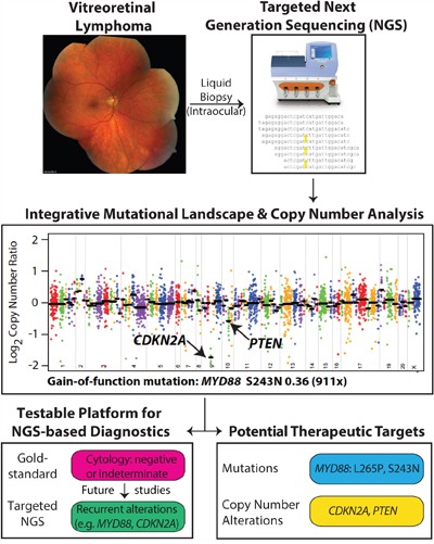
Background: Vitreoretinal lymphoma (VRL), the most common lymphoma of the eye, is a rare form of primary CNS lymphoma (PCNSL). Most frequently a high-grade diffuse large B cell lymphoma, VRL can cause vision loss and its prognosis remains dismal: the overall survival time is 3 years after diagnosis. Radiotherapy and chemotherapy are used but remain frequently ineffective, and no standardized treatment regimen exists. Furthermore, no biologically targeted treatments, based on the genetic profile of the tumor, are available, as VRL has hitherto not comprehensively been profiled. To address these unmet needs, we hypothesized that a next generation sequencing (NGS)-based, National Cancer Institute (NCI) MATCH Trial-modified panel would be able to identify actionable genomic alterations from small-volume, intraocular liquid biopsies.
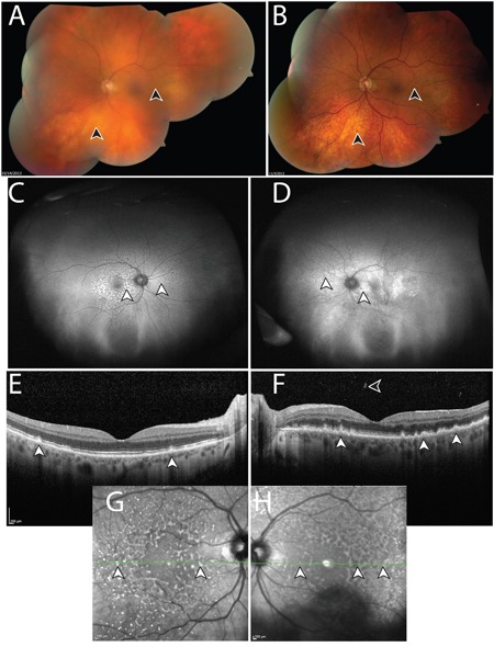
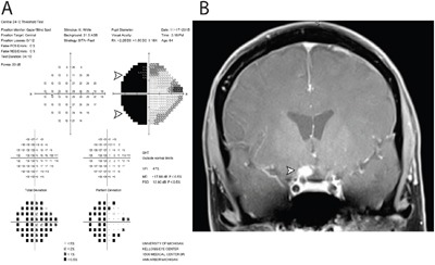
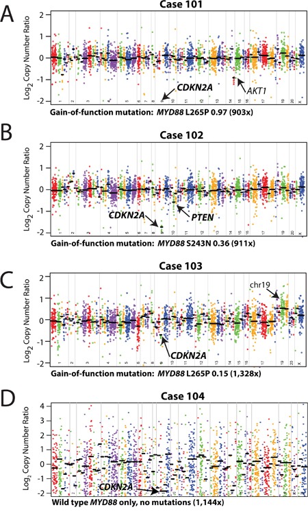
Methods and findings: In this retrospective study, we collected diluted vitreous biopsies from 4 patients with a high suspicion for VRL. Following cytological confirmation of lymphoma (all were diffuse large B cell lymphomas), we subjected genomic DNA from the biopsies to NGS, using a panel containing 126 genes (3,435 amplicons across several hotspots per gene), which was modified from that of the NCI MATCH Trial, a new trial that has matched patients with cancers that have not responded (or never responded), to investigational therapeutics based on their prioritized mutation profile rather than site of tumor origin. Using a validated bioinformatics pipeline, we assessed for the presence of actionable mutations and copy number alterations. In all four small-volume, intraocular liquid biopsies, we obtained sufficient genomic DNA for analysis, even in diluted samples in which the undiluted vitreous was used for cytology and flow cytometry. Using NGS, we found targetable heterozygous gain-of-function mutations in the MYD88 oncogene, and confirmed in our cohort the presence the L265 mutations, previously described using PCR-based assays. For the first time in VRL, we also identified the MYD88 S243N mutation. We also identified two-copy copy number losses in the tumor suppressor CDKN2A in all four cases, and one copy loss of the tumor suppressor PTEN in one sample. In one case, in which vitreous biopsies were originally read as cytologically negative, but which was confirmed as lymphoma when a lesion appeared in the brain two years later, our NGS-based approach detected tumoral DNA in the banked, original liquid biopsy.
Conclusions: We performed the first systematic exploration of the actionable cancer genome in VRL. Our NGS-based approach identified exploitable genomic alterations such as gain-of-function MYD88 oncogene mutations and loss of the tumor suppressor CDKN2A, and thus illuminates new routes to biologically targeted therapies for VRL, a cancer with a dismal prognosis. This precision medicine strategy could be used to nominate novel, targeted therapies in lymphomas and other blinding and deadly ocular, orbital, and ocular adnexal diseases for which few treatments exist.
Keywords: biopsy; next generation sequencing; precision medicine; primary central nervous system lymphoma; vitreoretinal lymphoma.
Conflict of interest statement
SAT has received travel support and had a separate sponsored research agreement with Compendia Bioscience/Life Technologies/ThermoFisher Scientific, which provided access to an earlier version of the sequencing panel used herein. No other aspect of the study described herein was supported by Compendia Bioscience/Life Technologies/ThermoFisher Scientific. S.A.T. has consulted and received grant support from Astellas/Medivation, and has consulted for Ventana Medical Systems, Janssen, Abbvie. S.A.T. is co-founder, equity holder and consultant for Strata Oncology. All other authors declare no conflicts of interest. S.A.T. and R.C.R. had full access to all the data in the study and take responsibility for the integrity of the data and the accuracy of the data analysis.
Figures
References
-
- Chan CC, Rubenstein JL, Coupland SE, Davis JL, Harbour JW, Johnston PB, Cassoux N, Touitou V, Smith JR, Batchelor TT, Pulido JS. Primary vitreoretinal lymphoma: a report from an International Primary Central Nervous System Lymphoma Collaborative Group symposium. Oncologist. 2011;16:1589–1599. - PMC - PubMed
-
- Fend F, Ferreri AJ, Coupland SE. How we diagnose and treat vitreoretinal lymphoma. Br J Haematol. 2016;173:680–692. - PubMed
-
- Hovelson DH, McDaniel AS, Cani AK, Johnson B, Rhodes K, Williams PD, Bandla S, Bien G, Choppa P, Hyland F, Gottimukkala R, Liu G, Manivannan M, Schageman J, Ballesteros-Villagrana E, Grasso CS, et al. Development and validation of a scalable next-generation sequencing system for assessing relevant somatic variants in solid tumors. Neoplasia. 2015;17:385–399. - PMC - PubMed
-
- Cani AK, Soliman M, Hovelson DH, Liu CJ, McDaniel AS, Haller MJ, Bratley JV, Rahrig SE, Li Q, Briceno CA, Tomlins SA, Rao RC. Comprehensive genomic profiling of orbital and ocular adnexal lymphomas identifies frequent alterations in MYD88 and chromatin modifiers: new routes to targeted therapies. Mod Pathol. 2016;29:685–697. - PMC - PubMed
MeSH terms
Substances
Grants and funding
LinkOut - more resources
Full Text Sources
Other Literature Sources
Research Materials
Miscellaneous
