. 2017 Jan 17;8(3):4888-4900.
doi: 10.18632/oncotarget.13982.

Sprouty2 correlates with favorable prognosis of gastric adenocarcinoma via suppressing FGFR2-induced ERK phosphorylation and cancer progression

Affiliations

Sprouty2 correlates with favorable prognosis of gastric adenocarcinoma via suppressing FGFR2-induced ERK phosphorylation and cancer progression

Yunfei Xu et al. Oncotarget. .

Abstract

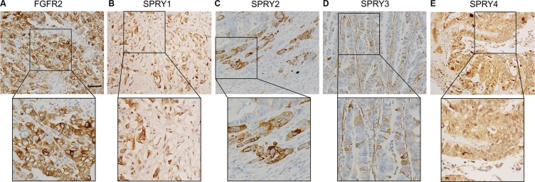
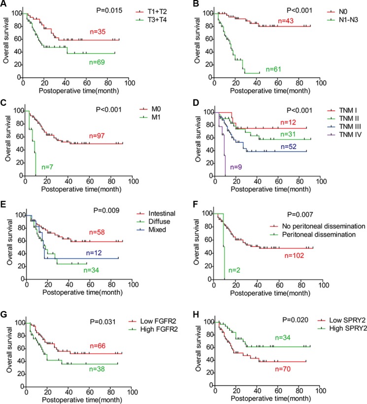
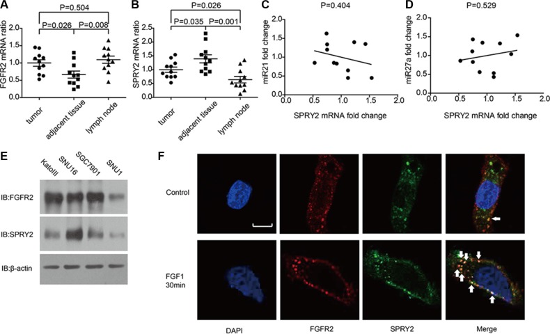
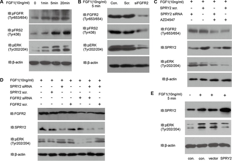
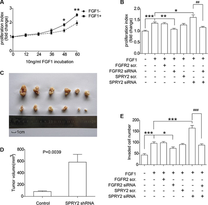
Fibroblast growth factor receptor 2 (FGFR2) has been identified as a predictive biomarker for unfavorable prognosis of gastric adenocarcinoma. As a well-defined antagonist in FGFR2-induced RAS/ERK activation, ectopic expression of sprouty (SPRY) family was reported in several kinds of cancers except gastric cancer. To explore the clinical significance of SPRY family and its correlation with FGFR2, we detected the expression of FGFR2 and SPRY family in 104 cases of gastric adenocarcinoma and subsequently analyzed their correlations with clinicopathological factors and overall survival rates by univariate and multivariate analysis. As the result, we demonstrated that both FGFR2 high-expression and SPRY2 low-expression indicated poorer prognosis of gastric adenocarcinoma. SPRY2 low-expression was significantly associated with FGFR2 high-expression, positive lymphatic invasion and metastasis. We further proved that SPRY2 could suppress FGFR2-induced ERK phosphorylation, cell proliferation and invasion with experiments in vitro and in vivo. In conclusion, we demonstrated that SPRY2 low-expression is a biomarker for unfavorable prognosis in gastric adenocarcinoma. SPRY2 can antagonize FGFR2-induced proliferation and invasion via suppressing ERK phosphorylation in gastric cancer cells, indicating SPRY2 as a potential therapeutic target for gastric adenocarcinoma treatment.

Keywords: FGFR2; SPRY2; gastric adenocarcinoma; invasion; proliferation.


Conflict of interest statement

CONFLICTS OF INTEREST

The authors have no conflicts of interest.

Figures

Figure 1
Figure 1. Representative figure of immunohistochemistry staining of high-expression of FGFR2 and SPRY family
Expression of FGFR2 and SPRY family was detected by immunohistochemistry in formalin-fixed and paraffin-bedded gastric adenocarcinoma tissues. The cohort was divided into low-expression and high-expression according to the immunohistochemistry score. Representative immunohistochemistry image of the high-expression of (A) FGFR2, (B) SPRY1, (C) SPRY2, (D) SPRY3 and (E) SPRY4 were displayed. Scale bar: 50 μm.
Figure 2
Figure 2. Correlations between overall survival rate and clinicopathologic factors
The survival curves were graphed with Kaplan-Meier method and the statistical difference was analyzed with log-rank test. This figure displays the overall survival curves of (A) T1/T2 and T3/T4; (B) N0 and N1-3; (C) M0 and M1; (D) TNM stage; (E) Intestinal type, diffuse type and mixed type; (F) Negative and positive peritoneal dissemination; (G) FGFR2 low-expression and high-expression; (H) SPRY2 low-expression and high-expression.
Figure 3
Figure 3. The mRNA level of FGFR2, SPRY2 and relative miRNAs in gastric adenocarcinoma tissues and cell lines
(A and B)The mRNA levels of (A) FGFR2 and (B) SPRY2 in gastric adenocarcinoma tissues, adjacent tumor tissues and lymph nodes of 11 patients were detected by quantitative PCR with GAPDH as an internal control. The mRNA levels were standardized by ΔΔCt method. FGFR2 mRNA in adjacent tumor tissues was significantly lower than in tumor tissues (P = 0.026) and lymph nodes (P = 0.008), while SPRY2 mRNA in adjacent tumor tissues was significantly higher than in tumor tissues (P = 0.035) and invaded lymph node (P = 0.001). (C and D) The correlation between miR21, miR27a and SPRY2 mRNA. The quantification of miR21, miR27a and SPRY2 was realized by real-time PCR and standardized with the meanvalue set as 1. The correlation between miR21 and SPRY2 (C), between miR27a and SPRY2 (D) was analyzed by Pearson correlation coefficient. (E) FGFR2 and SPRY2 expression in gastric cell line KatoIII, SNU16, SGC7901 and SNU1 were detected by Western blotting. (F) The locations of FGFR2 and SPRY2 in SNU16 cells were indicated by immunofluorescence with/without FGF1 stimulation. Cell nucleus was stained by DAPI, while FGFR2 or SPRY2 was visualized after incubation with corresponding primary antibodies and secondary antibodies after starvation in serum-free medium overnight. Without FGF1 stimulation, FGFR2(red) was distributed around cell membrane and scattered in cytoplasm, and SPRY2 (green) expressed mostly in cytoplasm. After 10 ng/ml FGF1 stimulation for 30 minutes, FGFR2 and SPRY2 formed stimulation cluster in both membrane and cytoplasm. FGFR2 and SPRY2 had more co-localizations after FGF1 stimulation. Arrows pointed representative co-localizations. Scale bar: 5 μm.
Figure 4
Figure 4. SPRY2 could antagonize FGFR2-induced ERK phosphorylation
(A) The phosphorylation of FGFR (Tyr653/654), FRS2 (Tyr436) and ERK (Tyr202/204) increased along with FGF1 stimulation in KatoIII cells. Cells were starved in serum-free medium for 12 hours and then stimulated with 10 ng/ml FGF1 for 0–20 minutes before test. (B) Effect of FGFR2 knockdown on FRS and ERK phosphorylation. SNU16 cells were transfected with FGFR2 siRNA 48 hours before 10 ng/ml FGF1 stimulation for 5 minutes. Total FGFR2, phosphorylation of FRS2 (Tyr436) and ERK (Tyr202/204) was detected. Under FGF1 stimulation, phosphorylation of FRS2 and ERK decreased significantly after FGFR2 successful knockdown. (C) Effects of FGFR inhibitor AZD4547 on ERK phosphorylation with/without SPRY2 knockdown. SNU16 cells were incubated in AZD4547 (300 nM) for 2 hours before FGF1 (10 ng/ml) stimulation. SPRY2 siRNA knockdown could increase ERK (Tyr202/204) phosphorylation, and AZD4547 incubation suppressed ERK phosphorylation significantly by inhibiting phosphorylation of pFGFR (Tyr653/654). (D) ERK phosphorylation was attenuated by FGFR2 and enhanced by SPRY2 knockdown. Cells were transfected by SPRY2 siRNA and/or FGFR2 siRNA 48 hours before FGF1 stimulation. SPRY2 knockdown significantly elevated ERK phosphorylation and FGFR2 knockdown could reverse this effect. (E) SPRY2 overexpression could suppress ERK phosphorylation under FGF1 stimulation. SUN16 cells were transfected with FLAG-SPRY2 plasmid with empty pFLAG-CMV-2 vector as control. Cells were starved in serum-free medium overnight and then stimulated with 10 ng/ml FGF1. Con. means control group with only transfection agent. Vector represents the group transfected with empty pFLAG-CMV-2 vector. All results were confirmed with at least 3 independent experiments.
Figure 5
Figure 5. SPRY2 could suppress FGFR2-induced cell proliferation and invasion of gastric adenocarcinoma
(A) SNU16 cells were incubated in FGF1 for 0 to 60 hours and the proliferation rate was quantified by MTT assay. Cells with FGF1 stimulation for 48 and 60 hours had remarkably higher proliferation rate than cells without FGF1 stimulation. *represents P < 0.05 and **represents P < 0.01 between FGF+ and FGF1- group at the same time. Data were from 3 independent experiments, analyzed by Student-t test. Error bar displays ± SEM. (B) Effect of FGFR2 and SPRY2 expression on gastric cancer cell proliferation. SNU16 cell proliferation was evaluated after 10 ng/ml FGF1 incubation for 48 hours and FGFR2 with/without SPRY2 knockdown. FGF1 could promote SNU16 proliferation via activating FGFR2, and SPRY2 could decrease FGFR2-promoted cell proliferation. *, ** and ***represents P < 0.05, P < 0.01 and P < 0.001 compared with the group only with FGF1 stimulation. ##means P < 0.01 between the linked columns. Data were from 3 independent experiments, analyzed by Student-t test. Error bar displays ± SEM. (C) Comparison of excised tumors volumn from xenograft model. SNU16 cells were first transfected with GV248-SPRY2 shRNA and selected with 1μg/ml puromycin before tumor injection. 106 Stable SNU16/NC or SN U16/shRNA cells were injected subcutaneously into the left flank or right flank of BALB/C male nude mice. Upper row: tumors with SPRY2 knockdown; Lower row: tumors without SPRY2 knockdown. (D) Volume of tumor with/without SPRY2 knockdown 28 days after injection. The tumor volume (V) were calculated using the following formula: V (mm3) = V (mm3) = π × width (mm) × width (mm) × length (mm)/6. The P value was analyzed by Student's t test. (E) Cell invasion ability was promoted by SPRY2 knockdown and impaired by FGFR2 knockdown. SNU16 cell invasion was detected by transwell assay after 10 ng/ml FGF1 incubation for 48 hours and FGFR2 and/or SPRY2 knockdown. FGFR2 could promote cell invasion with FGF1 stimulation and SPRY2 could suppress this effect. * and ***represents P < 0.05 and P < 0.001 compared with the group with FGF1 stimulation and without other interferences. ###means P < 0.001 between the 6 and 7 columns. Data were from 3 independent experiments, analyzed by Student-t test. Error bar displays ± SEM.

Similar articles

Cited by

References

    1. Torre LA, Bray F, Siegel RL, Ferlay J, Lortet-Tieulent J, Jemal A. Global cancer statistics, 2012. CA Cancer J Clin. 2015;65:87–108. - PubMed
    1. Torpy JM, Lynm C, Glass RM. JAMA patient page. Stomach cancer. JAMA. 2010;303:1771. - PubMed
    1. Brooks AN, Kilgour E, Smith PD. Molecular pathways: fibroblast growth factor signaling: a new therapeutic opportunity in cancer. Clin Cancer Res. 2012;18:1855–1862. - PubMed
    1. Katoh M, Nakagama H. FGF receptors: cancer biology and therapeutics. Med Res Rev. 2014;34:280–300. - PubMed
    1. Wu YM, Su F, Kalyana-Sundaram S, Khazanov N, Ateeq B, Cao X, Lonigro RJ, Vats P, Wang R, Lin SF, Cheng AJ, Kunju LP, Siddiqui J, et al. Identification of targetable FGFR gene fusions in diverse cancers. Cancer Discov. 2013;3:636–647. - PMC - PubMed

MeSH terms

Substances