. 2017 Mar 7;8(10):16170-16189.
doi: 10.18632/oncotarget.14016.

Sub-lethal oxidative stress induces lysosome biogenesis via a lysosomal membrane permeabilization-cathepsin-caspase 3-transcription factor EB-dependent pathway

Affiliations

Sub-lethal oxidative stress induces lysosome biogenesis via a lysosomal membrane permeabilization-cathepsin-caspase 3-transcription factor EB-dependent pathway

San Min Leow et al. Oncotarget. .

Abstract

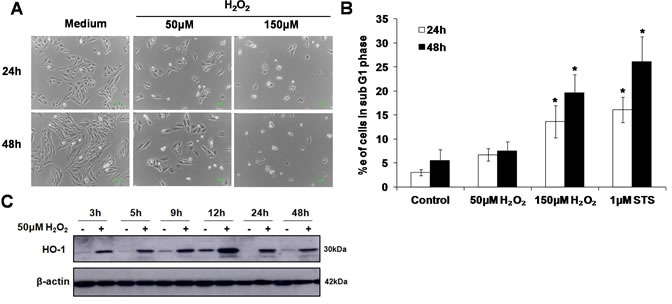
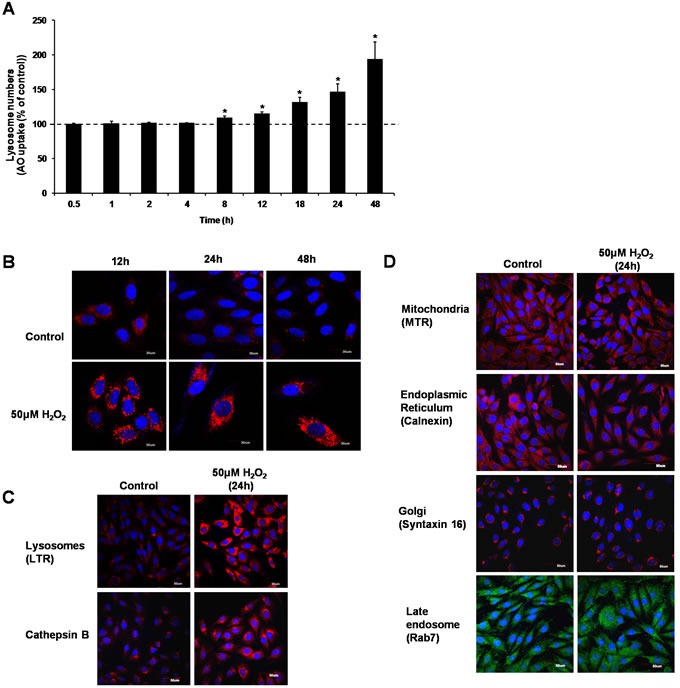
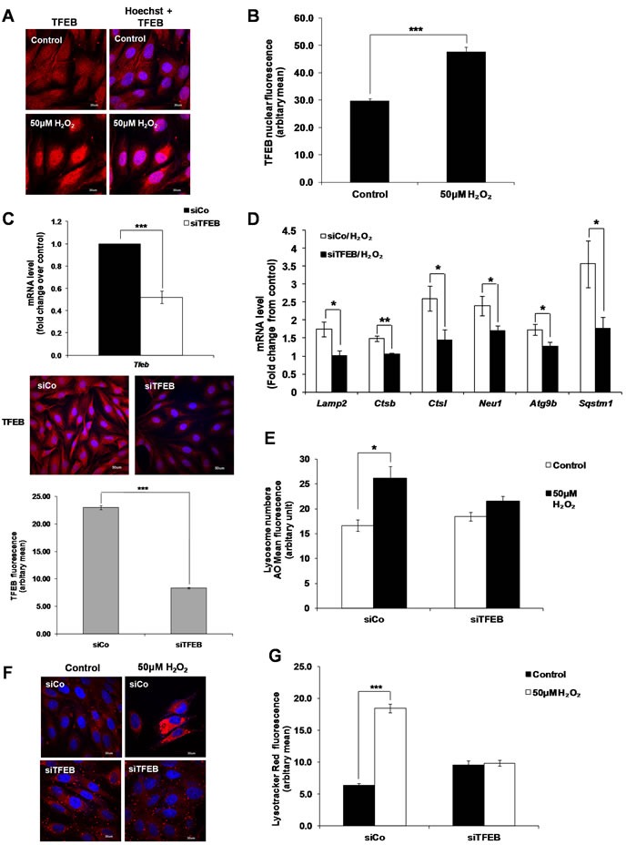
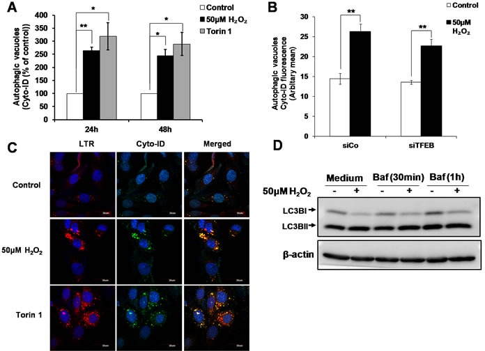
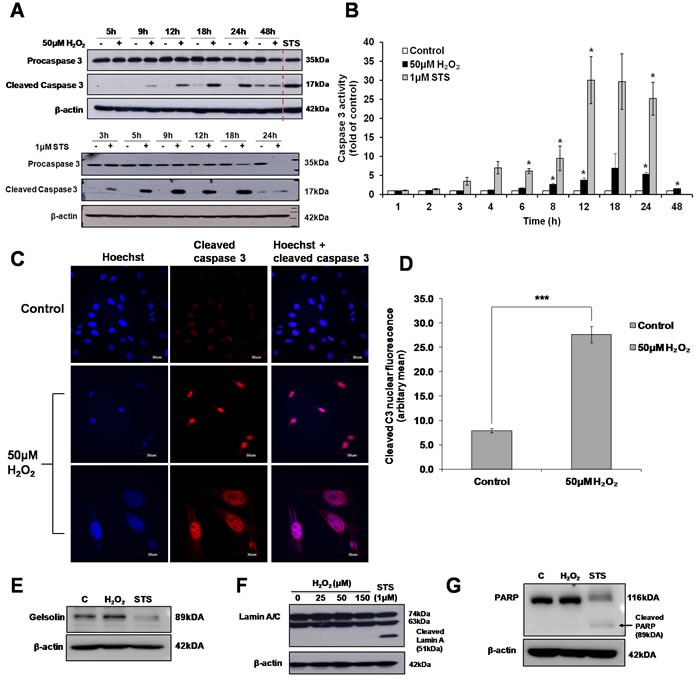
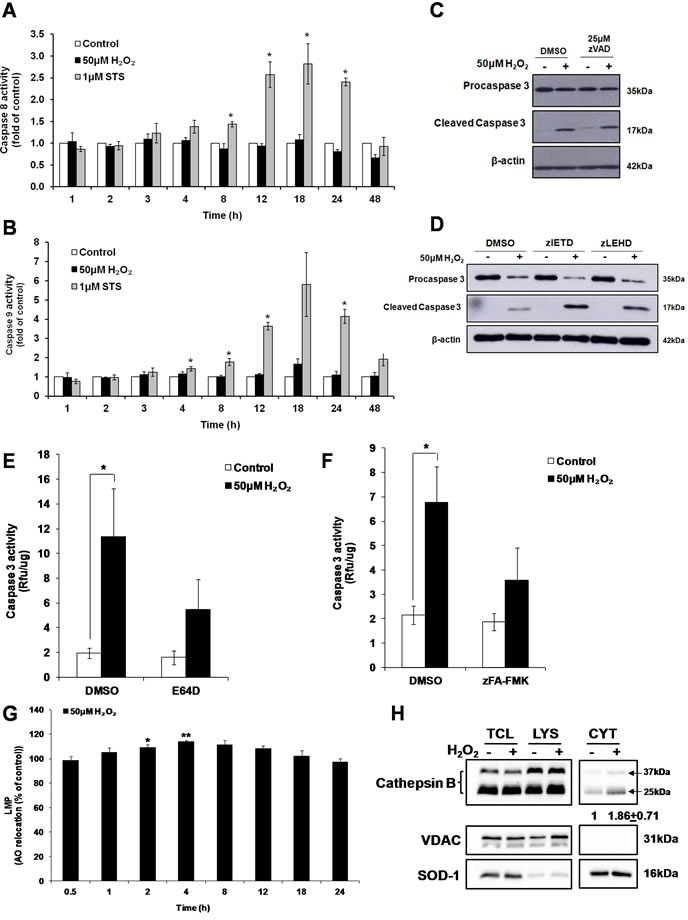
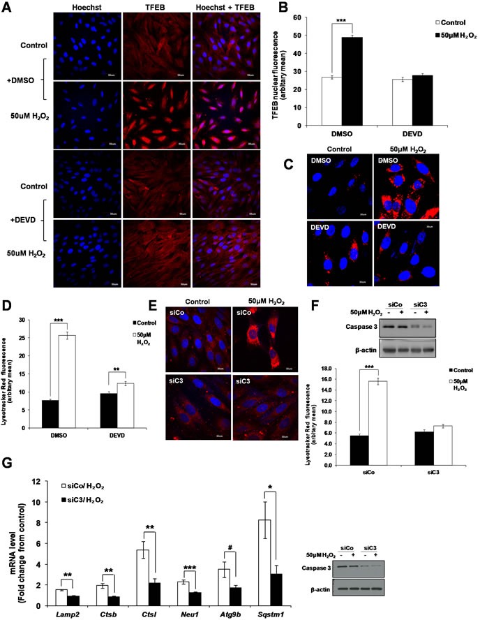
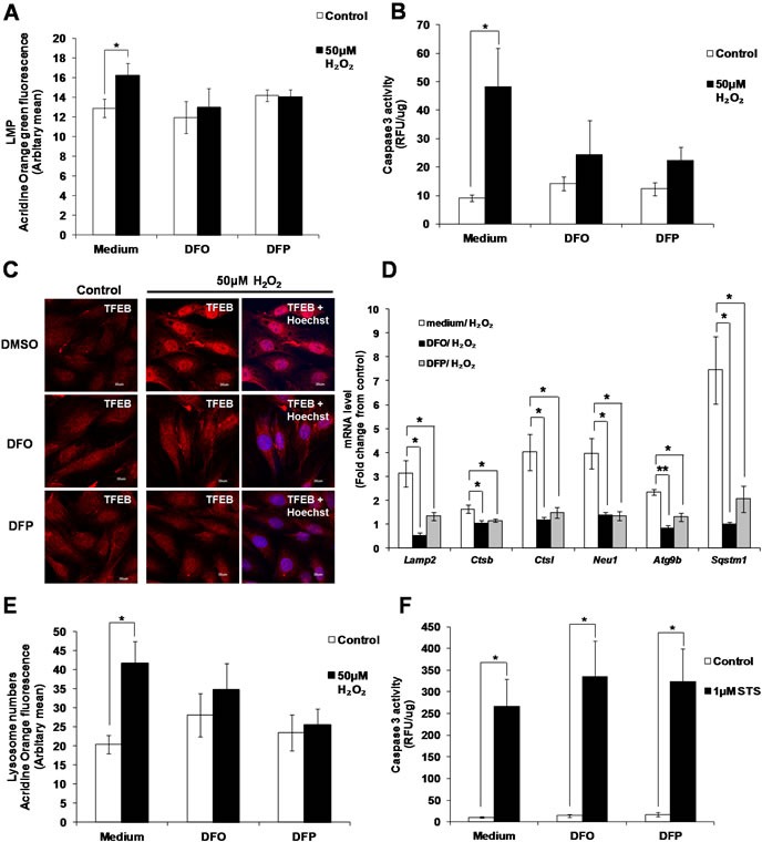
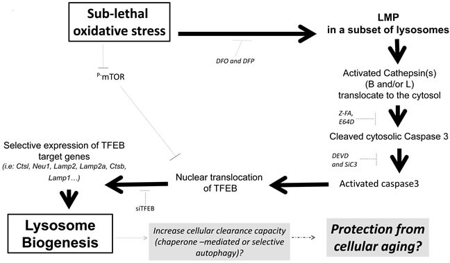
Here we provide evidence to link sub-lethal oxidative stress to lysosome biogenesis. Exposure of cells to sub-lethal concentrations of exogenously added hydrogen peroxide resulted in cytosol to nuclear translocation of the Transcription Factor EB (TFEB), the master controller of lysosome biogenesis and function. Nuclear translocation of TFEB was dependent upon the activation of a cathepsin-caspase 3 signaling pathway, downstream of lysosomal membrane permeabilization and accompanied by a significant increase in lysosome numbers as well as induction of TFEB-dependent lysosome-associated genes expression such as Ctsl, Lamp2 and its spliced variant Lamp2a, Neu1and Ctsb and Sqstm1 and Atg9b. The effects of sub-lethal oxidative stress on lysosomal gene expression and biogenesis were rescued upon gene silencing of caspase 3 and TFEB. Notably, caspase 3 activation was not associated with phenotypic hallmarks of apoptosis, evidenced by the absence of caspase 3 substrate cleavage, such as PARP, Lamin A/C or gelsolin. Taken together, these data demonstrate for the first time an unexpected and non-canonical role of a cathepsin-caspase 3 axis in the nuclear translocation of TFEB leading to lysosome biogenesis under conditions of sub-lethal oxidative stress.

Keywords: Autophagy; caspase 3; lysosomal membrane permeabilization; lysosomes; sub-lethal oxidative stress; transcription factor EB.


Conflict of interest statement

CONFLICTS OF INTEREST

Authors declare that there is no conflict of interest.

Figures

Figure 1
Figure 1. Sub-lethal oxidative stress does not induce apoptotic cell death
A. L6 myoblasts were exposed to 50μM or 150μM of exogenous H2O2 for 24 and 48h before cell morphology was observed under a phase contrast microscope. B. L6 myoblasts were treated with 50μM or 150μM of exogenous H2O2, or 1μM STS, for 24h and 48h.Percentage of cells in sub-G1 phase was assessed using the propidium iodide (PI) staining, statistical analysis was done by comparing treatment (H2O2 or STS) to untreated control at respective time points (24h or 48h), values represent mean +/- SEM. *P < 0.05; n = 4 (t-test). C. HO-1 protein expression in L6 cells exposed to 50μM H2O2 for the indicated time points.
Figure 2
Figure 2. Sub-lethal oxidative stress induces lysosome biogenesis
L6 cells were exposed to 50μM H2O2 for various time points before increase in lysosome numbers was assessed using A. AO uptake assay, values represent mean of the % of AO uptake compared to control cells at the same time point +/-SEM. *P < 0.05; n = 4 (t-test), or B. LTR uptake assay. Results are shown as confocal microscopy image showing cells with an increase in red fluorescence. Scale = 30μm. C. L6 cells were treated with 50μM H2O2 for 24h before being stained with LTR or with an antibody specific to Cathepsin B. Results are shown as confocal microscopy image showing cells with an increase in red fluorescence for LTR staining and for cathepsin B expression. Scale = 50μm. D. L6 cells were treated with 50μM H2O2 for 24h before being stained with organelle-specific antibodies or fluorescence dyes: mitochondria: MitoTracker Red CMXRos (MTR), endoplasmic reticulum: Calnexin, golgi: Syntaxin 16, late endosome: Rab7. Results are shown as confocal microscopy image. Scale = 50μm.
Figure 3
Figure 3. TFEB regulates lysosome biogenesis induced by a sub-lethal oxidative stress
A. TFEB translocation from the cytosol to the nucleus following cells’ exposure to 50μM H2O2 for 24 Hours. Results are shown as confocal microscopy image showing a translocation of the red fluorescence from the cytosol to the nucleus. Scale = 30μm. B. Analysis of TFEB positive nucleus in control and treated cells from (A) quantified by ImageJ. Values represent mean +/- SEM. ***P < 0.0005; control: n = 122, treated: n = 78 (t-test). C. mRNA expression of TFEB was assessed in L6 myoblasts transfected with a siRNA specific to TFEB for 48h (siTFEB) using SYBR Green Real-Time PCR, normalized to endogenous control 18s. Relative mRNA expression is expressed as fold change over cells transfected with a control siRNA (siCo). Values represent mean +/- SEM. ***P < 0.0005; n = 11 (t-test). Images show TFEB fluorescence (red) in cells transfected with siCo and siTFEB for 48h. TFEB fluorescence staining in siCo and siTFEB cells was then quantified by ImageJ. Values represent mean +/- SEM, ***P < 0.0005; siCo: n = 144, siTFEB: n = 169 (t-test). D.-F. L6 cells were transfected with siTFEB or negative control siRNA (siCo) and exposed to 50μM H2O2. (D) mRNA expression of Lamp2, Ctsb, Ctsl, Neu1, Atg9b and Sqstm1, as quantified by SYBR Green Real-Time PCR and normalized to endogenous control 18s. Relative mRNA expression is expressed as fold change over untreated control. Values represent mean+/- SEM. *P < 0.05, **P < 0.005, n = at least 4 (t-test). Lysosome numbers were determined using (E) AO uptake assay or (F) LTR immunofluorescence assay. (E) Values represent AO mean fluorescence in arbitrary units +/- SEM,*P < 0.05; n = 4 (t-test). The inhibitory effect of siTFEB on the increase of AO mean of fluorescence was statistically significant P < 0.05 (mixed model). (F) Results are shown as confocal microscopy image showing cells with an increase in red fluorescence as in Figure 2B. Scale = 30μm. G. Analysis of the LTR immunofluorescence assay shown in (F) using ImageJ. Values represent mean +/- SEM, ***P < 0.005; sico/control: n = 66, sico/H2O2; n = 66, siTFEB/control: n = 59, siTFEB/H2O2: n = 85 (t-test). The effect of siTFEB was statistically significant, P < 0.05 (mixed model).
Figure 4
Figure 4. TFEB is not involved in the increase in autophagic vacuoles induced by sub-lethal oxidative stress
A. Autophagic vacuoles in L6 cells exposed to 50μM H2O2 and 100nM Torin1 for 24h and 48h were detected using Cyto-ID staining, statistical analysis was done by comparing treatment (H2O2 or Torin1) to untreated control at respective time point (24h or 48h). Values represent mean +/- SEM, *P < 0.05, **P < 0.005; n = 4 (t-test). B. Detection of autophagic vacuoles in cells transfected with siTFEB or negative control siRNA (siCo), and exposed to 50μM H2O2 for 24h using Cyto-ID staining. Values represent mean +/- SEM, **P < 0.005; n = 5 (t-test). The effect of siTFEB was not statistically significant, P>0.05 (mixed model). C. Detection and co-localization of lysosomes and autophagic vacuoles using LTR and Cyto-ID respectively in L6 cells treated with 50μM H2O2 and 100nM Torin1 for 24h. Results are shown as confocal microscopy image. Scale = 30μm. D. L6 cells were treated with 50μM H2O2 for 24h. Cells were then incubated with 200nM Bafilomycin for 30min/1h before cells were harvested for LC3II Western Blot analysis.
Figure 5
Figure 5. Sub-lethal oxidative stress induces the activation of caspase 3
L6 cells were exposed to 50μM H2O2 or 1μM STS for the indicated time points before A. Western Blot analysis of caspase 3 cleavage and B. Caspase 3 activity was assessed, statistical analysis was done by comparing treatment (H2O2 or STS) to untreated control at respective time point (24h or 48h). Values represent mean +/- SEM, *P < 0.05; n = 4 (t-test). C. Detection of cleaved caspase 3 by immunofluorescence analysis of cells exposed to 50μM H2O2 for 24h, as viewed under a confocal microscope with low magnification (scale = 50μm) and high magnification (scale = 30μm). D. Analysis of the nuclear localization of cleaved caspase 3 shown in (C). Values represent mean +/- SEM, ***P < 0.0005; control: n = 49, treated: n = 73 (t-test). E.-G. Western Blot analysis of the cleavage of classical caspase 3 substrate in cells exposed to 50μM H2O2 or 1μM STS: (E) Gelsolin, (F) Lamin A/C and (G) PARP.
Figure 6
Figure 6. Cathepsin(s) induces the activation of caspase 3 by sub-lethal oxidative stress
A. Caspase 8 and B. Caspase 9 activity in cells exposed to 50μM H2O2 or 1μM STS for indicated time points, statistical analysis was done by comparing treatment (H2O2 or STS) to untreated control at respective time point (24h or 48h). Values represent mean+/- SEM, *P < 0.05; n = 4 (t-test). C.-D. Western Blot analysis of caspase 3 cleavage by 50μM H2O2, in the presence of (C) pan caspase inhibitor (zVAD-FMK), or (D) caspase 8 (zIETD-FMK) or caspase 9 (zLEHD-FMK) inhibitor. E.-F. Prior to treatment with 50μM H2O2, cells were pre-treated with (E) E64D (50μM) or (F) zFA-FMK (50μM). Cell lysate collected was assayed for caspase 3 activity with the fluorogenic substrate Ac-DEVD-AFC. Values represent mean + /- SEM, *P < 0.05; n = 4 (t-test). The inhibitory effect of E64D and zFA-FMK on the increase in caspase 3 activity was statistically significant P < 0.05 (mixed model). G. LMP in cells treated with 50μM H2O2 for indicated time points, measured by AO relocation assay. Values represent mean of the % of AO relocation compared to control cells at the same time point, mean +/-SEM, *P < 0.05; n = 4 (t-test). H. L6 cells were treated with 50μM H2O2 for 4h and cathepsin B was blotted in different fractions. TCL: total cell lysate (35μg); LYS: lysosome/membrane (70μg); CYT: cytosol (20μg). Band intensity of cathepsin B was quantified after normalization to loading control. Changes in cathepsin B protein level in the cytosol fraction is shown as the fold difference relative to control cells. Values represent the means +/-SEM of two independent experiments.
Figure 7
Figure 7. Caspase 3 is involved in the activation of TFEB leading to lysosome biogenesis
A. L6 cells were pretreated with 20μM zDEVD-FMK for 2h before treatment with 50μM H2O2 for 24h and TFEB intracellular location was assessed using a TFEB specific antibody. Scale = 50μm. B. Analysis of the red nuclear fluorescence representing nuclear TFEB in control and treated cells shown in (A) quantified by ImageJ. Values represent mean +/- SEM, ***P < 0.0005; DMSO/control: n = 108, DMSO/H2O2: n = 103, DEVD/control: n = 103. DEVD/H2O2: n = 114 (t-test). The inhibitory effect of DEVD on the translocation of TFEB to the nucleus was statistically significant, P < 0.05 (mixed model). C.-F. Lysosome number was assessed using the LTR assay in (C) L6 cells pre-treated with 20μM zDEVD-FMK or (E) L6 cells transfected with a specific Caspase 3 siRNA (siC3) or negative control siRNA (siCo), before treatment with 50μM H2O2 for 24h. (D and F) Values represent mean +/- SEM, **P < 0.005, ***P < 0.005; (D) DMSO/control: n = 85, DMSO/H2O2: n = 78, DEVD/control: n = 78. DEVD/H2O2: n = 72 (t-test). (F) siCo/control: n = 69, siCo/H2O2: n = 95, siC3/control: n = 61. siC3/H2O2: n = 104 (t-test). The inhibitory effect of DEVD and siC3 on lysosome number were statistically significant P < 0.05 (mixed model). G. mRNA expression of TFEB target genes, Lamp2, Ctsb, Ctsl, Neu1, Atg9b and Sqstm1, as quantified by SYBR Green Real-Time PCR and normalized to endogenous control 18s in cells transfected with a specific Caspase 3 siRNA (siC3) or negative control siRNA (siCo). Relative mRNA expression is expressed as fold change over untreated control. Values represent mean +/- SEM,*P < 0.05, **P < 0.005, ***P < 0.0005, #P = 0.069; n = at least 3 independent experiments (t-test).
Figure 8
Figure 8. Activation of caspase 3 and lysosome biogenesis induced by subtoxic oxidative stress involves iron
L6 cells were incubated with the iron chelators DFO (50μM) and DFP (50μM), before being exposed to 50μM H2O2. A. Values represent mean +/- SEM, *P < 0.05; n = 9 (t-test). The inhibitory effect of DFO and DFP on LMP was statistically significant, P < 0.05 (mixed model). B. Values represent mean +/- SEM, *P < 0.05; n = 5 (t-test). The inhibitory effect of DFO and DFP on caspase 3 activity was statistically significant, P < 0.05 (mixed model). C. TFEB cellular location in presence or absence of DFO and DFP (scale = 30μm), D. mRNA expression of TFEB target genes, Lamp2, Ctsb, Ctsl, Neu1, Atg9b and Sqstm1, and E. lysosome number in presence or absence of DFO and DFP was assessed as described in materials and methods. Values represent mean+/- SEM, *P < 0.05, **P < 0.005; n = at least 3 independent experiments (t-test). The inhibitory effect of DFO and DFP was statistically significant for P < 0.05 (mixed model). F. Caspase 3 activity in cells exposed to 1μM STS, pre-treated with DFO and DFP. Values represent mean+/- SEM, *P < 0.05; n = 5, (t-test). The effect of DFO and DFP was not statistically significant, P>0.05 (mixed model).
Figure 9
Figure 9. Sublethal oxidative stress activates an mTOR-independent signaling pathway involving the activation of a LMP-cathepsin-caspase 3 axis leading to the transcription of TFEB-target genes involved in lysosome biogenesis
We propose that caspase 3-dependent activation of TFEB might be a protective mechanism from cellular aging through activation of chaperone-mediated or selective autophagy following an increase in lysosome biogenesis in absence of autophagic flux.

Similar articles

Cited by

References

    1. Davies KJ. The broad spectrum of responses to oxidants in proliferating cells: a new paradigm for oxidative stress. IUBMB Life. 1999;48(1):41–47. - PubMed
    1. Mates JM, Sanchez-Jimenez FM. Role of reactive oxygen species in apoptosis: implications for cancer therapy. Int J Biochem Cell Biol. 2000;32(2):157–170. - PubMed
    1. Jurk D, Wilson C, Passos JF, Oakley F, Correia-Melo C, Greaves L, Saretzki G, Fox C, Lawless C, Anderson R, Hewitt G, Pender SL, Fullard N, Nelson G, Mann J, van de Sluis B, et al. Chronic inflammation induces telomere dysfunction and accelerates ageing in mice. Nature communications. 2014;2:4172. - PMC - PubMed
    1. Lu T, Finkel T. Free radicals and senescence. Experimental cell research. 2008;314(9):1918–1922. - PMC - PubMed
    1. Starke-Reed PE, Oliver CN. Protein oxidation and proteolysis during aging and oxidative stress. Archives of biochemistry and biophysics. 1989;275(2):559–567. - PubMed

MeSH terms