. 2017 Jul;45(5):529-538.
doi: 10.1111/ceo.12908. Epub 2017 Jan 29.

Lutein facilitates physiological revascularization in a mouse model of retinopathy of prematurity

Affiliations

Lutein facilitates physiological revascularization in a mouse model of retinopathy of prematurity

Zhongjie Fu et al. Clin Exp Ophthalmol. 2017 Jul.

Abstract

Background: Retinopathy of prematurity is one of the leading causes of childhood blindness worldwide, with vessel growth cessation and vessel loss in phase I followed by neovascularization in phase II. Ischaemia contributes to its pathogenesis, and lutein protects against ischaemia-induced retinal damages. We aimed to investigate the effects of lutein on a murine model of oxygen-induced retinopathy.

Methods: Mouse pups were exposed to 75% oxygen for 5 days and returned to room air for another 5 days. Vascular obliteration, neovascularization and blood vessel leakage were examined. Immunohistochemistry for glial cells and microglia were performed.

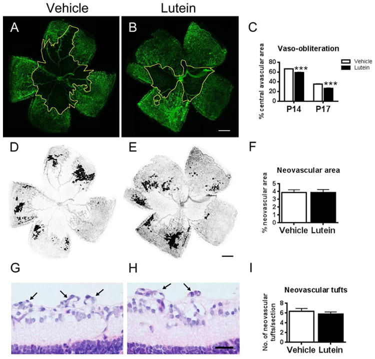
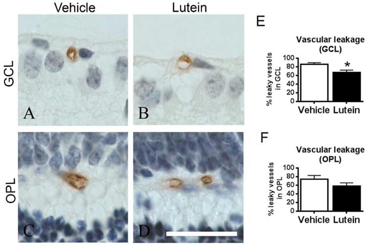
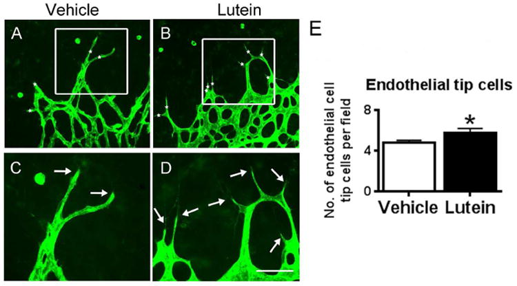
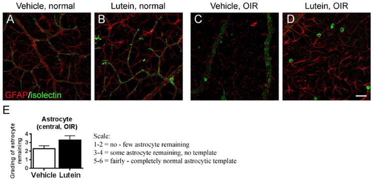
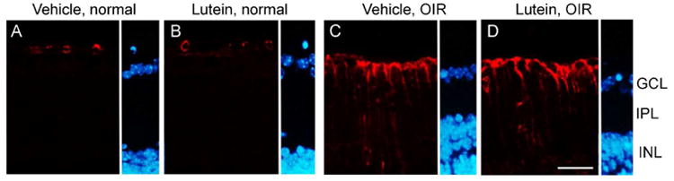
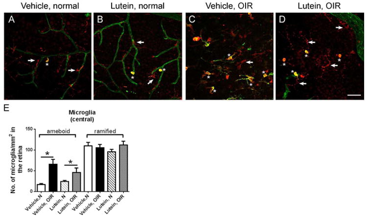
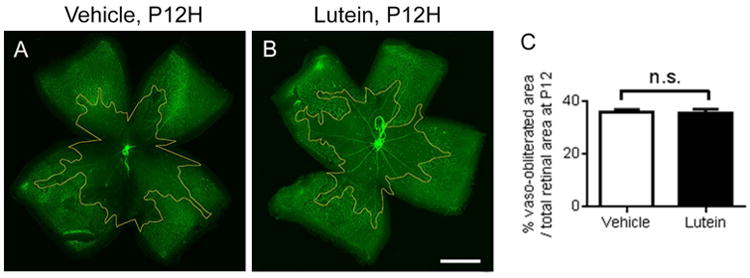
Results: Compared with vehicle controls, mouse pups receiving lutein treatment displayed smaller central vaso-obliterated area and reduced blood vessel leakage. No significant difference in neovascular area was found between lutein and vehicle controls. Lutein promoted endothelial tip cell formation and maintained the astrocytic template in the avascular area in oxygen-induced retinopathy. No significant changes in Müller cell gliosis and microglial activation in the central avascular area were found in lutein-treated pups.

Conclusions: Our observations indicated that lutein significantly promoted normal retinal vascular regrowth in the central avascular area, possibly through promoting endothelial tip cell formation and preserving astrocytic template. Our results indicated that lutein might be considered as a supplement for the treatment of proliferative retinopathy of prematurity because of its role in facilitating the revascularization of normal vasculature.

Keywords: oxygen-induced retinopathy; retinopathy of prematurity; vascular development.


Conflict of interest statement

Conflict of interest: None

Figures

Figure 1
Figure 1
Lutein promoted the normal vessel regrowth in the central avascular area in oxygen-induced retinopathy (OIR). Flat-mounted retinae were stained with isolectin GS-IB4 for blood vessels (green, A-B). After OIR, a central avascular area was observed (outlined in yellow). (C) Percentage of the central avascular area over the total retinal area at different postnatal days was estimated. Significantly reduced avascular area was observed in the lutein- versus vehicle-treated retinae on P14 and P17. In addition, bucketed flat-mounted retinae (D-E) and hematoxyline-and-eosin-stained retinal sagittal sections (G-H) were used to analyze neovascularization on P17. No significant difference in neovascular area and number of neovascular tufts was shown between vehicle- and lutein- treated OIR retinae (F, I). n = 6 to 9 for each group. ***p<0.001, Unpaired t test. Scale bar, 500μm (B, E); 25μm (H).
Figure 2
Figure 2
Lutein reduced the retinal vascular leakage along ganglion cell layer (GCL) in OIR. Representative images of IgG-stained retinal sagittal sections in GCL (A-B) and outer plexiform layer (OPL) (C-D) on P17. After OIR, intact vessel lumen and IgG staining outside the lumen were observed (A-D). The number of leaky vessels was quantified and expressed as a percentage of total number of retinal vessels (E-F). Significantly reduced retinal leaky vessels in GCL and a trend of decrease in OPL were found in lutein- versus vehicle-treated mice. The sections were counterstained with hematoxylin (blue) for nucleus. n = 5 to 8 for each group. *p<0.05, Unpaired t test. Scale bar, 10μm.
Figure 3
Figure 3
Lutein increased the endothelial cell tip formation in the retinal avascular area at P14. Flat-mounted retinae were stained with isolectin GS-IB4 for blood vessels (green, A-H). Representative images of retinal endothelial cell tip cells (asterisks, A-B) and filopodia (arrows, C-D) were shown. The number of the endothelial cell tip cells was quantified and compared between vehicle- and lutein- treated groups at P14. Significantly increased endothelial tip cells were observed with lutein treatment (E). n = 7 to 12 for each group. *p<0.05, Unpaired t test. Scale bar, 50μm.
Figure 4
Figure 4
Lutein maintained the astrocytic template in the retinal avascular area in OIR. Immunohistochemical staining of glial cells with GFAP antibody were conducted in retinal flat-mounts on P17 (A-D). In the normal retinae, GFAP-positive astrocytes (red) showed stellate/dendritic morphology and were tightly correlated with the superficial layer of retinal vasculature (isolectin GS-IB4, green). In OIR, the astrocytes lost their normal structure and network in avascular areas. Few astrocytes were present in the vehicle-treated retinae while more astrocytes were observed in lutein-treated retinae. n = 7 to 9 for each group. Scale bar, 50μm.
Figure 5
Figure 5
No significant differences were found in Müller cell gliosis between vehicle- and lutein- treated OIR retinas. GFAP immunoreactivity (red) was observed in astrocytes around the blood vessel lumen in room-air controls (A-B). Increased GFAP immunoreactivity along inner limiting membrane (ILM) and across inner plexiform layer (IPL) was observed in both OIR groups compared with their room-air controls respectively (C-D). The sections were counterstained with DAPI (blue) for nucleus. INL, inner nuclear layer. n = 7 to 9 for each group. Scale bar, 25μm.
Figure 6
Figure 6
No significant differences were found in microglial activation between vehicle- and lutein- treated OIR retinas. Representative images of retinal areas stained with Iba-1 antibody (red) and isolectin GS-IB4 (green) on P17. Generally, ramified microglia dominated (arrow, A-B) while occasionally ameboid microglia (asterisk, A-B) were observed in room-air retinae. In OIR, positive staining with both Iba-1 and isolectin GS-IB4 indicated activated microglia. In the retinal avascular area, ameboid microglia (asterisk, C-D) was significantly increased in superficial vascular layers (E). n = 5 to 9 for each group. *p<0.05, one-way ANOVA followed by Bonferroni's Multiple Comparison Test. Scale bar, 50μm.
Figure 7
Figure 7
No significant differences were found in hyperoxia-induced vessel loss at P12. Representative images of retinal whole mount stained with isolectin GS-IB4 (green) on P12. The vaso-obliterated area was outlined in yellow. n = 7 to 8 for each group. Unpaired t test. Scale bar, 1mm.

References

    1. Terry TL. Fibroblastic Overgrowth of Persistent Tunica Vasculosa Lentis in Infants Born Prematurely: II. Report of Cases-Clinical Aspects. Transactions of the American Ophthalmological Society. 1942;40:262–84. - PMC - PubMed
    1. Li SY, Fu ZJ, Lo AC. Hypoxia-induced oxidative stress in ischemic retinopathy. Oxidative medicine and cellular longevity. 2012;2012:426769. - PMC - PubMed
    1. Terry TL. Retrolental Fibroplasia in the Premature Infant: V. Further Studies on Fibroplastic Overgrowth of the Persistent Tunica Vasculosa Lentis. Transactions of the American Ophthalmological Society. 1944;42:383–96. - PMC - PubMed
    1. Kijlstra A, Tian Y, Kelly ER, Berendschot TT. Lutein: more than just a filter for blue light. Progress in retinal and eye research. 2012;31:303–15. - PubMed
    1. Li SY, Fu ZJ, Ma H, Jang WC, So KF, Wong D, Lo AC. Effect of lutein on retinal neurons and oxidative stress in a model of acute retinal ischemia/reperfusion. Investigative ophthalmology & visual science. 2009;50:836–43. - PubMed

MeSH terms

LinkOut - more resources