Review
. 2017 Feb;152(3):533-545.
doi: 10.1053/j.gastro.2016.10.047. Epub 2016 Dec 19.

Hippo Signaling in the Liver Regulates Organ Size, Cell Fate, and Carcinogenesis

Affiliations
Review

Hippo Signaling in the Liver Regulates Organ Size, Cell Fate, and Carcinogenesis

Sachin H Patel et al. Gastroenterology. 2017 Feb.

Abstract

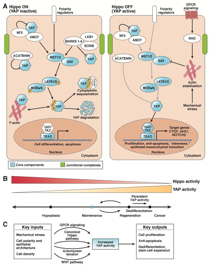
The Hippo signaling pathway, also known as the Salvador-Warts-Hippo pathway, is a regulator of organ size. The pathway takes its name from the Drosophila protein kinase, Hippo (STK4/MST1 and STK3/MST2 in mammals), which, when inactivated, leads to considerable tissue overgrowth. In mammals, MST1 and MST2 negatively regulate the transcriptional co-activators yes-associated protein 1 and WW domain containing transcription regulator 1 (WWTR1/TAZ), which together regulate expression of genes that control proliferation, survival, and differentiation. Yes-associated protein 1 and TAZ activation have been associated with liver development, regeneration, and tumorigenesis. How their activity is dynamically regulated in these contexts is just beginning to be elucidated. We review the mechanisms of Hippo signaling in the liver and explore outstanding questions for future research.

Keywords: TAZ; WWTR1; YAP1; YES-Associated Protein.


Figures

Figure 1
Figure 1. Regulation of the Mammalian Hippo Signaling Pathway
A. The Hippo pathway (mammalian) consists of the core components STK3 and STK4, SAV, LATS1 and 2, MOB1A and B, YAP, TAZ, and TEAD. Upon activation of the canonical Hippo pathway, STK3/4 phosphorylates and activates Lats1/2, which subsequently phosphorylates cytoplasmic Yap. During homeostasis, Hippo signaling is ON resulting in Yap phosphorylation (S112 in mice, S127 in humans) causing 14-3-3 binding and cytoplasmic sequestration. Phosphorylation of Yap can also lead to proteasomal degradation. When Hippo is off, YAP (in the unphosphorylated for) translocates to the nucleus and binds to the TEAD family of transcription factors, leading to the transcription of genes involved in cell survival, growth, and proliferation. Proposed cell activities for each state are found beneath the gene in italics. Arrows indicate positive relationships, while bars indicate negative activity. B. YAP activity at various levels and for various time periods differentially modulates cell state and phenotype. C. Multiple physiologic and pathologic inputs modulate YAP activity. Mechanical stress, cell polarity, and cell density are all factors that have been shown to modulate Yap activity. Additionally, knowledge of these different states is communicated via various signaling modalities, including the aforementioned canonical Hippo pathway, the Wnt pathway, GPCRs, and changes in cytoskeletal tension.
Figure 2
Figure 2. Yap Expression During Homeostasis and Regeneration
A. YAP is present in the epithelial cells of mouse liver (hepatocytes and biliary cells). YAP expression and nuclear-localization is more prominent in biliary cells (arrowhead) as compared to hepatocytes. Ad-Cre Yap fl/fl illustrates that YAP is present in hepatocytes as documented by mosaic Yap staining after deletion. B. Schematic of YAP activity in the liver. YAP activity is highest in the biliary cells/portal hepatocytes, diminishing in the hepatocytes toward the central vein. C. Hippo/Yap activity dynamically changes after partial hepatectomy. Yap levels increase with an associated decrease in MST1, MST2, LATS1 and LATS2 activity. These return to their normal levels as the liver reaches its appropriate size. Partial hepatectomy in mice results in YAP enrichment and an increase in nuclear localization (Day 2). After 8 days of recovery, YAP expression is reduced to below baseline levels.
Figure 3
Figure 3. Effects of Yap Overexpression in the Liver
A. Liver-specific overexpression of YAP leads to massive hepatomegaly with livers approaching 4–5x their original size. Upon restoration of endogenous levels of YAP, the liver returns to its usual size. Persistent YAP activation for 2 months frequently results in HCC development (Arrowheads). B. Increased overall YAP and nuclear YAP is a feature of several liver cancers, including HCC, CCA, and HB. C. YAP can mediate its tumorigenic effects either autonomously or through synergy with other pathways. YAP can be activated through canonical Hippo inactivation (1) or non-canonical membrane-associated signaling (2). YAP can also interact with the PI3K–Akt–mTOR pathway through a microRNA-mediated mechanism or via upregulation of lysosomal SLC transporters (3). Finally, YAP can interact with the NOTCH and Wnt pathways, as evidenced through upregulation of NOTCH ligands and receptors (4) and YAP’s stabilization by the Wnt target gene TRIB2 (5).

References

    1. Michalopoulos GK, DeFrances MC. Liver regeneration. Science. 1997;276:60–6. - PubMed
    1. Taub R. Liver regeneration: from myth to mechanism. Nat Rev Mol Cell Biol. 2004;5:836–47. - PubMed
    1. Dutkowski P, Linecker M, DeOliveira ML, et al. Challenges to liver transplantation and strategies to improve outcomes. Gastroenterology. 2015;148:307–23. - PubMed
    1. Emre S, Umman V. Split liver transplantation: an overview. Transplant Proc. 2011;43:884–7. - PubMed
    1. Tanaka A, Tanaka K, Tokuka A, et al. Graft size-matching in living related partial liver transplantation in relation to tissue oxygenation and metabolic capacity. Transpl Int. 1996;9:15–22. - PubMed

MeSH terms

Substances