Review
. 2017 Mar 1;9(3):a027847.
doi: 10.1101/cshperspect.a027847.

Directional Fluid Transport across Organ-Blood Barriers: Physiology and Cell Biology

Affiliations
Review

Directional Fluid Transport across Organ-Blood Barriers: Physiology and Cell Biology

Paulo S Caceres et al. Cold Spring Harb Perspect Biol. .

Abstract

Directional fluid flow is an essential process for embryo development as well as for organ and organism homeostasis. Here, we review the diverse structure of various organ-blood barriers, the driving forces, transporters, and polarity mechanisms that regulate fluid transport across them, focusing on kidney-, eye-, and brain-blood barriers. We end by discussing how cross talk between barrier epithelial and endothelial cells, perivascular cells, and basement membrane signaling contribute to generate and maintain organ-blood barriers.


Figures

Figure 1.
Figure 1.
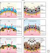
Organ–blood barriers in kidney, brain, and eye. Organ–blood barriers consist of a layer of parenchymal cells (top), a basement membrane (middle), and a layer of endothelial cells (bottom). (A) Kidney glomerular barrier, (B) kidney proximal tubule barrier, (C) brain–blood barrier, (D) choroid plexus barrier, (E) inner retina–blood barrier, and (F) outer retina–blood barrier. Net fluid transport (represented by a red arrow) can follow a transcellular route (C, D, E, and F) a paracellular route (A) or both (B). RPE, Retinal pigment epithelium; TJs, tight junctions.
Figure 2.
Figure 2.
Directional fluid transport across epithelia. (A) Effect of osmotic gradients on the direction of fluid transport by gall bladder epithelium. (Panel is based on data in Diamond 1962.) Fluid flow was measured in gall bladder (y axis) to determine whether the epithelium was absorbing (red part of the trace) or secreting (blue part of the trace). The transepithelial osmotic gradient was manipulated to establish a driving force toward the lumen or the basolateral side (x axis). When the osmotic gradient was neutral (dashed line) isosmotic absorption was observed, and when the osmotic gradient reversed, the epithelium continued to absorb against the driving force until it eventually reversed the direction of the fluid toward secretion at higher osmotic gradients. (B) Three-compartment model (Curran and Macintosh 1962). The three compartments A, B, and C are separated by two membranes with a reflection coefficient σ ≈ 1 (impermeable to solutes) and σ ≈ 0 (permeable to solutes). Water moves from A to B by osmosis and from B to C because of hydrostatic pressure. (C) Standing osmotic gradient model (Diamond and Bossert 1967). Active solute pumping into the blind end of the lateral intercellular space (LIS) drives water flow into the compartment and exit from it through the open end. The model assumes an impermeable blind end of the LIS. (D) Three-compartment model compatible with epitelial architecture (Weinstein and Stephenson 1981). Active solute pumping by the Na+, K+-ATPase into the intermediate compartment (LIS) generates the osmotic force for water absorption from the lumen through the intracelular space. The increase in hydrostatic pressure inside the LIS drives fluid exit through the basement membrane (BM) into the interstitium. The model assumes a higher reflection coefficient of the tight junctions (TJs) relative to the BM. (E) Osmotic model (Spring 1998). A small transepithelial osmotic gradient is sufficient to drive fluid flow (secretion in the illustration) thanks to the very high water permeability of some epithelia. This model applies to absorptive and secretory epithelia depending on the direction of the osmotic gradient. (F) Water pump (Zeuthen and Stein 1994). Water is transported against an osmotic gradient coupled with one or more solutes (X) using the favorable electrochemical gradient for the cotransported solutes. In all figures, the osmotic gradient is represented by the red shading, in which lighter indicates lower and darker represents higher osmolality.
Figure 3.
Figure 3.
Variable configuration of transporter polarity in secretory and absorptive epithelia. Different epithelial cells vary the localization of Na+, K+-ATPase, Na+, K+, Cl, bicarbonate, and water transporters and channels to perform functions specific for each organ and tissue. (A) Kidney proximal tubule: The electrochemical gradient generated by the basolateral Na+, K+-ATPase is used by apical Na+/H+ exchanger NHE3 and Na+-coupled transporters (SCTs) to absorb Na+ from the lumen. Apical anion exchangers (AEs) transport Clinside the cell. Sodium and Cl exit through basolateral transporters and channels, producing net absorption of NaCl, which provides the driving force for water absorption via aquaporin-1. (B) Choroid plexus: Uses apical Na+, K+-ATPase and a different combination of Na+, Cl, K+, and bicarbonate channels and transporters to secrete cerebrospinal fluid (CSF) into the brain ventricle. NaCl secretion into the CSF provides the driving force for water exit through aquaporin-1. (C) Retinal pigment epithelium: The apical Na+, K+-ATPase pumps Na+ into the cell and removes K+ from the subretinal space, which is necessary for the dark current of the photoreceptors. However, net transport of fluid occurs in the apical to basal direction driven by KCl absorption via apical Na+, K+, 2Cl cotransport and basolateral exit via K+ and Cl channels. Candidates are Maxi-K, CFTR, Bestrophin, and probably additional Cl channels.
Figure 4.
Figure 4.
Contributions of protein trafficking to establishing epithelial polarity. Epithelial cells are polarized with apical and basolateral membrane domains separated by tight junctions (TJs). Selective trafficking of domain-specific proteins in and out of these domains allows epithelia to perform their many functions. Apically and basolaterally targeted proteins use various sorting signals (right panel). Apical sorting signals can be associated with lipid rafts and basolateral sorting signals are often dependent on clathrin and the clathrin adaptors AP-1 and AP-2. Different routes are indicated in colored arrows and can traverse several endosomal compartments. Biosynthetic route (black): It originates in the endoplasmic reticulum (ER), then proteins traffic to the Golgi and they are sorted in the trans-Golgi network (TGN). Then proteins are delivered directly to the basolateral or apical membranes, they can traverse apical sorting endosomes (ASEs) and apical recycling endosomes (AREs) en route to the apical membrane or they can reach the basolateral membrane via common recycling endosomes (CREs). Apical recycling route (blue): Proteins are endocytosed from the apical membrane via clathrin, AP-2, and the small GTPases Rab4 and Rab5. They transit to the ASE, CRE, and ARE before reaching the apical membrane again via Rab11a. Basolateral recycling route (red): It originates in the basolateral membrane via endocytosis mediated by clathrin, AP-2, and Rab4 and 5. Proteins transit through the CRE before reaching the basolateral membrane again. Transcytotic route (green): Similar to the basolateral recycling route, it originates in the basolateral membrane, but from the CRE proteins are directed to the ARE and reach the apical membrane. BB, Basal body.

Similar articles

Cited by

References

    1. Abbott NJ. 2004. Evidence for bulk flow of brain interstitial fluid: Significance for physiology and pathology. Neurochem Int 45: 545–552. - PubMed
    1. Adelman JL, Sheng Y, Choe S, Abramson J, Wright EM, Rosenberg JM, Grabe M. 2014. Structural determinants of water permeation through the sodium-galactose transporter vSGLT. Biophys J 106: 1280–1289. - PMC - PubMed
    1. Adijanto J, Banzon T, Jalickee S, Wang NS, Miller SS. 2009. CO2-induced ion and fluid transport in human retinal pigment epithelium. J Gen Physiol 133: 603–622. - PMC - PubMed
    1. Alper SL, Stuart-Tilley A, Simmons CF, Brown D, Drenckhahn D. 1994. The fodrin-ankyrin cytoskeleton of choroid plexus preferentially colocalizes with apical Na+K+-ATPase rather than with basolateral anion exchanger AE2. J Clin Invest 93: 1430–1438. - PMC - PubMed
    1. Amorena C, Castro AF. 1997. Control of proximal tubule acidification by the endothelium of the peritubular capillaries. Am J Physiol 272: R691–R694. - PubMed

LinkOut - more resources