. 2017 Jan;5(1):17-28.
doi: 10.1158/2326-6066.CIR-16-0206. Epub 2016 Dec 21.

Angiopoietin-2 as a Biomarker and Target for Immune Checkpoint Therapy

Affiliations

Angiopoietin-2 as a Biomarker and Target for Immune Checkpoint Therapy

Xinqi Wu et al. Cancer Immunol Res. 2017 Jan.

Abstract

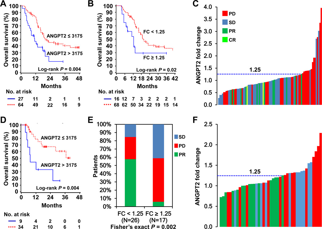
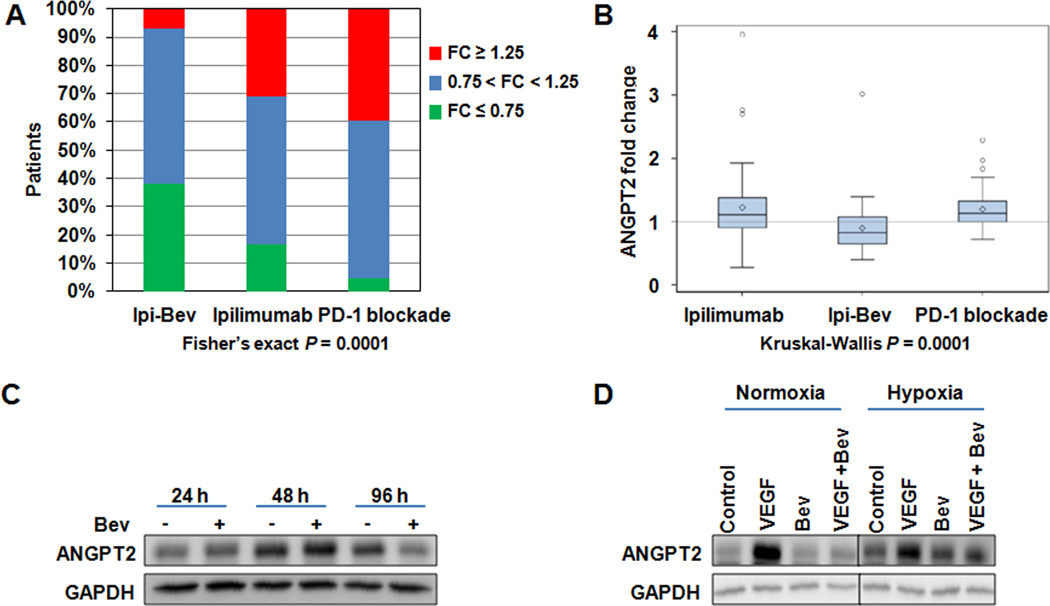
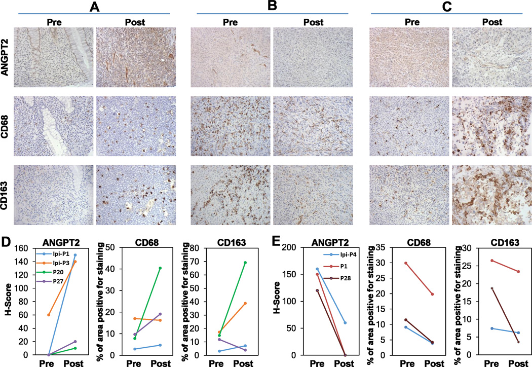
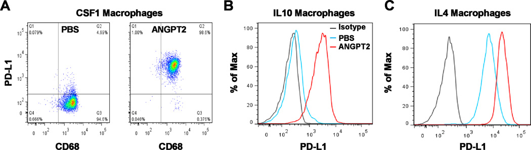
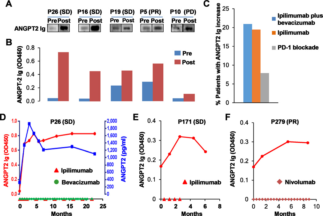
Immune checkpoint therapies targeting CTLA-4 and PD-1 have proven effective in cancer treatment. However, the identification of biomarkers for predicting clinical outcomes and mechanisms to overcome resistance remain as critical needs. Angiogenesis is increasingly appreciated as an immune modulator with potential for combinatorial use with checkpoint blockade. Angiopoietin-2 (ANGPT2) is an immune target in patients and is involved in resistance to anti-VEGF treatment with the monoclonal antibody bevacizumab. We investigated the predictive and prognostic value of circulating ANGPT2 in metastatic melanoma patients receiving immune checkpoint therapy. High pretreatment serum ANGPT2 was associated with reduced overall survival in CTLA-4 and PD-1 blockade-treated patients. These treatments also increased serum ANGPT2 in many patients early after treatment initiation, whereas ipilimumab plus bevacizumab treatment decreased serum concentrations. ANGPT2 increases were associated with reduced response and/or overall survival. Ipilimumab increased, and ipilimumab plus bevacizumab decreased, tumor vascular ANGPT2 expression in a subset of patients, which was associated with increased and decreased tumor infiltration by CD68+ and CD163+ macrophages, respectively. In vitro, bevacizumab blocked VEGF-induced ANGPT2 expression in tumor-associated endothelial cells, whereas ANGPT2 increased PD-L1 expression on M2-polarized macrophages. Treatments elicited long-lasting and functional antibody responses to ANGPT2 in a subset of patients receiving clinical benefit. Our findings suggest that serum ANGPT2 may be considered as a predictive and prognostic biomarker for immune checkpoint therapy and may contribute to treatment resistance via increasing proangiogenic and immunosuppressive activities in the tumor microenvironment. Targeting ANGPT2 provides a rational combinatorial approach to improve the efficacy of immune therapy. Cancer Immunol Res; 5(1); 17-28. ©2016 AACR.


Figures

Fig. 1
Fig. 1
High pretreatment ANGPT2 concentrations and increases in serum ANGPT2 were associated with poor clinical outcomes to immune checkpoint therapy in metastatic melanoma. A and B, Kaplan-Meier survival curves of pooled data from patients receiving ipilimumab or ipilimumab plus bevacizumab, based on ANGPT2 pretreatment concentrations (A, n = 91) and fold changes (B, n = 84). C, ANGPT2 fold changes and clinical responses in pooled patients receiving ipilimumab or ipilimumab plus bevacizumab (n = 84). Each bar represents a patient and its color indicates clinical response of the patient. D, Kaplan-Meier survival curves of PD-1 blockade-treated patients by pretreatment ANGPT2 levels (n = 43). E, Proportions of PD-1 blockade-treated patients with PR, SD and PD by ANGPT2 fold changes (n = 43). F, ANGPT2 fold changes and clinical responses to PD-1 blockade (n =43).
Fig. 2
Fig. 2
High pretreatment serum ANGPT2 concentrations followed by treatment-induced increases were associated with the worst OS and progressive disease. Data sets from patients receiving ipilimumab, ipilimumab plus bevacizumab or PD-1 blockade were combined and analyzed. A, Kaplan-Meier survival curves based on pretreatment ANGPT2 levels (n = 134). B, Kaplan-Meier survival curves by ANGPT2 fold changes (n = 127). C, Proportions of patients with complete remission/partial remission (CR/PR), stable disease (SD) and progressive disease (PD) according to ANGPT2 fold changes (n = 127). D, Kaplan-Meier survival curves based on pretreatment ANGPT2 concentrations and fold changes (n = 127). E, Proportions of patients with CR/PR, SD, and PD by the combination of pretreatment ANGPT2 levels and fold changes (n = 127).
Fig. 3
Fig. 3
PD-1 blockade and ipilimumab increased, whereas ipilimumab plus bevacizumab (Ipi-Bev) decreased serum ANGPT2 in significant proportions of patients. A, Proportions of patients displayed increase (fold change ≥ 1.25), decrease (fold change ≤ 0.75) or no change (0.75 < fold change < 1.25) in ANGPT2 in response to immune checkpoint therapy. B, Ipilimumab plus bevacizumab-treated patients (n = 43) displayed smaller fold changes than patients receiving ipilimumab (n = 41) or PD-1 blockade (n = 43). The diamonds, horizontal lines, and upper and lower boundaries of the boxes represent the sample average, median, 75th and 25th percentiles, respectively. C, Bevacizumab (Bev) downregulated ANGPT2 expression in TEC. D. VEGF promoted ANGPT2 expression and bevacizumab blocked VEGF-induced ANGPT2 expression in TEC. Representative imgaes of two experiments are shown.
Fig. 4
Fig. 4
Ipilimumab and ipilimumab plus bevacizumab influenced tumor ANGPT2 expression and macrophage infiltration. Paired and sequential pre- and posttreatment tumor biopsies were stained with anti-ANGPT2, anti-CD68, and anti-CD163, respectively. A, ANGPT2 upregulation was accompanied by increased infiltration of CD68+ and CD163+ macrophages in posttreatment tumor of an ipilimumab-treated patient. B and C, ANGPT2 down- and up-regulation in posttreatment tumor vasculature of ipilimumab plus bevacizumab-treated patients was respectively accompanied by decreased and increased infiltration of CD68+ and CD163+ macrophages. D and E, Semi-quantitative analysis of macrophage infiltration in tumors with increased (D, n = 4) and decreased (E, n = 3) vascular ANGPT2 expression.
Fig. 5
Fig. 5
ANGPT2 induces PD-L1 expression on M2-polarized monocyte-derived macrophages (MDM). A–C, MDM were differentiated from monocytes with CSF1 and then treated with ANGPT2 (300 ng/ml) for 3 days in the presence of CSF1 (A) or for 2 days in the presence of IL10 (B) or IL4 (C). MDM were sequentially stained with PE-conjugated PD-L1 antibody and FITC-conjugated CD68 antibody. Macrophages were gated on FSC/SSC and analyzed for CD68 and PD-L1 expression (A) or gated on CD68+ cells and analyzed for PD-L1 expression (B and C). Represntative results of at least 4 independent experiments are shown.
Fig. 6
Fig. 6
Immune checkpoint therapy elicited antibody responses to ANGPT2. A and B, ANGPT2 antibodies were detected in pre- and posttreatment plasma samples of ipilimumab plus bevacizumab-treated patients by immunoblot analysis (A) and ELISA (B). Clinical responses are also indicated. C, Proportions of patients receiving ipilimumab plus bevacizumab (n =43), ipilimumab (n = 36), and PD-1 blockade (n = 38) displayed an increase by 40% or more in ANGPT2 antibody concentrations. D–F, Longitudinal analysis of serum ANGPT2 and/or ANGPT2 antibodies in patients receiving ipilimumab plus bevacizumab (D), ipilimumab (E), or PD-1 blockade (F). Dosing of ipilimumab, bevacizumab, or nivolumab was indicated on the x-axis. Day 0 is pretreatment.

References

    1. Hodi FS, O’Day SJ, McDermott DF, Weber RW, Sosman JA, Haanen JB, et al. Improved survival with ipilimumab in patients with metastatic melanoma. N Engl J Med. 2010;363(8):711–723. - PMC - PubMed
    1. Robert C, Thomas L, Bondarenko I, O’Day S, Weber J, Garbe C, et al. Ipilimumab plus dacarbazine for previously untreated metastatic melanoma. N Engl J Med. 2011;364(26):2517–2526. - PubMed
    1. Hodi FS, Lawrence D, Lezcano C, Wu X, Zhou J, Sasada T, et al. Bevacizumab plus ipilimumab in patients with metastatic melanoma. Cancer Immunol Res. 2014;2(7):632–642. - PMC - PubMed
    1. Topalian SL, Hodi FS, Brahmer JR, Gettinger SN, Smith DC, McDermott DF, et al. Safety, activity, and immune correlates of anti-PD-1 antibody in cancer. N Engl J Med. 2012;366(26):2443–2454. - PMC - PubMed
    1. Hamid O, Robert C, Daud A, Hodi FS, Hwu WJ, Kefford R, et al. Safety and tumor responses with lambrolizumab (anti-PD-1) in melanoma. N Engl J Med. 2013;369(2):134–144. - PMC - PubMed

MeSH terms