. 2017 Feb 1;312(2):F284-F296.
doi: 10.1152/ajprenal.00271.2016. Epub 2016 Dec 21.

Insulin-like growth factor binding protein 7 and tissue inhibitor of metalloproteinases-2: differential expression and secretion in human kidney tubule cells

Affiliations

Insulin-like growth factor binding protein 7 and tissue inhibitor of metalloproteinases-2: differential expression and secretion in human kidney tubule cells

David R Emlet et al. Am J Physiol Renal Physiol. .

Abstract

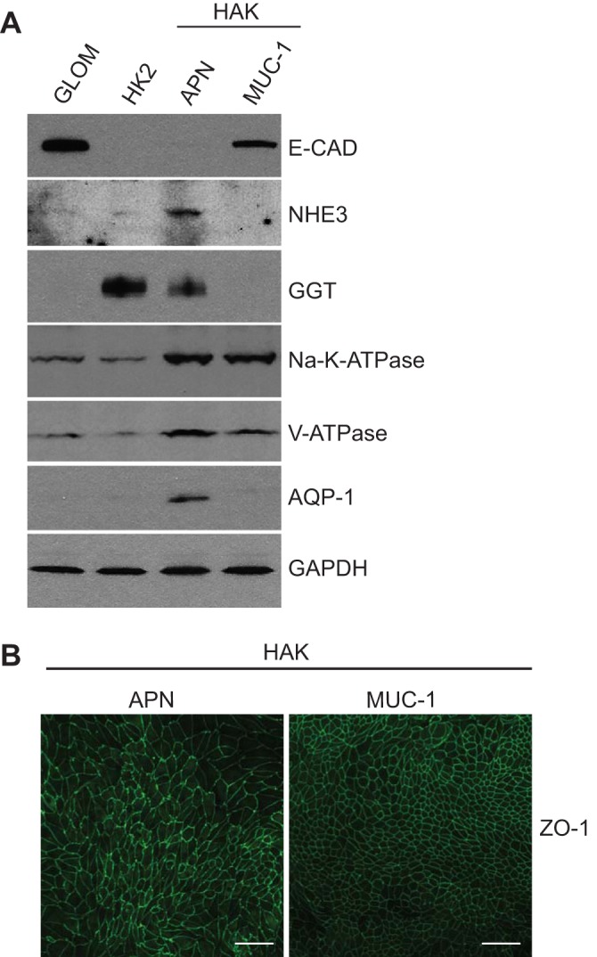
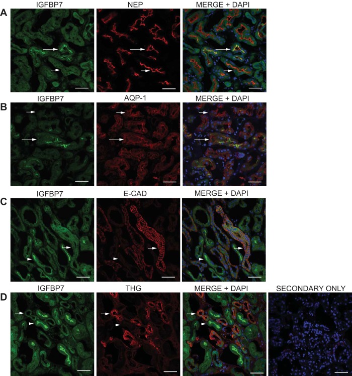
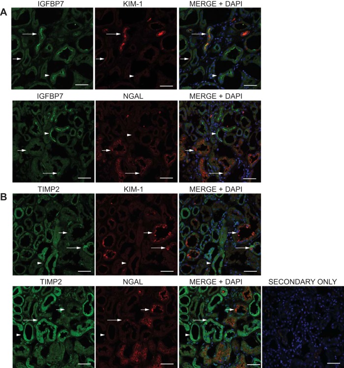
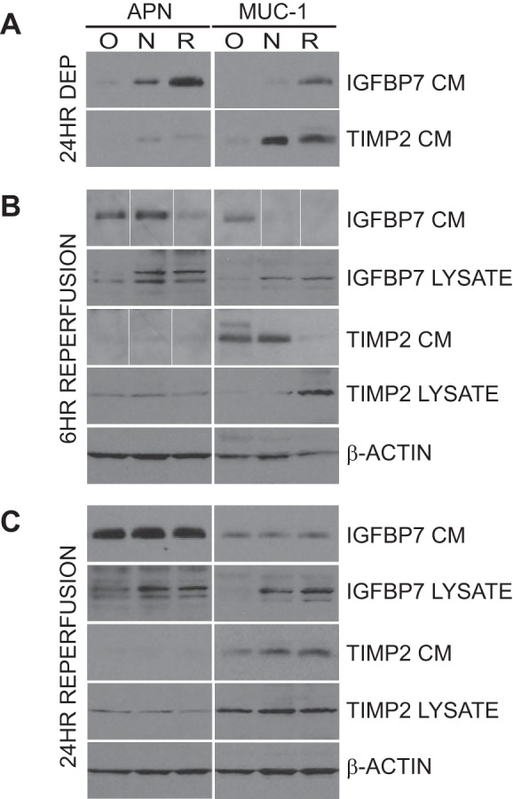
We have characterized the expression and secretion of the acute kidney injury (AKI) biomarkers insulin-like growth factor binding protein 7 (IGFBP7) and tissue inhibitor of metalloproteinases-2 (TIMP-2) in human kidney epithelial cells in primary cell culture and tissue. We established cell culture model systems of primary kidney cells of proximal and distal tubule origin and observed that both proteins are indeed expressed and secreted in both tubule cell types in vitro. However, TIMP-2 is both expressed and secreted preferentially by cells of distal tubule origin, while IGFBP7 is equally expressed across tubule cell types yet preferentially secreted by cells of proximal tubule origin. In human kidney tissue, strong staining of IGFBP7 was seen in the luminal brush-border region of a subset of proximal tubule cells, and TIMP-2 stained intracellularly in distal tubules. Additionally, while some tubular colocalization of both biomarkers was identified with the injury markers kidney injury molecule-1 and neutrophil gelatinase-associated lipocalin, both biomarkers could also be seen alone, suggesting the possibility for differential mechanistic and/or temporal profiles of regulation of these early AKI biomarkers from known markers of injury. Last, an in vitro model of ischemia-reperfusion demonstrated enhancement of secretion of both markers early after reperfusion. This work provides a rationale for further investigation of these markers for their potential role in the pathogenesis of acute kidney injury.

Keywords: IGFBP7; acute kidney injury; and TIMP-2; biomarkers.


Figures

Fig. 1.
Fig. 1.
Characterization of immunoaffinity isolated primary human kidney epithelial cells of proximal and distal tubule origin. Heterogeneous pools of cultured human kidney epithelial cells were immunoaffinity isolated using antibodies directed against the proximal tubule marker aminopeptidase N (APN) or the distal tubule marker Muc-1 (MUC-1). A: representative immunoblot characterization of lysates from isolated human adult kidney (HAK) cells of proximal origin (APN), and distal origin (MUC-1), compared with cells from glomeruli isolated by retention on an 180-µm sieve (GLOM), and HK2 cells (HK2). Lysates were assessed for expression of the proximal tubule markers sodium/hydrogen exchanger 3 (NHE3), γ-glutamyl-transpeptidase (GGT), and aquaporin-1 (AQP1), the distal tubule marker E-cadherin (E-CAD), as well as the ion transporters Na+-K+-ATPase (Na-K-ATPase) and the vacuolar H+-ATPase (V-ATPase). GAPDH was used for protein loading. B: HAK-APN and HAK-MUC-1 cells were cultured for at least 6 days on Transwell permeable supports, fixed with 2% paraformaldehyde, and subjected to immunofluorescence with the antibody against zonula occludens (ZO-1) for the identification of epithelial monolayer development. Scale bar = 50 µm.
Fig. 3.
Fig. 3.
Tubule-specific expression of IGFBP7 and TIMP-2 in primary human kidney cortical tissue. Paraformaldehyde-fixed human kidney sections were subjected to double-label immunofluorescent staining for IGFBP7 and TIMP2 vs. the markers used for immunoaffinity isolation, APN and the sialomucin Muc-1 (MUC-1). Single-stain micrographs are shown along with a 3-color micrograph (MERGE+DAPI) to identify localization and nuclei. A: comparison of IGFBP7 staining (green) to the proximal tubule marker APN (red). The long arrows show an example of tubule colocalization of brush-border staining of IGFBP7 with APN, and the short arrows show examples of tubules stained with APN alone. B: comparison of IGFBP7 staining (green) to the distal tubule marker MUC-1 (red). Short arrows show examples of tubules stained with MUC-1 alone, and the arrowheads show an example of tubule staining of IGFBP7 alone. C: comparison of TIMP-2 staining (green) to the proximal tubule marker APN (red). Short arrows show an example of a tubule stained with APN alone, and the arrowheads show tubule staining of TIMP-2 alone. D: comparison of TIMP-2 staining (green) to the distal tubule marker MUC-1 (red). The long arrows show an example of tubule colocalization of TIMP-2 with MUC-1, and the arrowheads show tubule staining of TIMP-2 alone. E: representative staining of IGFBP7 and TIMP2 (green) in glomeruli. Glomeruli are identified by brackets, and TIMP2 staining was present in glomeruli (long arrows), while IGFBP7 staining was very low (short arrows) compared with IGFBP7 positive tubules (arrowheads). The representative secondary only control (E; SECONDARY ONLY) was imaged at the highest laser power, gain, and offset used for all images. Scale bar = 50 µm in all images.
Fig. 2.
Fig. 2.
Expression and secretion of insulin-like growth factor binding protein 7 (IGFBP7) and tissue inhibitor of metalloproteinases-2 (TIMP-2) from immunoaffinity isolated primary human kidney epithelial cells of proximal and distal tubule origin. A: representative immunoblot analysis of IGFBP7, TIMP-2, and β-actin (loading control) expression in lysates from HAK isolated primary cells of proximal origin (HAK-APN) and distal origin (HAK-MUC-1), compared with cells from glomeruli isolated by retention on a 180-µm sieve (GLOM) and HK2 cells (HK2). B: representative immunoblot analysis of IGFBP7 and TIMP-2 secretion in conditioned media (24 h) from HAK-APN and HAK-MUC-1 cells compared with conditioned media from GLOM and HK2 cells and unconditioned media (UM). C: IGFBP7 and TIMP-2 densitometry values from 4 genetically separate HAK samples were adjusted to lysate protein concentrations and plotted normalized to the HAK-APN levels for both proteins.
Fig. 4.
Fig. 4.
Additional analysis of proximal tubule-specific expression of IGFBP7 in primary human kidney cortical tissue. Kidney sections were prepared, stained, and analyzed as in Fig. 3, using the IGFBP7 antibody compared with antibodies directed against the proximal tubule markers neprilysin (NEP) and AQP1 and the distal tubule markers E-CAD and Tamm-Horsfall glycoprotein (THG). Single-stain micrographs are shown along with a three color micrograph (MERGE+DAPI) to identify localization and nuclei. A and B: IGFBP7 staining (green) was compared with the proximal tubule markers NEP and AQP1 (red). C and D: IGFBP7 staining (green) was compared with the distal tubule markers E-CAD and THG (red). This analysis supports the findings with APN and MUC-1 that the bright, luminal, brush-border staining of IGFBP7 in human tissue samples localizes in tubules of proximal origin and does not localize with tubules of distal origin. The representative secondary only control was imaged the highest laser power, gain, and offset used for all images. Scale bar = 50 µm.
Fig. 5.
Fig. 5.
Additional analysis of distal tubule-specific expression of TIMP-2 in primary human kidney cortical tissue. Kidney sections were prepared, stained, and analyzed as in Fig. 3, using the TIMP-2 antibody compared with antibodies directed against the proximal tubule markers NEP and AQP1 and the distal tubule markers E-CAD and THG. Single-stain micrographs are shown along with a 3-color micrograph (MERGE+DAPI) to identify localization and nuclei. A and B: TIMP-2 staining (green) was compared with the proximal tubule markers NEP and AQP-1 (red). C and D: TIMP-2 staining (green) was compared with the distal tubule markers E-CAD and THG (red). This analysis supports the findings with APN and MUC-1 that the bright cytoplasmic staining of TIMP-2 in human tissue samples localizes in tubules of distal origin and does not localize with tubules of proximal origin. The representative secondary only control was imaged the highest laser power, gain, and offset used for all images. Scale bar = 50 µm.
Fig. 6.
Fig. 6.
Comparison of IGFBP7 and TIMP-2 with kidney injury molecule 1 (KIM-1) and NGAL staining in human kidney tissue. Kidney sections were prepared, stained, and analyzed as in Fig. 3, using the IGFBP7 and TIMP-2 antibodies compared with antibodies directed against the kidney injury markers KIM-1 and NGAL. Single-stain micrographs are shown along with a 3-color micrograph (MERGE+DAPI) to identify localization and nuclei. A: comparison of IGFBP7 staining (green) with KIM-1 and NGAL (red). The long arrows show examples of tubules with tubule colocalization of IGFBP7 with KIM-1 or NGAL, the short arrows show examples of tubules with KIM-1 or NGAL staining only, and the arrowheads demonstrate tubules with brush-border staining of IGFBP7 alone. B: comparison of TIMP-2 staining with KIM-1 and NGAL. The long arrows show examples of tubules with tubule colocalization of TIMP-2 with KIM-1 or NGAL, the short arrows show examples of tubules with KIM-1 or NGAL staining only, and the arrowheads demonstrate tubules with cytoplasmic staining of TIMP-2 alone. The representative secondary only was imaged at the highest laser power, gain, and offset used for all images. Scale bar = 50 µm for all images.
Fig. 7.
Fig. 7.
Effect of oxygen-nutrient deprivation and reperfusion on the expression and secretion of IGFBP7 and TIMP2 in cells of proximal and distal tubule origin. Proximal tubule (APN) and distal tubule (MUC-1) cells were prepared as in Fig. 1 and subjected to 24 h of oxygen-nutrient, nutrient deprivation alone, or no deprivation (24HR DEP; O, N, R, respectively) followed by 6 or 24 h of reperfusion by culture in regular culture media (6 HR REPERFUSION and 24 HR REPERFUSION). A: conditioned media (CM) after 24 h of deprivation (24HR DEP) were compared with media from cells cultured under normal culture conditions for the same period to assess the secretion of IGFBP7 and TIMP2. B: all conditions were reperfused in regular culture media for 6 h, and expression (LYSATE) and secretion (CM) of IGFBP7 and TIMP2 were assessed by immunoblot analysis. β-Actin was used as a loading control. The white lines between the lanes in some CM images denote noncontiguous lanes. These blots are composite images with individual lanes reorganized for the sake of presentation. The lanes in each image are compiled from the same blot and same film exposure, and no modification to individual lanes was performed. C: all conditions were reperfused in regular culture media for 24 h, and expression (LYSATE) and secretion (CM) of IGFBP7 and TIMP2 were assessed by immunoblot analysis. β-Actin was used as a loading control. Media and lysate from HAK4 are shown.

Similar articles

Cited by

References

    1. Al-bataineh MM, Gong F, Marciszyn AL, Myerburg MM, Pastor-Soler NM. Regulation of proximal tubule vacuolar H+-ATPase by PKA and AMP-activated protein kinase. Am J Physiol Renal Physiol 306: F981–F995, 2014. doi:10.1152/ajprenal.00362.2013. - DOI - PMC - PubMed
    1. Amemiya M, Loffing J, Lötscher M, Kaissling B, Alpern RJ, Moe OW. Expression of NHE-3 in the apical membrane of rat renal proximal tubule and thick ascending limb. Kidney Int 48: 1206–1215, 1995. doi:10.1038/ki.1995.404. - DOI - PubMed
    1. Amemiya Y, Yang W, Benatar T, Nofech-Mozes S, Yee A, Kahn H, Holloway C, Seth A. Insulin like growth factor binding protein-7 reduces growth of human breast cancer cells and xenografted tumors. Breast Cancer Res Treat 126: 373–384, 2011. doi:10.1007/s10549-010-0921-0. - DOI - PubMed
    1. Asanuma K, Shirato I, Ishidoh K, Kominami E, Tomino Y. Selective modulation of the secretion of proteinases and their inhibitors by growth factors in cultured differentiated podocytes. Kidney Int 62: 822–831, 2002. doi:10.1046/j.1523-1755.2002.00539.x. - DOI - PubMed
    1. Aubert S, Fauquette V, Hémon B, Lepoivre R, Briez N, Bernard D, Van Seuningen I, Leroy X, Perrais M. MUC1, a new hypoxia inducible factor target gene, is an actor in clear renal cell carcinoma tumor progression. Cancer Res 69: 5707–5715, 2009. doi:10.1158/0008-5472.CAN-08-4905. - DOI - PubMed

MeSH terms

Substances