. 2017 Feb;173(2):1059-1074.
doi: 10.1104/pp.16.01600. Epub 2016 Dec 21.

Identification and Characterization of Arabidopsis Seed Coat Mucilage Proteins

Affiliations

Identification and Characterization of Arabidopsis Seed Coat Mucilage Proteins

Allen Yi-Lun Tsai et al. Plant Physiol. 2017 Feb.

Abstract

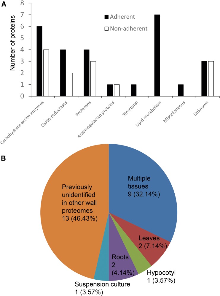
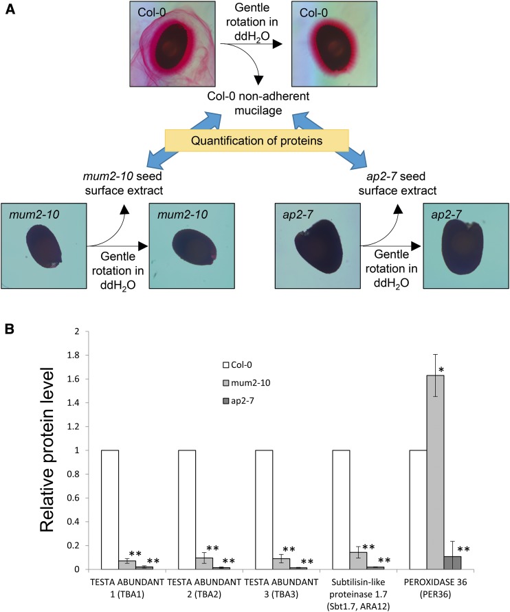
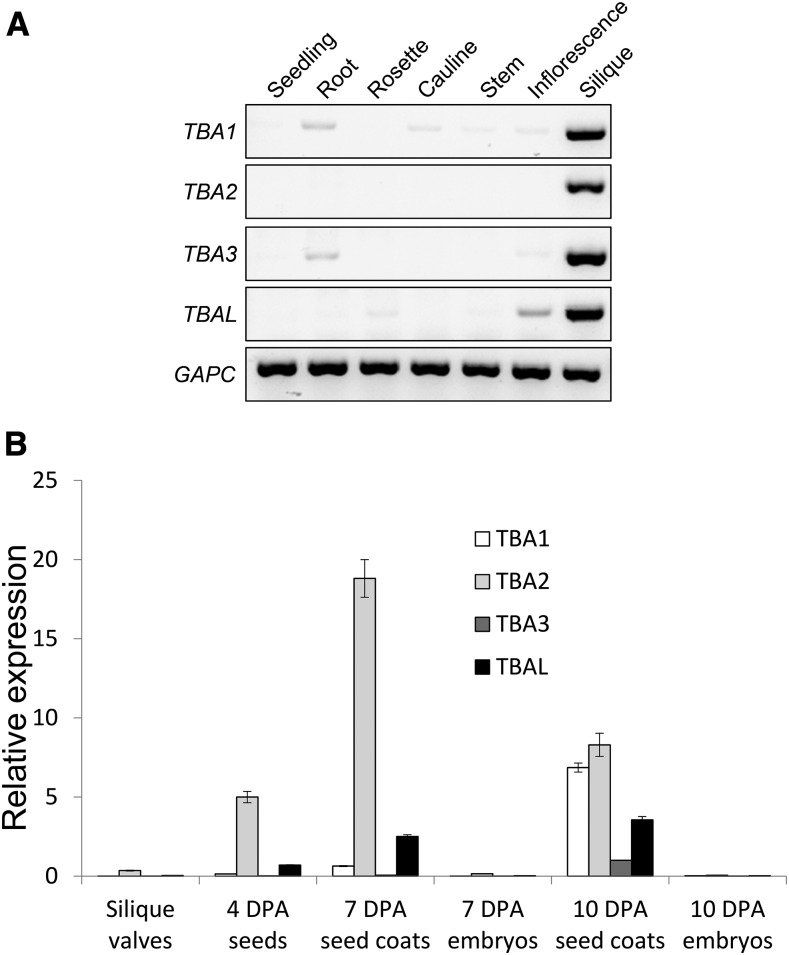
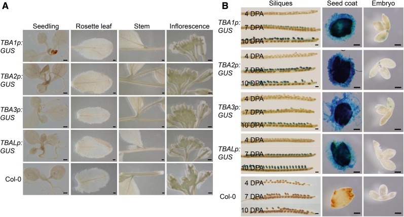
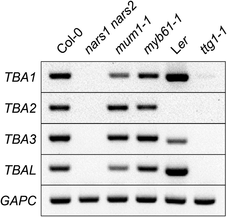
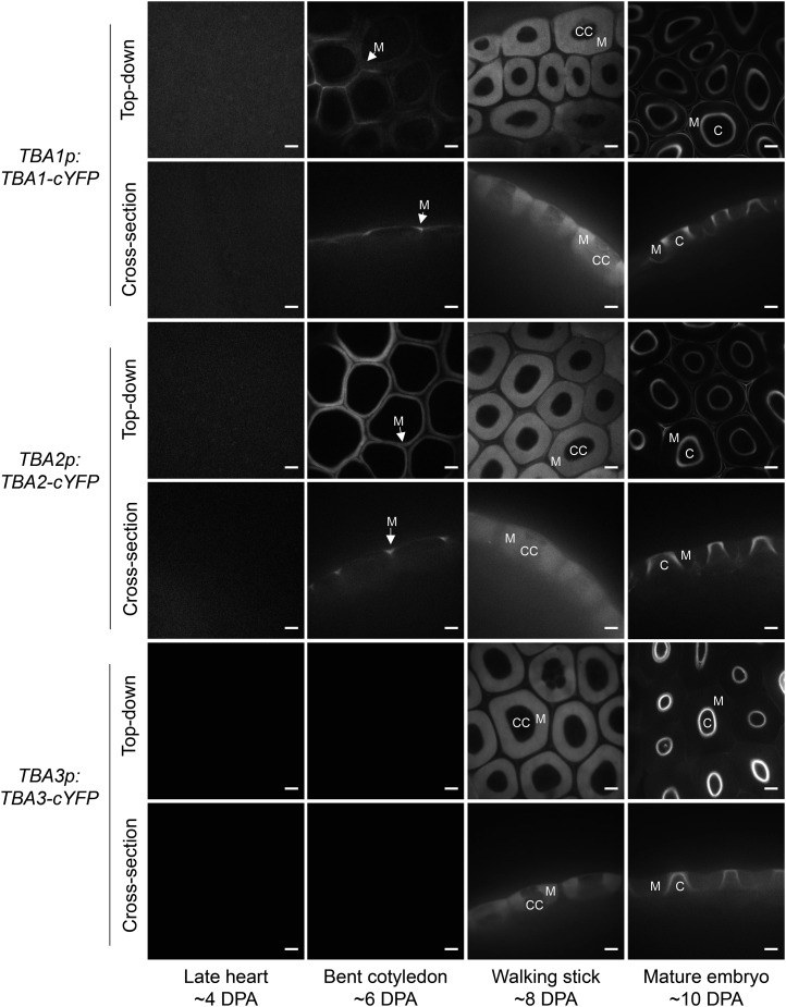
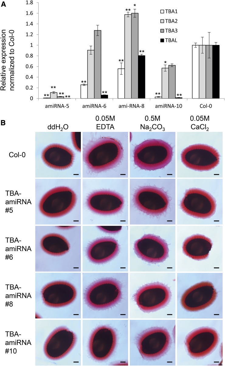
Plant cell wall proteins are important regulators of cell wall architecture and function. However, because cell wall proteins are difficult to extract and analyze, they are generally poorly understood. Here, we describe the identification and characterization of proteins integral to the Arabidopsis (Arabidopsis thaliana) seed coat mucilage, a specialized layer of the extracellular matrix composed of plant cell wall carbohydrates that is used as a model for cell wall research. The proteins identified in mucilage include those previously identified by genetic analysis, and several mucilage proteins are reduced in mucilage-deficient mutant seeds, suggesting that these proteins are genuinely associated with the mucilage. Arabidopsis mucilage has both nonadherent and adherent layers. Both layers have similar protein profiles except for proteins involved in lipid metabolism, which are present exclusively in the adherent mucilage. The most abundant mucilage proteins include a family of proteins named TESTA ABUNDANT1 (TBA1) to TBA3; a less abundant fourth homolog was named TBA-LIKE (TBAL). TBA and TBAL transcripts and promoter activities were detected in developing seed coats, and their expression requires seed coat differentiation regulators. TBA proteins are secreted to the mucilage pocket during differentiation. Although reverse genetics failed to identify a function for TBAs/TBAL, the TBA promoters are highly expressed and cell type specific and so should be very useful tools for targeting proteins to the seed coat epidermis. Altogether, these results highlight the mucilage proteome as a model for cell walls in general, as it shares similarities with other cell wall proteomes while also containing mucilage-specific features.


Figures

Figure 1.
Figure 1.
Strategy to isolate and identify seed coat mucilage proteins. A, Columbia-0 (Col-0) seed coat mucilage stained with Ruthenium Red. Double-headed arrows depict the two mucilage layers. Bar = 100 µm. B, Schematic depiction of the extraction and identification of mucilage proteins. The nonadherent mucilage and adherent mucilage were extracted sequentially. Proteins in each mucilage layer were identified by mass spectrometry (MS) after chemical deglycosylation and trypsin digestion. ddH2O, Distilled, deionized water.
Figure 2.
Figure 2.
Mucilage proteins are functionally similar to other cell wall proteins. A, Numbers of seed coat mucilage proteins from each mucilage layer sorted by the cell wall protein functional categories. B, Proportions of mucilage proteins previously identified in cell walls from other tissue types as documented by WallProtDB. Numbers denote the number of proteins in each category, while percentages denote the proportion of proteins that occupy each category.
Figure 3.
Figure 3.
Proteins identified are genuinely associated with mucilage. A, Schematic depiction of the mucilage protein quantification in mum2-10 and ap2-7 seed surface extracts relative to Col-0 nonadherent mucilage. ddH2O, Distilled, deionized water. B, Relative levels of mucilage proteins in Col-0 nonadherent mucilage, mum2-10 seed surface extract, and ap2-7 seed surface extract. Values are normalized to Col-0. Averages ± sd are shown; n = 3. *, P < 0.01; and **, P < 0.001.
Figure 4.
Figure 4.
Amino acid sequences of the TBA proteins. Amino acid sequence alignment is shown for TBA1, TBA2, and TBA3. Dark gray highlights amino acid residues that are identical, and light gray highlights amino acid residues that are similar. Underlined residues denote signal peptides predicted by SignalP (http://www.cbs.dtu.dk/services/SignalP/). Boldface S and T residues are predicted by NetOGlyc (http://www.cbs.dtu.dk/services/NetOGlyc/) to be O-glycosylated.
Figure 5.
Figure 5.
TBA and TBAL transcripts are found predominantly in the developing seed coat. A, RT-PCR detection of TBA1, TBA2, TBA3, and TBAL transcripts in seedlings, roots, rosette and cauline leaves, stem, inflorescence, and siliques. CYTOSOLIC GLYCERALDEHYDE-3-PHOSPHATE DEHYDROGENASE (GAPC) transcripts are shown as cDNA loading controls. B, Quantitative RT-PCR results showing the relative expression levels of TBA1, TBA2, TBA3, and TBAL in empty silique valves, 4-DPA seeds, 7-DPA seed coats, 7-DPA embryos, 10-DPA seed coats, and 10-DPA embryos. Expression levels were relative to GAPC transcript levels. n = 3, and error bars denote sd. A second biological replicate was processed with similar results.
Figure 6.
Figure 6.
TBA and TBAL promoters are active exclusively in the seed coat. A, TBA1p:GUS, TBA2p:GUS, TBA3p:GUS, TBALp:GUS, and Col-0 seedlings, rosette leaves, stems, and inflorescences stained for GUS activities. Bars = 500 µm. B, TBA1p:GUS, TBA2p:GUS, TBA3p:GUS, TBALp:GUS, and Col-0 siliques and developing seeds at 4, 7, and 10 DPA stained for GUS activities. The 10-DPA seed coats and embryos were dissected and stained separately, as shown in the two columns at right. Bars = 500 µm for siliques and 100 µm for dissected seed coats and embryos.
Figure 7.
Figure 7.
TBA and TBAL expression requires NARS1, NARS2, and TTG1. RT-PCR detection is shown for TBA and TBAL transcripts in 7-DPA seeds of nars1 nars2, mum1-1, myb61-1, ttg1-1, and their respective ecotype backgrounds. GAPC transcripts are shown as cDNA loading controls. Ler, Landsberg erecta.
Figure 8.
Figure 8.
TBA proteins are secreted to the seed coat epidermis apoplast. Confocal microscopy images denote the localization of cYFP-tagged TBA1, TBA2, and TBA3 in developing seed coats driven by their respective endogenous promoters. C, Columella; CC, cytoplasmic column; M, mucilage pockets. Bars = 10 µm.
Figure 9.
Figure 9.
Down-regulation of TBA and TBAL does not affect mucilage extrusion. A, Quantitative RT-PCR analysis of the expression of TBA and TBAL in 10-DPA siliques from four independent UBQ1p:TBA-amiRNA lines and Col-0. All expression levels were relative to GAPC and were then normalized to Col-0 expression levels. n = 3, and error bars denote sd. Asterisks denote transcript levels significantly different from the wild type at P < 0.05 (*) and P < 0.01 (**). B, Ruthenium Red-stained seed coat mucilage from the four independent UBQ1p:TBA-amiRNA lines shown in A. Seeds were imbibed in water, 0.05 m EDTA, 0.5 m Na2CO3, or 0.05 m CaCl2 prior to staining. Bars = 100 µm.

References

    1. Albenne C, Canut H, Jamet E (2013) Plant cell wall proteomics: the leadership of Arabidopsis thaliana. Front Plant Sci 4: 111. - PMC - PubMed
    1. Ambrose C, Allard JF, Cytrynbaum EN, Wasteneys GO (2011) A CLASP-modulated cell edge barrier mechanism drives cell-wide cortical microtubule organization in Arabidopsis. Nat Commun 2: 430. - PMC - PubMed
    1. Arsovski AA, Haughn GW, Western TL (2010) Seed coat mucilage cells of Arabidopsis thaliana as a model for plant cell wall research. Plant Signal Behav 5: 796–801 - PMC - PubMed
    1. Arsovski AA, Popma TM, Haughn GW, Carpita NC, McCann MC, Western TL (2009) AtBXL1 encodes a bifunctional β-D-xylosidase/α-L-arabinofuranosidase required for pectic arabinan modification in Arabidopsis mucilage secretory cells. Plant Physiol 150: 1219–1234 - PMC - PubMed
    1. Burton RA, Gidley MJ, Fincher GB (2010) Heterogeneity in the chemistry, structure and function of plant cell walls. Nat Chem Biol 6: 724–732 - PubMed

Publication types

MeSH terms

LinkOut - more resources