. 2017 Feb 1;37(5):1162-1175.
doi: 10.1523/JNEUROSCI.2181-16.2016. Epub 2016 Dec 21.

KCTD Hetero-oligomers Confer Unique Kinetic Properties on Hippocampal GABAB Receptor-Induced K+ Currents

Affiliations

KCTD Hetero-oligomers Confer Unique Kinetic Properties on Hippocampal GABAB Receptor-Induced K+ Currents

Thorsten Fritzius et al. J Neurosci. .

Abstract

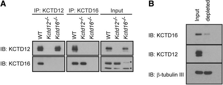
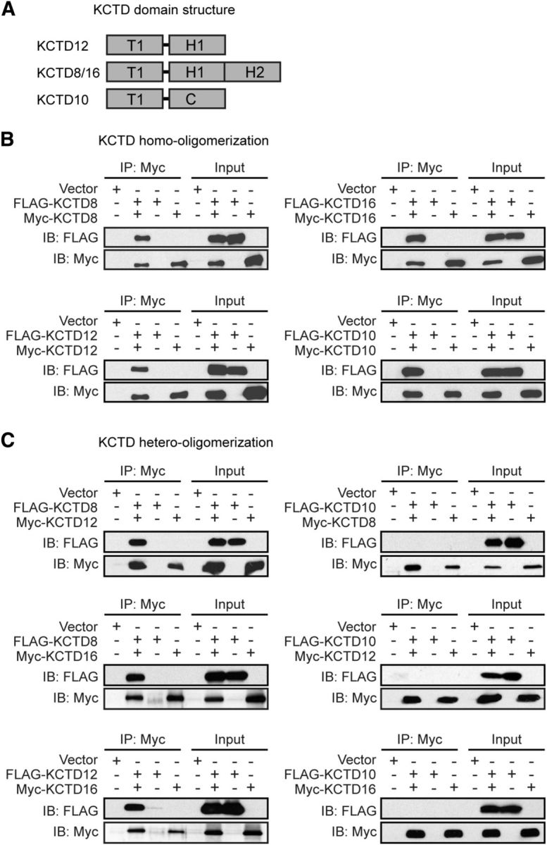
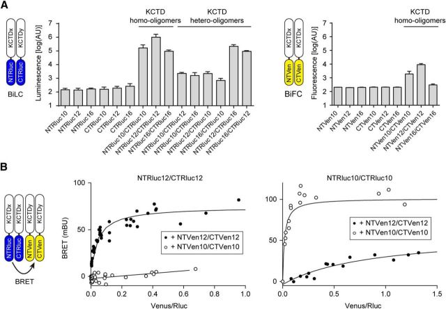
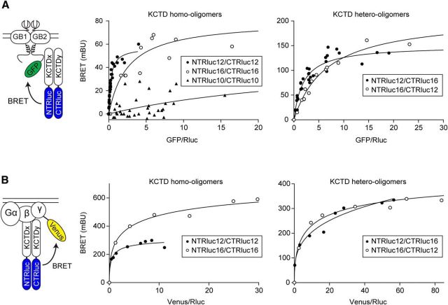
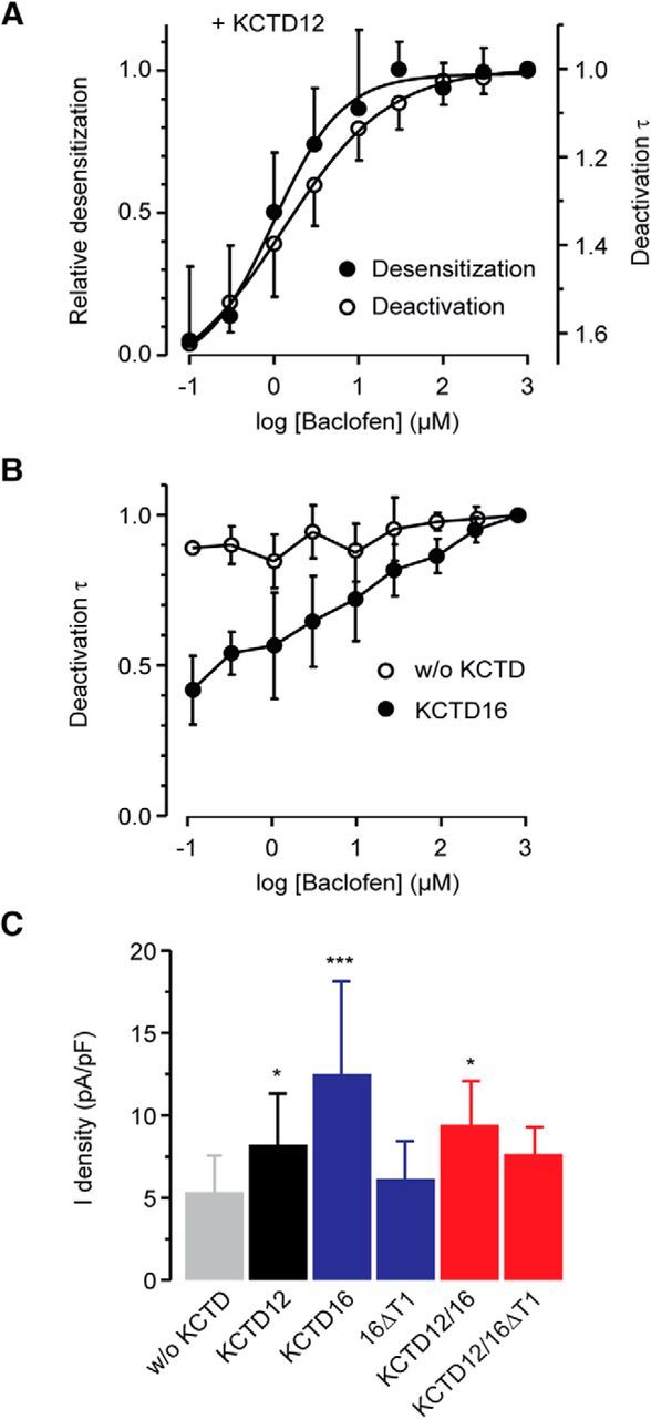
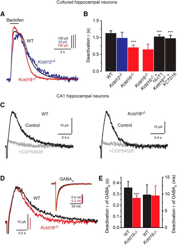
GABAB receptors are the G-protein coupled receptors for the main inhibitory neurotransmitter in the brain, GABA. GABAB receptors were shown to associate with homo-oligomers of auxiliary KCTD8, KCTD12, KCTD12b, and KCTD16 subunits (named after their T1 K+-channel tetramerization domain) that regulate G-protein signaling of the receptor. Here we provide evidence that GABAB receptors also associate with hetero-oligomers of KCTD subunits. Coimmunoprecipitation experiments indicate that two-thirds of the KCTD16 proteins in the hippocampus of adult mice associate with KCTD12. We show that the KCTD proteins hetero-oligomerize through self-interacting T1 and H1 homology domains. Bioluminescence resonance energy transfer measurements in live cells reveal that KCTD12/KCTD16 hetero-oligomers associate with both the receptor and the G-protein. Electrophysiological experiments demonstrate that KCTD12/KCTD16 hetero-oligomers impart unique kinetic properties on G-protein-activated Kir3 currents. During prolonged receptor activation (one min) KCTD12/KCTD16 hetero-oligomers produce moderately desensitizing fast deactivating K+ currents, whereas KCTD12 and KCTD16 homo-oligomers produce strongly desensitizing fast deactivating currents and nondesensitizing slowly deactivating currents, respectively. During short activation (2 s) KCTD12/KCTD16 hetero-oligomers produce nondesensitizing slowly deactivating currents. Electrophysiological recordings from hippocampal neurons of KCTD knock-out mice are consistent with these findings and indicate that KCTD12/KCTD16 hetero-oligomers increase the duration of slow IPSCs. In summary, our data demonstrate that simultaneous assembly of distinct KCTDs at the receptor increases the molecular and functional repertoire of native GABAB receptors and modulates physiologically induced K+ current responses in the hippocampus.

Significance statement: The KCTD proteins 8, 12, and 16 are auxiliary subunits of GABAB receptors that differentially regulate G-protein signaling of the receptor. The KCTD proteins are generally assumed to function as homo-oligomers. Here we show that the KCTD proteins also assemble hetero-oligomers in all possible dual combinations. Experiments in live cells demonstrate that KCTD hetero-oligomers form at least tetramers and that these tetramers directly interact with the receptor and the G-protein. KCTD12/KCTD16 hetero-oligomers impart unique kinetic properties to GABAB receptor-induced Kir3 currents in heterologous cells. KCTD12/KCTD16 hetero-oligomers are abundant in the hippocampus, where they prolong the duration of slow IPSCs in pyramidal cells. Our data therefore support that KCTD hetero-oligomers modulate physiologically induced K+ current responses in the brain.

Keywords: G-protein coupled receptor; GABA-B; GPCR; KCTD12; KCTD16; Kir3.


Figures

Figure 1.
Figure 1.
KCTD hetero-oligomers are abundant in the adult mouse brain. A, Anti-KCTD12 and anti-KCTD16 antibodies (Schwenk et al., 2010) copurify KCTD12 and KCTD16 proteins in immunoprecipitation (iP) experiments with brain lysates of WT mice, as shown on immunoblots (IB). Control IPs with brain lysates of Kctd12−/− and Kctd16−/− mice (Metz et al., 2011) show that the antibodies are specific for the KCTD12 and 16 proteins. Asterisks indicate cross-reactions of the anti-KCTD16 antibody in the Input samples. B, Affinity depletion of KCTD12 from hippocampi of adult mouse brains using anti-KCTD12 antibodies leads to a codepletion of KCTD16 engaged in KCTD12/KCTD16 hetero-oligomers (depleted). Control IBs of β-tubulin III show that the neuron-specific marker protein is not depleted by anti-KCTD12 antibodies.
Figure 2.
Figure 2.
KCTD8, KCTD12, and KCTD16 form homo- and hetero-oligomers in transfected HEK293. A, KCTD domain structure. KCTDs contain T1, H1, and H2 homology domains that are not closely sequence-related with the T1 and C-terminal (C) domains of KCTD10. The H2 domains are selectively present in KCTD8 and KCTD16. B, KCTDs form homo-oligomeric complexes. Input lanes indicate the expression of FLAG- or Myc-tagged KCTD proteins in the cell lysates used for IP with anti-Myc antibodies. Proteins in the IPs were revealed by immunoblotting (IB) with anti-FLAG and anti-Myc antibodies. C, KCTD8, KCTD12, and KCTD16 form hetero-oligomers with each other, but not with KCTD10. Input lanes indicate expression of the FLAG- or Myc-tagged KCTD proteins in the cell lysates used for IPs with anti-Myc antibodies. Proteins in the IPs were revealed by IB with anti-FLAG and anti-Myc antibodies.
Figure 3.
Figure 3.
Self-interacting T1 and H1 domains mediate KCTD homo- and hetero-oligomerization in HEK293 cells. IP of FLAG-KCTD12 (A) or FLAG-KCTD16 (B) with the Myc-tagged T1 and H1 domains of KCTD12 or KCTD16. IPs were performed with anti-Myc antibodies from total cell lysates. Proteins in the IPs were revealed by immunoblotting (IB) using anti-FLAG and anti-Myc antibodies. Input lanes indicate expression of the tagged proteins in the cell lysates used for the IPs. The Myc-tagged T1 and H1 domains of KCTD12 and KCTD16, but not the Myc-tagged H2 domain of KCTD16, coprecipitate FLAG-KCTD12 and FLAG-KCTD16. KCTD16 lacking the T1 domain (Myc-16ΔT1) interacts with FLAG-KCTDs via its H1 domain. Myc-KCTD12 and Myc-KCTD16 were used as positive controls, Myc-KCTD10 as a negative control. C, Myc-16ΔT1 copurifies GABAB2 (GB2-YFP) in the presence of KCTD16 or KCTD12, showing that Myc-16ΔT1 can homo- and hetero-oligomerize with KCTD16 and KCTD12 at GABAB2. Myc-16ΔT1 does not directly interact with GABAB2 because it lacks the T1 domain that is a prerequisite necessary for binding to GABAB2. IPs were performed with anti-Myc antibodies from total cell lysates. Proteins in the IPs were revealed by IB using anti-GABAB2, anti-FLAG, and anti-Myc antibodies. Input lanes (bottom) indicate expression of the tagged proteins in the cell lysates used for the IPs.
Figure 4.
Figure 4.
KCTD oligomerization in live HEK293 cells. A, BiLC or BiFC through oligomerization of split Rluc- or split Venus-tagged KCTDs, respectively. Cells were transfected with NTRlucKCTD and CTRlucKCTD or NTVenKCTD and CTVenKCTD constructs, and oligomerization between Rluc- or Venus-fragment-tagged KCTDs was monitored by measuring reconstituted luciferase or fluorescence activity, respectively. KCTD10, KCTD12, and KCTD16 all form homo-oligomers. KCTD12 and KCTD16 additionally form hetero-oligomers together. KCTD10 has a weak propensity to also form hetero-oligomers with KCTD12 or KCTD16. Labels in the bar graphs indicate the transfected Rluc- or Venus-fragment-tagged KCTD proteins. Data are mean ± SEM of 3 or 4 independent experiments. B, BRET between reconstituted Rluc and Venus in KCTD oligomers. Cells were transfected with fixed amounts of NTRlucKCTD and CTRlucKCTD constructs and increasing amounts of NTVenKCTD and CTVenKCTD constructs. BRET donor saturation curves were generated by expressing the net BRET signal detected as a function of the ratio between the total fluorescence signal and the luminescence signal (acceptor/donor ratio; expressed in milliBRET units, mBU) and demonstrate that both KCTD12 and KCTD10 can assemble into homo-tetramers or higher-order oligomers from the individually tagged monomers. KCTD12 and KCTD10 have a much weaker propensity to form hetero-tetramers. Data points represent the mean of technical duplicates combined from 4 independent experiments.
Figure 5.
Figure 5.
KCTD homo- and hetero-oligomers bind to both GABAB receptors and G-proteins in live HEK293 cells. A, BRET between KCTD oligomers and GABAB receptors in living cells. Cells were transfected with fixed amounts of NTRlucKCTD and CTRlucKCTD constructs and increasing amounts of Myc-GABAB1 and HA-GABAB2-GFP constructs. BRET was measured between reconstituted Rluc and GABAB2-GFP. Data points represent the mean of technical duplicates combined from 6 or 7 independent experiments. Donor/acceptor ratios needed to reach half BRET saturation (BRET50) were as follows: KCTD12 homo-oligomer: 0.27 ± 0.03; KCTD16 homo-oligomer: 2.55 ± 0.97; KCTD12/KCTD16 hetero-oligomer: 1.91 ± 0.29; KCTD16/KCTD12 hetero-oligomer: 6.65 ± 0.71 (mean ± SEM, n = 6 or 7). B, BRET between KCTD oligomers and the G-protein in live cells. Cells were transfected with fixed amounts of NTRlucKCTD and CTRlucKCTD constructs and increasing amounts of Gβ2 and Gγ2-YFP constructs. BRET was measured between reconstituted Rluc and Gγ2-YFP. Data points represent the mean of technical quadruplicates from a representative experiment (n = 3).
Figure 6.
Figure 6.
Bidirectional modulation of GABAB-activated Kir3 currents by KCTD hetero-oligomers. A, Representative baclofen-activated K+ current traces recorded at −50 mV from CHO cells expressing GABAB receptors and Kir3.1/3.2 channels either without KCTD (without [w/o], gray trace), with KCTD12 alone (black), with KCTD16 alone (blue), or with both KCTDs (red). KCTD12, but not KCTD16, induces pronounced and rapid desensitization of K+ currents. Coexpression of KCTD12 and KCTD16 results in intermediate current desensitization. B, Baclofen-activated K+ currents recorded from CHO cells either expressing KCTD12 (black), a KCTD16 mutant lacking the T1 domain (16ΔT1, blue), or both KCTD isoforms (red). Expression of KCTD12 together with 16ΔT1, which only binds to the receptor in a complex with KCTD12, reduces KCTD12-induced desensitization. C, Bar graph summarizing the relative desensitization of baclofen-activated K+ currents. The relative desensitization was calculated as follows: (1 − (ratio of current amplitude after 60 s vs peak current)) × 100. Values are mean ± SD of 26 (w/o KCTD), 17 (KCTD12), 14 (KCTD16), 11 (16ΔT1), 6 (KCTD12/KCTD16), and 13 (KCTD12/KCTD16ΔT1) cells. D, Normalized traces represent a slower time course of K+ current desensitization in CHO cells coexpressing KCTD12 and 16ΔT1 (red) compared with CHO cells expressing KCTD12 alone (black). Traces are fitted to a double exponential function (gray solid line) with time constants τ1 = 1.0 s (relative contribution to desensitization 71.7%) and τ2 = 9.4 s for KCTD12 and τ1 = 3.9 s (33.8%) and τ2 = 28.4 s for KCTD12/KCTD16ΔT1. E, Bar graph showing mean amplitude-weighted time constants obtained from fits of a double exponential function to K+ current deactivation. F, Superimposed traces of the deactivation phase of K+ currents activated by application of baclofen to CHO cells for 1 min as in A or B displayed with an expanded time scale. KCTD12 and KCTD16 have opposite effects on the time course of the deactivation, with KCTD12 being dominant when coexpressed with KCTD16. G, Bar graph summarizing the time constants obtained from a fit of the K+ current deactivation to a single exponential function. H, Representative traces of K+ currents activated by baclofen (2 s) to CHO cells expressing no KCTD (w/o, gray), KCTD12 alone (black), 16ΔT1 alone (blue), or both KCTDs (red). KCTD12 neither reduces the effect of 16ΔT1 nor accelerates deactivation of brief current responses. I, Bar graph showing mean time constants obtained from fits of current deactivation to a one exponential function. Data are collected from 12 (w/o KCTD), 11 (KCTD12), 6 (16ΔT1), and 6 (KCTD12/KCTD16ΔT1) experiments. *p < 0.05 (Dunnett's multiple-comparison test and Bonferroni pairwise comparison test). **p < 0.01 (Dunnett's multiple-comparison test and Bonferroni pairwise comparison test). ***p < 0.001 (Dunnett's multiple-comparison test and Bonferroni pairwise comparison test).
Figure 7.
Figure 7.
Influence of KCTD12 and KCTD16 on desensitization, deactivation, and amplitude of GABAB-induced Kir3 currents in CHO cells. A, Agonist concentration dependence of the desensitization and deactivation of Kir3 currents in the presence of KCTD12. Each point in the curve represents the relative desensitization (mean ± SD, data from 9 cells) or deactivation time constant (8 cells) of 25-s-long baclofen responses, normalized in each cell to the value obtained at the highest baclofen concentration (1 mm). The log(baclofen)-response curves (solid black lines) were obtained by fitting the experimental data to a sigmoidal function (GraphPad Prism, RRID:SCR_002798). The half-maximal desensitization or deactivation time constant reduction was observed at 1.3 or 1.0 μm of baclofen, respectively. B, Agonist concentration dependence of the deactivation of Kir3 currents in the absence (without [w/o] KCTD) or presence of KCTD16. C, Bar graph summarizing the effects of KCTD12 and KCTD16 on K+ current densities (current normalized to cell capacitance) evoked by 100 μm baclofen. Data are the mean ± SD of 23 (w/o KCTD), 15 (KCTD12), 12 (KCTD16), 11 (16ΔT1), 5 (KCTD12/KCTD16), and 7 (KCTD12/KCTD16ΔT1) cells. KCTD12 and KCTD16 expressed alone or in combination significantly increased the amplitudes of Kir3 currents. *p < 0.05 (Dunnett's multiple-comparison test). ***p < 0.001 (Dunnett's multiple-comparison test). Data are mean ± SD of 10 (w/o KCTD) and 6 (KCTD16) cells.
Figure 8.
Figure 8.
KCTD12/KCTD16 hetero-oligomers modulate baclofen-activated K+ current responses in cultured hippocampal neurons. A, Representative traces of baclofen-evoked K+ currents recorded neurons of WT (black), Kctd12−/− (blue), Kctd16−/− (red), or Kctd12/16−/− (gray) mice. B, Bar graph summarizing K+ current desensitization in neurons of different genotypes. Data are mean ± SD of 18 (WT), 14 (Kctd12−/−), 13 (Kctd16−/−), and 12 (Kctd12/16−/−) neurons. Genetic ablation of KCTD12 or KCTD16 leads to decreased or increased current desensitization, respectively. C, Representative traces of baclofen-evoked K+ currents recorded from Kctd16−/− neurons expressing exogenous 16ΔT1. D, Bar graph summarizing K+ current desensitization in neurons with and without 16ΔT1 or KCTD16. Data are collected of 6 (Kctd16−/−), 7 (Kctd16−/− + 16ΔT1), and 6 (Kctd16−/− + KCTD16) neurons. E, Superimposed traces of the deactivation phase of baclofen-evoked K+ currents shown in A displayed with an expanded time scale. F, Bar graph summarizing the time constants obtained from a fit of the current deactivation to a single exponential function. The current deactivation is similar in Kctd16−/− neurons and WT neurons. However, current deactivation in Kctd12−/− neurons reveals a KCTD16-mediated slowing compared with Kctd12/16−/− neurons. Expression of KCTD16, but not 16ΔT1, in Kctd16−/− neurons significantly prolonged the K+ current deactivation phase, suggesting a competition of exogenous KCTD16 with endogenous KCTD12 at GABAB receptor. Data of 18 (WT), 14 (Kctd12−/−), 12 (Kctd16−/−), 12 (Kctd12/16−/−), 6 (Kctd16−/− + GFP), 7 (Kctd16−/− + 16ΔT1), and 4 (Kctd16−/− + KCTD16) neurons. *p < 0.05 (Dunnett's multiple-comparison test or Bonferroni pairwise comparison test). **p < 0.01 (Dunnett's multiple-comparison test or Bonferroni pairwise comparison test). ***p < 0.001 (Dunnett's multiple-comparison test or Bonferroni pairwise comparison test).
Figure 9.
Figure 9.
Rapid deactivation kinetics of sIPSCs in Kctd16−/− neurons. A, Superimposed traces showing K+ currents evoked by application of baclofen for 1 s to cultured hippocampal neurons of WT, Kctd12−/−, or Kctd16−/− mice. B, Deactivation time constants obtained from fitting the deactivation phase to a single exponential function. The deactivation time constant was similar in WT and Kctd12−/− neurons but significantly reduced in Kctd16−/− neurons. The slow deactivation of the currents was restored in Kctd16−/− neurons transfected with 16ΔT1 or KCTD16. Data of 6 (WT), 7 (Kctd12−/−), 8 (Kctd16−/−), 5 (Kctd16−/− + 16ΔT1), and 5 (Kctd16−/− + KCTD16) neurons. ***p < 0.001 (Dunnett's multiple-comparison test). C, Examples of sIPSCs recorded from CA1 hippocampal neurons of WT or Kctd16−/− mice in the absence (Control) or presence of the specific GABAB receptor antagonist CGP54626 (holding potential −60 mV). Traces are averages of 10 sIPSCs. D, Superimposed traces of control sIPSCs shown in C displayed with an expanded time scale. Note the faster deactivation kinetics obtained from Kctd16−/− neurons. Inset, GABAA receptor-mediated IPSCs recorded from CA1 hippocampal neurons as in C (in the absence of gabazine), clamped at −60 mV. E, Bar graph summarizing the time constants obtained from a fit of the sIPSC deactivation phase to a single exponential function (left) and the amplitude-weighted mean time constants obtained from a fit of GABAA IPSC deactivation phase to a double exponential function (right). Data are of 8 (WT) or 8 (Kctd16−/−) experiments. ***p < 0.001 (unpaired Student's t test).

Similar articles

Cited by

References

    1. Adelfinger L, Turecek R, Ivankova K, Jensen AA, Moss SJ, Gassmann M, Bettler B (2014) GABAB receptor phosphorylation regulates KCTD12-induced K+ current desensitization. Biochem Pharmacol 91:369–379. 10.1016/j.bcp.2014.07.013 - DOI - PMC - PubMed
    1. Armando S, Quoyer J, Lukashova V, Maiga A, Percherancier Y, Heveker N, Pin JP, Prezeau L, Bouvier M (2014) The chemokine CXC4 and CC2 receptors form homo- and heterooligomers that can engage their signaling G-protein effectors and βarrestin. FASEB J 10:4509–4523. 10.1096/fj.13-242446 - DOI - PubMed
    1. Arora D, Hearing M, Haluk DM, Mirkovic K, Fajardo-Serrano A, Wessendorf MW, Watanabe M, Luján R, Wickman K (2011) Acute cocaine exposure weakens GABAB receptor-dependent G-protein-gated inwardly rectifying K+ signaling in dopamine neurons of the ventral tegmental area. J Neurosci 31:12251–12257. 10.1523/JNEUROSCI.0494-11.2011 - DOI - PMC - PubMed
    1. Bartoi T, Rigbolt KT, Du D, Köhr G, Blagoev B, Kornau HC (2010) GABAB receptor constituents revealed by tandem affinity purification from transgenic mice. J Biol Chem 285:20625–20633. 10.1074/jbc.M109.049700 - DOI - PMC - PubMed
    1. Benes FM. (2010) Amygdalocortical circuitry in schizophrenia: from circuits to molecules. Neuropsychopharmacology 35:239–257. 10.1038/npp.2009.116 - DOI - PMC - PubMed

Publication types

LinkOut - more resources