

Edge-Related Activity Is Not Necessary to Explain Orientation Decoding in Human Visual Cortex
- PMID: 28003346
- PMCID: PMC6596854
- DOI: 10.1523/JNEUROSCI.2690-16.2016
Edge-Related Activity Is Not Necessary to Explain Orientation Decoding in Human Visual Cortex
Abstract
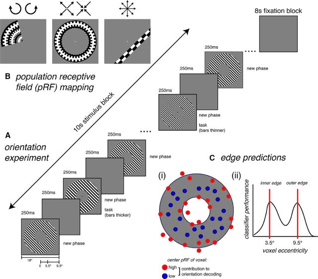
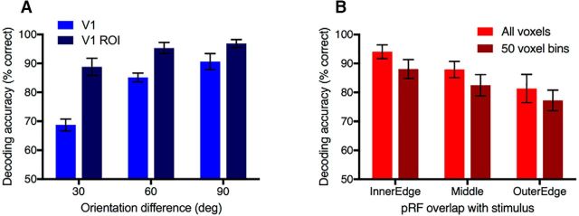
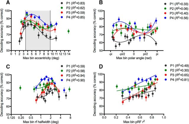
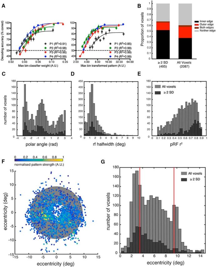
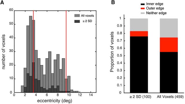
Multivariate pattern analysis is a powerful technique; however, a significant theoretical limitation in neuroscience is the ambiguity in interpreting the source of decodable information used by classifiers. This is exemplified by the continued controversy over the source of orientation decoding from fMRI responses in human V1. Recently Carlson (2014) identified a potential source of decodable information by modeling voxel responses based on the Hubel and Wiesel (1972) ice-cube model of visual cortex. The model revealed that activity associated with the edges of gratings covaries with orientation and could potentially be used to discriminate orientation. Here we empirically evaluate whether "edge-related activity" underlies orientation decoding from patterns of BOLD response in human V1. First, we systematically mapped classifier performance as a function of stimulus location using population receptive field modeling to isolate each voxel's overlap with a large annular grating stimulus. Orientation was decodable across the stimulus; however, peak decoding performance occurred for voxels with receptive fields closer to the fovea and overlapping with the inner edge. Critically, we did not observe the expected second peak in decoding performance at the outer stimulus edge as predicted by the edge account. Second, we evaluated whether voxels that contribute most to classifier performance have receptive fields that cluster in cortical regions corresponding to the retinotopic location of the stimulus edge. Instead, we find the distribution of highly weighted voxels to be approximately random, with a modest bias toward more foveal voxels. Our results demonstrate that edge-related activity is likely not necessary for orientation decoding.
Significance statement: A significant theoretical limitation of multivariate pattern analysis in neuroscience is the ambiguity in interpreting the source of decodable information used by classifiers. For example, orientation can be decoded from BOLD activation patterns in human V1, even though orientation columns are at a finer spatial scale than 3T fMRI. Consequently, the source of decodable information remains controversial. Here we test the proposal that information related to the stimulus edges underlies orientation decoding. We map voxel population receptive fields in V1 and evaluate orientation decoding performance as a function of stimulus location in retinotopic cortex. We find orientation is decodable from voxels whose receptive fields do not overlap with the stimulus edges, suggesting edge-related activity does not substantially drive orientation decoding.
Keywords: fMRI decoding; hyperacuity; multivariate pattern analysis; orientation columns; population receptive field mapping; visual cortex.
Copyright © 2017 the authors 0270-6474/17/371187-10$15.00/0.
Figures
Similar articles
-
Orientation decoding in human visual cortex: new insights from an unbiased perspective.J Neurosci. 2014 Jun 11;34(24):8373-83. doi: 10.1523/JNEUROSCI.0548-14.2014. J Neurosci. 2014. PMID: 24920640 Free PMC article.
-
Spatial scale and distribution of neurovascular signals underlying decoding of orientation and eye of origin from fMRI data.J Neurophysiol. 2017 Feb 1;117(2):818-835. doi: 10.1152/jn.00590.2016. Epub 2016 Nov 30. J Neurophysiol. 2017. PMID: 27903637 Free PMC article.
-
Decoupling of BOLD amplitude and pattern classification of orientation-selective activity in human visual cortex.Neuroimage. 2018 Oct 15;180(Pt A):31-40. doi: 10.1016/j.neuroimage.2017.09.046. Epub 2017 Sep 22. Neuroimage. 2018. PMID: 28951159
-
Modeling correlated noise is necessary to decode uncertainty.Neuroimage. 2018 Oct 15;180(Pt A):78-87. doi: 10.1016/j.neuroimage.2017.08.015. Epub 2017 Aug 8. Neuroimage. 2018. PMID: 28801251 Review.
-
Encoding and decoding in fMRI.Neuroimage. 2011 May 15;56(2):400-10. doi: 10.1016/j.neuroimage.2010.07.073. Epub 2010 Aug 4. Neuroimage. 2011. PMID: 20691790 Free PMC article. Review.
Cited by
-
Improving the validity of neuroimaging decoding tests of invariant and configural neural representation.PLoS Comput Biol. 2023 Jan 23;19(1):e1010819. doi: 10.1371/journal.pcbi.1010819. eCollection 2023 Jan. PLoS Comput Biol. 2023. PMID: 36689555 Free PMC article.
-
Framing orientation selectivity.Elife. 2018 Aug 14;7:e39762. doi: 10.7554/eLife.39762. Elife. 2018. PMID: 30106374 Free PMC article.
-
Stimulus vignetting and orientation selectivity in human visual cortex.Elife. 2018 Aug 14;7:e37241. doi: 10.7554/eLife.37241. Elife. 2018. PMID: 30106372 Free PMC article.
-
Natural scene sampling reveals reliable coarse-scale orientation tuning in human V1.Nat Commun. 2022 Oct 29;13(1):6469. doi: 10.1038/s41467-022-34134-7. Nat Commun. 2022. PMID: 36309512 Free PMC article.
-
Representation, Pattern Information, and Brain Signatures: From Neurons to Neuroimaging.Neuron. 2018 Jul 25;99(2):257-273. doi: 10.1016/j.neuron.2018.06.009. Neuron. 2018. PMID: 30048614 Free PMC article. Review.
References
Publication types
MeSH terms
LinkOut - more resources
Full Text Sources
Other Literature Sources