. 2017 Feb 10;292(6):2457-2469.
doi: 10.1074/jbc.M116.752535. Epub 2016 Dec 21.

A Single Amino Acid Mutation Converts (R)-5-Diphosphomevalonate Decarboxylase into a Kinase

Affiliations

A Single Amino Acid Mutation Converts (R)-5-Diphosphomevalonate Decarboxylase into a Kinase

Kento Motoyama et al. J Biol Chem. .

Abstract

The biosynthesis of isopentenyl diphosphate, a fundamental precursor for isoprenoids, via the mevalonate pathway is completed by diphosphomevalonate decarboxylase. This enzyme catalyzes the formation of isopentenyl diphosphate through the ATP-dependent phosphorylation of the 3-hydroxyl group of (R)-5-diphosphomevalonate followed by decarboxylation coupled with the elimination of the 3-phosphate group. In this reaction, a conserved aspartate residue has been proposed to be involved in the phosphorylation step as the general base catalyst that abstracts a proton from the 3-hydroxyl group. In this study, the catalytic mechanism of this rare type of decarboxylase is re-investigated by structural and mutagenic studies on the enzyme from a thermoacidophilic archaeon Sulfolobus solfataricus The crystal structures of the archaeal enzyme in complex with (R)-5-diphosphomevalonate and adenosine 5'-O-(3-thio)triphosphate or with (R)-5-diphosphomevalonate and ADP are newly solved, and theoretical analysis based on the structure suggests the inability of proton abstraction by the conserved aspartate residue, Asp-281. Site-directed mutagenesis on Asp-281 creates mutants that only show diphosphomevalonate 3-kinase activity, demonstrating that the residue is required in the process of phosphate elimination/decarboxylation, rather than in the preceding phosphorylation step. These results enable discussion of the catalytic roles of the aspartate residue and provide clear proof of the involvement of a long predicted intermediate, (R)-3-phospho-5-diphosphomevalonate, in the reaction of the enzyme.

Keywords: archaea; crystal structure; decarboxylase; diphosphomevalonate decarboxylase; enzyme catalysis; enzyme mechanism; isoprenoid; kinase; mevalonate pathway; mutagenesis.


Conflict of interest statement

The authors declare that they have no conflicts of interest with the contents of this article

Figures

FIGURE 1.
FIGURE 1.
Reaction mechanisms of DMD (A) and M3K (B).
FIGURE 2.
FIGURE 2.
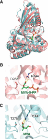
Substrate-complex structures of DMD and M3K. A, superposed structures of DMD (MVA-5-PP complex, PDB code 4DU7) from S. epidermidis and M3K (MVA complex, PDB code 4RKS) from T. acidophilum. DMD (pink) and M3K (light blue) are shown as ribbon figures. MVA-5-PP in DMD and MVA in M3K are shown as stick figures. B and C, active site structures of DMD (B) and M3K (C). Residues (Arg-144 and Asp-283 in DMD and Arg-144 and Thr-275 in T3K) and the ligand molecules are shown as stick figures. The viewing directions of B and C are in accordance.
FIGURE 3.
FIGURE 3.
Substrate·complex structures of SsoDMD. A and B, complex structures (monomer, colored in deep blue) of SsoDMD·ATPγS·MVA-5-PP (A) and SsoDMD·ADP·MVA-5-PP (B) are superimposed with a substrate-free structure (PDB code 4Z7Y, gray). MVA-5-PP (green), ATPγS (yellow), ADP (pink), and AMP (yellow) molecules that are bound to SsoDMD and the Tyr-72 residue are represented by stick models. The FoFc omit electron density maps for ligands bound to SsoDMD are shown as mesh. The omit maps are contoured at 2.2σ. C and D, hydrogen-bonding interactions around α1-helix in SsoDMD·ATPγS·MVA-5-PP (C) and SsoDMD·ADP·MVA-5-PP (D). The dashed lines denote hydrogen bonds. E and F, stereo view of the active site structures of SsoDMD·ATPγS·MVA-5-PP (E) and SsoDMD·ADP·MVA-5-PP (F). The ligands and the amino acid residues involved in the ligand binding with hydrogen-bonding interactions are represented as stick models. Probable hydrogen bond-forming interactions (in proximity less than ∼3 Å) are indicated by yellow dotted lines.
FIGURE 4.
FIGURE 4.
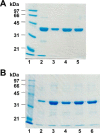
SDS-PAGE analysis of the purified enzymes. A, wild type and Asp-281 mutants of SsoDMD. Lane 1, molecular marker; lane 2, wild type (37.0 kDa); lane 3, D281N; lane 4, D281T; and lane 5, D281V. B, wild type, Thr-275 single, and Leu-18/Thr-275 double mutants of TacM3K. Lane 1, molecular marker; lane 2, wild type (35.2 kDa); lane 3, T275D; lane 4, T275E; lane 5, L18K/T275D; and lane 6, L18K/T275E. The proteins were separated on 12.5% SDS-PAGE.
FIGURE 5.
FIGURE 5.
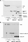
Radio-TLC analyses of the products of the wild type and mutants of SsoDMD (A) and TacM3K (B). The asterisk indicates an unknown product from the reaction with MVA-5-P and the D281N mutant, which seems distinct from unknown compounds synthesized by the reaction with MVA-5-PP and the D281T or D281V mutant. ori., origin; s.f., solvent front.
FIGURE 6.
FIGURE 6.
Semi-quantitative radio-TLC assays of the wild type SsoDMD and the D281T and D281V mutants. 2.5, 25, or 250 ng (0.068, 0.68, and 6.8 pmol, respectively) of each enzyme was reacted for 15 min at 60 °C with 25 pmol of [2-14C]MVA-5-PP in the presence of ATP and Mg2+. ori., origin; s.f., solvent front.
FIGURE 7.
FIGURE 7.
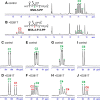
13C NMR analysis of the product of the D281T mutant. Parts of the 13C NMR spectra after reaction without (A, C, E, G, and I) or with (B, D, F, H, and J) the D281T mutant are shown. Green asterisks indicate the indistinguishable signals of [U-13C]MVA-5-P and [U-13C]MVA-5-PP, which are derived from the conversion of [U-13C]MVA by MVK and PMK from S. solfataricus. Black asterisks in A represent the signals of [U-13C]MVA-5-P, which are distinguishable from those of [U-13C]MVA-5-PP indicated by green asterisks. Red asterisks indicate the signal of the product of the D281T mutant. The spectra corresponding to the signals of C3 (A and B), C1 (C and D), C2 (E and F), C4 (G and H), and C6 (I and J) of MVA-5-PP and the new product are shown. The reaction with the D281V mutant gave almost the same results as those from the reaction with D281T (data not shown).
FIGURE 8.
FIGURE 8.
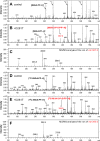
Negative ESI-MS analyses of the product of the D281T mutant from MVA-5-PP. The samples from the reaction with the non-labeled substrate (A–C) or the 13C-labeled substrate (D and E) were analyzed by ESI-MS. A and D, negative control; B and E, the sample after the reaction of the D281T mutant; C and F, MS/MS analysis of the ion of m/z 387.2 and 393.0 from B and E, respectively. *1, *2, and *3 indicate the ion peaks of [AMP-H] (m/z ∼346), [ADP-H] (m/z ∼426), and [ATP-H] (m/z ∼506), respectively. The reaction with the D281V mutant gave almost the same results as those from the reaction with D281T (data not shown).
FIGURE 9.
FIGURE 9.
Newly proposed roles of Asp-281 residue in the reaction catalyzed by SsoDMD. A, stabilization of the carbocation intermediate formed by the elimination of inorganic phosphate from MVA-3-P-5-PP. The preceding phosphotransfer reaction from ATP to the non-ionized 3-hydroxyl group of MVA-5-PP is supposedly stimulated by the interactions with Mg2+ ion and Lys-190 residue, which are not shown in the figure. OPP represents the diphosphate group of MVA-5-PP. B, formation of the repulsive force that pushes the 3-phosphate group of MVA-3-P-5-PP away and changes the conformation of the intermediate from gauche to anti.

Similar articles

Cited by

References

    1. Kuzuyama T., Hemmi H., and Takahashi S. (2010) in Comprehensive Natural Products II Chemistry and Biology (Mander L., and Liu H.-W., eds) pp. 493–516, Elsevier, Oxford, United Kingdom
    1. Miziorko H. M. (2011) Enzymes of the mevalonate pathway of isoprenoid biosynthesis. Arch. Biochem. Biophys. 505, 131–143 - PMC - PubMed
    1. Li T., Huo L., Pulley C., and Liu A. (2012) Decarboxylation mechanisms in biological system. Bioorg. Chem. 43, 2–14 - PubMed
    1. Froese D. S., Forouhar F., Tran T. H., Vollmar M., Kim Y. S., Lew S., Neely H., Seetharaman J., Shen Y., Xiao R., Acton T. B., Everett J. K., Cannone G., Puranik S., Savitsky P., et al. (2013) Crystal structures of malonyl-coenzyme A decarboxylase provide insights into its catalytic mechanism and disease-causing mutations. Structure 21, 1182–1192 - PMC - PubMed
    1. Bertoldi M. (2014) Mammalian dopa decarboxylase: structure, catalytic activity and inhibition. Arch. Biochem. Biophys. 546, 1–7 - PubMed

Publication types

MeSH terms

Substances

LinkOut - more resources