. 2017 Feb 10;292(6):2470-2484.
doi: 10.1074/jbc.M116.766824. Epub 2016 Dec 21.

UBE3B Is a Calmodulin-regulated, Mitochondrion-associated E3 Ubiquitin Ligase

Affiliations

UBE3B Is a Calmodulin-regulated, Mitochondrion-associated E3 Ubiquitin Ligase

Andrea Braganza et al. J Biol Chem. .

Abstract

Recent genome-wide studies found that patients with hypotonia, developmental delay, intellectual disability, congenital anomalies, characteristic facial dysmorphic features, and low cholesterol levels suffer from Kaufman oculocerebrofacial syndrome (KOS, also reported as blepharophimosis-ptosis-intellectual disability syndrome). The primary cause of KOS is autosomal recessive mutations in the gene UBE3B However, to date, there are no studies that have determined the cellular or enzymatic function of UBE3B. Here, we report that UBE3B is a mitochondrion-associated protein with homologous to the E6-AP Cterminus (HECT) E3 ubiquitin ligase activity. Mutating the catalytic cysteine (C1036A) or deleting the entire HECT domain (amino acids 758-1068) results in loss of UBE3B's ubiquitylation activity. Knockdown of UBE3B in human cells induces changes in mitochondrial morphology and physiology, a decrease in mitochondrial volume, and a severe suppression of cellular proliferation. We also discovered that UBE3B interacts with calmodulin via its N-terminal isoleucine-glutamine (IQ) motif. Deletion of the IQ motif (amino acids 29-58) results in loss of calmodulin binding and a significant increase in the in vitro ubiquitylation activity of UBE3B. In addition, we found that changes in calcium levels in vitro disrupt the calmodulin-UBE3B interaction. These studies demonstrate that UBE3B is an E3 ubiquitin ligase and reveal that the enzyme is regulated by calmodulin. Furthermore, the modulation of UBE3B via calmodulin and calcium implicates a role for calcium signaling in mitochondrial protein ubiquitylation, protein turnover, and disease.

Keywords: HECT; Kaufman oculocerebrofacial syndrome; calcium; mitochondria; oxidative stress; protein degradation; reactive oxygen species (ROS); super-resolution microscopy; ubiquitin/proteosome system; ubiquitylation (ubiquitination).


Figures

FIGURE 1.
FIGURE 1.
Alignment of UBE3B with select IQ motif proteins and HECT E3 ubiquitin ligases. A, schematic of UBE3B showing the IQ domain (amino acids 29–58) and the HECT domain (amino acids 757–1068). The proposed 3D structures of the IQ and HECT domains using Phyre2 are shown above the schematic. The N terminus of HECT domains are known to bind to substrate. The HECT domain is composed of two lobes as follows: the N-lobe binds the E2(s), and the C-lobe contains the catalytic cysteine that binds ubiquitin. B, alignment of UBE3B with calmodulin binding domains as predicted by Phyre2 and using ClustalW2. C, alignment of UBE3B with HECT E3 ligase domains as predicted by Phyre2 and using ClustalW2. The conserved catalytic cysteine is highlighted in red. * denotes a single fully conserved residue; : denotes conservation between groups of strongly similar properties, . denotes conservation between groups of weakly similar properties.
FIGURE 2.
FIGURE 2.
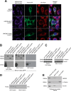
UBE3B is associated with mitochondria. A, LN428 cells were transduced with lentivirus to stably express UBE3B, UBE3BΔHECT, or UBE3B(C1036A), all with C-terminal copGFP tags, and then were fixed and imaged with a Nikon A1rsi confocal microscope. MitoTracker DeepRed (excitation wavelength, 647 nm; emission wavelength, 665 nm) was used to stain mitochondria before fixation; cells were then immunostained for PDI, a marker for the endoplasmic reticulum (excitation wavelength, 568 nm; emission wavelength, 602 nm). DAPI (excitation wavelength, 360 nm; emission wavelength, 460 nm) was used to counterstain nuclei, as seen in the merged images. B, to confirm the immunofluorescence results, subcellular fractionation of the stable cell lines was performed, resulting in isolation of mitochondrial, ER, and cytoplasmic fractions, which were then probed by immunoblot (IB). An antibody against TurboGFP was used to detect UBE3B-copGFP proteins. Full-length UBE3B-copGFP proteins were detected only in mitochondrial fractions and in whole cell lysate. To confirm the quality of fractionation, fractions were probed with markers for different cellular compartments. C, mitochondrial fractions lack the cytoplasmic marker α-tubulin and show enrichment of the mitochondrial marker Tom20. D, purity of the ER fraction was assessed by immunoblot probe for the ER marker PDI, showing no cross-contamination with the mitochondrial fraction. E, to show that endogenous UBE3B associates with mitochondria and the immunofluorescence and subcellular fractionation results in A–D are not artifacts of overexpression or of the copGFP tag, we performed subcellular fractionation and immunoblot analysis for endogenous UBE3B in LN428 cells, using the cytoplasmic marker α-tubulin and the mitochondrial marker Tom40 to confirm fractionation.
FIGURE 3.
FIGURE 3.
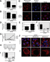
Knockdown of UBE3B compromises mitochondrial morphology and function and reduces cell proliferation and colony formation. LN428 cells were transfected with UBE3B siRNA or SCR siRNA (90 nm, final concentration) for 72 h before being used for subsequent experiments. A, qRT-PCR was performed to measure the siRNA-mediated knockdown of UBE3B mRNA expression. β-Actin was used as the endogenous control, and mRNA expression was normalized to SCR siRNA cells. RQ indicates the relative quantification. B, to determine cellular metabolic activity, as an indicator of decreased cellular survival, 2000 cells/well were plated 24 h after siRNA transfection. After 48 h of incubation, an MTS assay was performed. C, to determine whether there are changes in mitochondrial morphology after depletion of UBE3B protein, confocal imaging was performed on fixed cells 72 h after siRNA transfection. ATP synthase β is the mitochondrial marker detected by immunofluorescence (excitation wavelength, 647 nm; emission wavelength, 666 nm). D, Z-stacking using confocal microscopy on the fixed cells from C was performed to determine the changes in mitochondrial voxels in UBE3B-KD cells as compared with scrambled (negative control) treated cells. E, to determine whether UBE3B knockdown induced mitochondrial stress, cells were co-transfected with UBE3B siRNA, and the ratiometric reporter gene Mito-Timer. Confocal imaging was performed 72 h after transfection. Quantitation of the collected images reveal an increase in mitochondrial stress in the UBE3B knockdown cells. F, LN428 cells were individually transduced with lentivirus expressing a scrambled shRNA control or either of two lentiviral shRNAs (sh2 and sh3) targeting different sequences of UBE3B. 48 h later, the loss of UBE3B mRNA expression was measured by qRT-PCR and normalized to the SCR control. G, 48 h after lentiviral infection, the SCR, sh2, and sh3 cells were seeded at 5000 cells/well in 24-well plates in 500 μl/well growth medium. The cell numbers of each plate were counted at 2 h after seeding and then again on days 3, 5, 7, and 10. The relative proliferation for each cell line was calculated by normalizing the cell number each day to the cell number at 2 h after seeding. H, 48 h after lentiviral infection, the SCR, sh2, and sh3 cells were seeded at 500 cells/well in 6-well plates in 5 ml/well growth medium and incubated for 10 days. The colonies were stained with crystal violet, and images were generated. I, quantitation of the clonogenic assay from the results in H, using Celigo, showed a strong decrease of colony numbers in both shRNA knockdown cells. J, to validate changes in mitochondrial morphology, UBE3B-sh2 and UBE3B-sh3 cells were stained with MitoTracker CMX-ROS and fixed, and correlative microscopy was performed with confocal (top row) and SIM super-resolution microscopy (bottom row), showing changes induced by UBE3B knockdown.
FIGURE 4.
FIGURE 4.
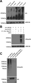
UBE3B exhibits ubiquitin ligase activity. Cell lysates from LN428 cells that stably express UBE3A, UBE3B, UBE3BΔHECT, or UBE3B(C1036A), all with N terminus HA tags, were immunoprecipitated using anti-HA affinity matrix and subjected to ubiquitylation activity assays. The ubiquitylation assays were carried out in 30-μl reactions with 0.1 μm E1, 0.25 μm of an E2 mixture, 1 μm ubiquitin aldehyde, 0.75 μg/μl His-ubiquitin, and 1× magnesium/ATP mixture, unless otherwise noted. A, ubiquitylation activity assay was performed in vitro using His-tagged wild type ubiquitin (His-Ub-WT). The expected size of HA-UBE3B is 125 kDa. Note the time-dependent increase in the high molecular mass signal. B, polyubiquitin chain formation was lost when ubiquitin or E1, E2s, or ATP were removed from the reaction mixture, or if 20 μg of the catalytic core of the deubiquitinating enzyme USP2 was added to a completed reaction for 30 min at 37 °C (63). C, polyubiquitin chains were lacking in reactions containing the catalytically inactive (HA-UBE3B(C1036A)) or HECT deleted (HA-UBE3BΔHECT) mutants of UBE3B. Completed reactions were applied to immunoblot for analysis of ubiquitylation with anti-hemagglutinin (HA) or ubiquitin (Ub) antibodies. Bottom panel shows comparable loading of the immunoprecipitated proteins. Representative immunoblots from three independent experiments are shown. Antibodies used for each of the immunoblots are listed on the side of the panels.
FIGURE 5.
FIGURE 5.
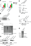
UBE3B associates with calmodulin through the IQ motif in a calcium-dependent manner. A, quantification results of HA-Ab affinity purification and high resolution LC-MS analysis for two proteotypic calmodulin peptides, VFDKDGNGYISAAELR and DTDSEEEIREAFR. Bar graphs represent the standardized mean values ± S.E. following affinity purification and differential mass spectrometry analysis of LN428 cells containing an empty vector control or expressing wild type HA-tagged UBE3B (HA-UBE3B) or the catalytic inactive UBE3B with an HA tag (HA-UBE3B(C1036A)). B, cells, indicated in the figure, were lysed, and the HA-tagged proteins were immunoprecipitated, as described under “Experimental Procedures,” to identify the proteins that interact with UBE3B, UBE3B(C1036A), or UBE3BΔHECT. Shown is an immunoblot to confirm the interaction of calmodulin with UBE3B as seen by LC-MS using anti-UBE3B and calmodulin antibodies. C, BioID analysis was performed using BirA-UBE3B and UBE3B-BirA with streptavidin immunoprecipitated to confirm that HA-UBE3B and calmodulin interact. Cells were treated with biotin (5 μm) for 24 h before harvest, as indicated. Streptavidin-purified proteins were then probed by immunoblot using streptavidin-HRP. Bottom right panel indicates the identification of calmodulin after streptavidin capture. D, 293FT cells were transiently transfected (48 h) with a plasmid expressing WT UBE3B-HA or UBE3BΔIQ-HA followed by immunoprecipitation with HA-affinity matrix beads. Deletion of the IQ motif demolished the interaction between UBE3B and calmodulin. E, 239FT cells were transiently transfected (48 h) with a plasmid expressing WT UBE3B-HA or UBE3BΔIQ-HA followed by immunoprecipitation with HA-affinity matrix beads. A ubiquitylation assay was then performed as described under “Experimental Procedures.” The results were analyzed by immunoblot using anti-ubiquitin, anti-His, and anti-HA antibodies to detect UBE3B and anti-CaM antibody to detect calmodulin. Ubiquitylation is increased in UBE3BΔIQ due to the loss of loss of the interaction with calmodulin. F, 239FT cells were transiently transfected (48 h) with a plasmid expressing WT UBE3B-HA or UBE3BΔIQ-HA. The expression of UBE3BΔIQ-HA triggered cell apoptosis as indicated by an elevation of the cleaved form of caspase3. G, calmodulin interaction with UBE3B is lost when immunoprecipitation beads are washed with CaCl2, and ubiquitylation is increased when calmodulin is not bound to UBE3B. Immunoprecipitations and the bead-based in vitro assay were performed as described above.
FIGURE 6.
FIGURE 6.
Model for UBE3B activation in response to calcium signaling/mitochondrial stress. We propose that the E3 ligase UBE3B associates with the mitochondria and that in the absence of stress UBE3B is bound to calmodulin through its N-terminal IQ motif in a calcium-dependent manner. However, in response to mitochondrial stress the intracellular calcium levels increase, thereby activating calmodulin and releasing it from UBE3B. Calmodulin-free UBE3B can now bind to its substrates (potentially damaged mitochondrial proteins) and ubiquitylate substrates for degradation by the UPS.

Similar articles

Cited by

References

    1. Komander D. (2009) The emerging complexity of protein ubiquitination. Biochem. Soc. Trans. 37, 937–953 - PubMed
    1. Tai H. C., and Schuman E. M. (2008) Ubiquitin, the proteasome and protein degradation in neuronal function and dysfunction. Nat. Rev. Neurosci. 9, 826–838 - PubMed
    1. Kawabe H., and Brose N. (2011) The role of ubiquitylation in nerve cell development. Nat. Rev. Neurosci. 12, 251–268 - PubMed
    1. Schmidt M., and Finley D. (2014) Regulation of proteasome activity in health and disease. Biochim. Biophys. Acta 1843, 13–25 - PMC - PubMed
    1. Varshavsky A. (2012) The ubiquitin system, an immense realm. Annu. Rev. Biochem. 81, 167–176 - PubMed

Publication types

LinkOut - more resources