. 2017 Apr 1;156(2):402-411.
doi: 10.1093/toxsci/kfw261.

From the Cover: Prolonged Exposure to Volatile Anesthetic Isoflurane Worsens the Outcome of Polymicrobial Abdominal Sepsis

Affiliations

From the Cover: Prolonged Exposure to Volatile Anesthetic Isoflurane Worsens the Outcome of Polymicrobial Abdominal Sepsis

Sophia Koutsogiannaki et al. Toxicol Sci. .

Abstract

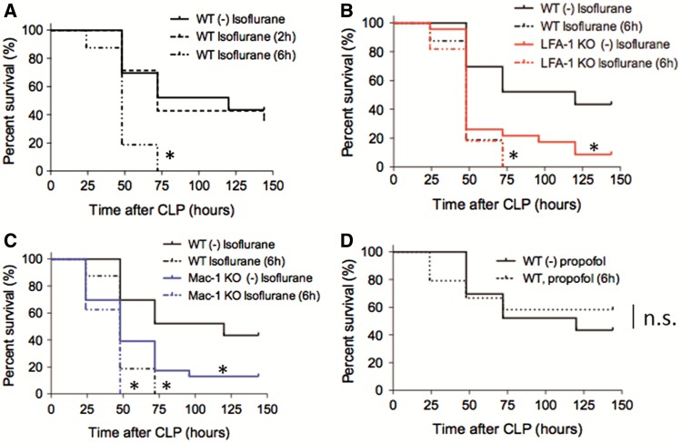
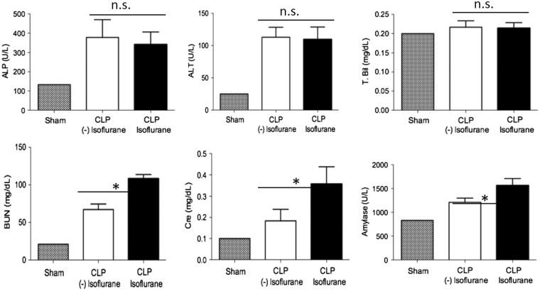
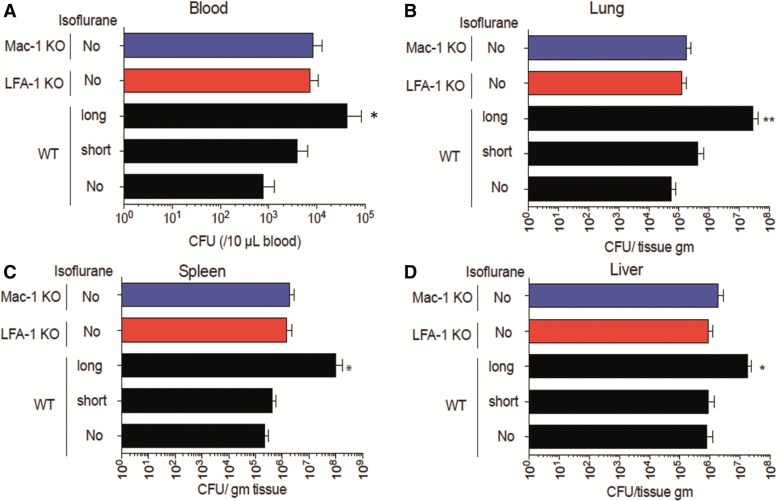
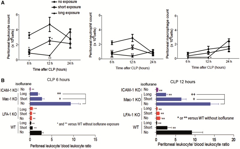
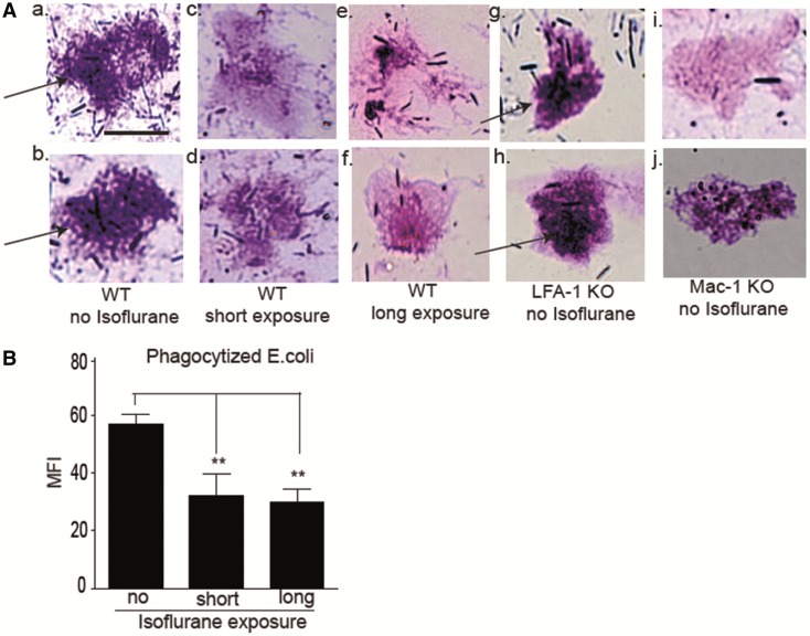
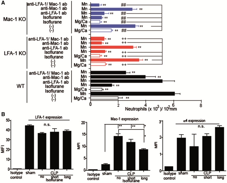
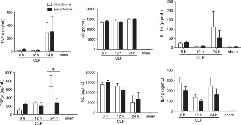
Sepsis continues to result in high morbidity and mortality. General anesthesia is often administered to septic patients, but the impacts of general anesthesia on host defense are not well understood. General anesthesia can be given by volatile and intravenous anesthetics. Our previous in vitro study showed that volatile anesthetic isoflurane directly inhibits leukocyte function-associated antigen-1 (LFA-1) and macrophage-1 antigen (Mac-1), critical adhesion molecules on leukocytes. Thus, the role of isoflurane exposure on in vivo LFA-1 and Mac-1 function was examined using polymicrobial abdominal sepsis model in mice. As a comparison, intravenous anesthetic propofol was given to a group of mice. Wild type, LFA-1, Mac-1, and adhesion molecule-1 knockout mice were used. Following the induction of polymicrobial abdominal sepsis by cecal ligation and puncture, groups of mice were exposed to isoflurane for either 2 or 6 h, or to propofol for 6 h, and their outcomes were examined. Bacterial loads in tissues and blood, neutrophil recruitment to the peritoneal cavity and phagocytosis were studied. Six hours of isoflurane exposure worsened the outcome of abdominal sepsis (P < .0001) with higher bacterial loads in tissues, but 2 h of isoflurane or 6 h of propofol exposure did not. Isoflurane impaired neutrophil recruitment to the abdominal cavity by inhibiting LFA-1 function. Isoflurane also impaired bacterial phagocytosis via complement receptors including Mac-1. In conclusion, prolonged isoflurane exposure worsened the outcome of experimental polymicrobial abdominal sepsis and was associated with impaired neutrophil recruitment and bacterial phagocytosis via reduced LFA-1 and Mac-1 function.

Keywords: anesthesia; leukocyte function-associated antigen-1; macrophage-1 antigen.; neutrophil; sepsis.


Figures

FIG. 1
FIG. 1
The impact of isoflurane exposure on mortality in experimental polymicrobial abdominal sepsis. A–C, The outcomes of polymicrobial abdominal sepsis induced by CLP in WT mice without isoflurane exposure (n = 25), with isoflurane exposure for 2 h (short exposure, n = 14) and 6 h (long exposure, n = 20), LFA-1 KO mice without isoflurane exposure (n = 24), LFA-1 KO mice with isoflurane exposure for 6 h (n = 16), Mac-1 KO mice without isoflurane exposure (n = 24) and Mac-1 KO mice with isoflurane exposure for 6 h (n = 15) are shown. D, The outcomes of mice after CLP in WT mice with propofol exposure (n = 20) or without propofol exposure (n = 20) for 6 h. Statistical significance was evaluated using Log-rank test. * and ** represent P < .05 and .01, respectively versus WT without isoflurane (or propfol). CLP, cecal ligation and puncture.
FIG. 2
FIG. 2
The effect of isoflurane on liver and kidney functions after CLP. Liver and kidney functions were assessed after CLP in no and long isoflurane exposure groups of mice. Data represent mean ± SD of 8 mice. Statistical analysis was performed using 1-way analysis of variance with Bonferroni post hoc analysis.
FIG. 3
FIG. 3
The effect of isoflurane exposure on tissue and blood bacterial levels after CLP. Bacterial loads in blood (A), lung (B), spleen (C), and liver (D) at 6 h after CLP was compared among WT mice in no, short and long isoflurane exposure groups, LFA-1 KO and Mac-1 KO mice. Blood and tissue bacterial loads were shown as mean ± SD of 8 mice. Statistical significance was evaluated using Kruskal Wallis test with Dunn’s multiple comparisons. In figures, * and ** represent P < .05 and .01, respectively versus WT mice without isoflurane exposure. CFU, colony forming unit.
FIG. 4
FIG. 4
The effect of isoflurane exposure on leukocyte migration to the abdominal cavity. A, The number of total leukocytes, neutrophils and macrophages in the peritoneal cavity at 6, 12, and 24 h after CLP. The data represent mean ± SD of 4 mice. Statistical analysis was performed using 2-way analysis of variance with Bonferroni post hoc analysis. * and ** represent P < .05 and .01, respectively versus WT mice without isoflurane exposure at the same time point. B, The peritoneal leukocyte/blood leukocyte ratio 12 h after CLP. Data represent mean ± SD of 4 mice. Statistical analysis was evaluated using 1-way analysis of variance with Bonferroni post hoc analysis versus WT mice without isoflurane exposure. In figures, * and ** represent P < .05 and .01 versus WT without isoflurane exposure. Separate, statistical analysis was also performed among Mac-1 KO mice. + and ++ denote P < .05 and .01, respectively versus Mac-1 KO mice without isoflurane exposure.
FIG. 5
FIG. 5
The effect of isoflurane exposure on bacterial phagocytosis. A, Representative images of peritoneal leukocytes from mice 12 h after CLP. (a) and (b), WT mice with no isoflurane exposure; (c) and (d), WT mice with isoflurane short exposure; (e) and (f), WT mice with long isoflurane exposure; (g) and (h), LFA-1 KO mice with no isoflurane exposure; (i) and (j), Mac-1 KO mice with no isoflurane exposure. Arrows indicate bacteria. Scale bar 10 µm. B, Whole blood phagocytosis assay using fluorescent E. coli. Mice in the short isoflurane exposure group received 1% isoflurane for 2 h and then were kept in cages for 4 h before blood collection. The long exposure group received 1% isoflurane for 6 h and then blood was collected immediately. Gated on neutrophil population. Data represent mean ± SD of 4 mice. Statistical significance was analyzed using 1-way analysis of variance with Bonferroni post hoc analysis. * and ** denote P < .05 and .01, respectively versus mice without isoflurane exposure. MFI, mean fluorescence intensity.
FIG. 6
FIG. 6
The effect of isoflurane on LFA-1 and Mac-1 on primary neutrophils. A, The binding of ICAM-1 to LFA-1 and Mac-1 was evaluated using neutrophils from WT, LFA-1 KO and Mac-1 KO mice with or without isoflurane exposure. As controls, LFA-1 blocking antibody M17/4 (15 µg/ml) and Mac-1 blocking antibody M1/70 (15 µg/ml) were used. Data are shown as mean ± SD of quadruplicates. Statistical analysis was performed using 1-way analysis of variance with Bonferroni post hoc analysis. * and ** denote P < .05 and .01, respectively versus WT neutrophils with Mn2+ activation group. Among LFA-1 KO neutrophils, + and ++ denote P < .05 and .01, respectively versus LFA-1 KO neutrophils with Mn2+ activation. Among Mac-1 KO neutrophils, # and ## denote P < .05 and .01, respectively versus Mac-1 KO neutrophils with Mn2+ activation. B, The expression of LFA-1, Mac-1, and α4β1 on neutrophils were examined at 12 h after CLP using flow cytometry. Data show mean ± SD of 4 mice. Statistical analysis was performed using 1-way analysis of variance with Bonferroni post hoc analysis. MFI, mean fluorescence intensity. * and ** denote P < .05 and .01, respectively.
FIG. 7
FIG. 7
The effect of prolonged isoflurane exposure on proinflammatory cytokines. The proinflammatory cytokine profiles in blood (A) and peritoneal fluid (B) were compared between non-isoflurane exposure group and prolonged isoflurane exposure group. The data represent mean ± SD of 8 mice. Statistical analysis was performed using 2-way analysis of variance with Bonferroni post hoc analysis.

References

    1. Alves-Filho J. C., de Freitas A., Spiller F., Souto F. O., Cunha F. Q. (2008). The role of neutrophils in severe sepsis. Shock 30(Suppl 1), 3–9. - PubMed
    1. Angus D. C., Linde-Zwirble W. T., Lidicker J., Clermont G., Carcillo J., Pinsky M. R. (2001). Epidemiology of severe sepsis in the United States: Analysis of incidence, outcome, and associated costs of care. Crit. Care Med. 29, 1303–1310. - PubMed
    1. Bullard D. C., Hu X., Schoeb T. R., Collins R. G., Beaudet A. L., Barnum S. R. (2007). Intercellular adhesion molecule-1 expression is required on multiple cell types for the development of experimental autoimmune encephalomyelitis. J. Immunol. 178, 851–857. - PubMed
    1. Burns J. A., Issekutz T. B., Yagita H., Issekutz A. C. (2001). The alpha 4 beta 1 (very late antigen (VLA)-4, CD49d/CD29) and alpha 5 beta 1 (VLA-5, CD49e/CD29) integrins mediate beta 2 (CD11/CD18) integrin-independent neutrophil recruitment to endotoxin-induced lung inflammation. J. Immunol. 166, 4644–4649. - PubMed
    1. Campagna J. A., Miller K. W., Forman S. A. (2003). Mechanisms of actions of inhaled anesthetics. N. Engl. J. Med. 348, 2110–2124. - PubMed

Publication types

MeSH terms