. 2017 Jan 10;114(2):206-214.
doi: 10.1073/pnas.1617020114. Epub 2016 Dec 21.

Structural basis of influenza virus fusion inhibition by the antiviral drug Arbidol

Affiliations

Structural basis of influenza virus fusion inhibition by the antiviral drug Arbidol

Rameshwar U Kadam et al. Proc Natl Acad Sci U S A. .

Abstract

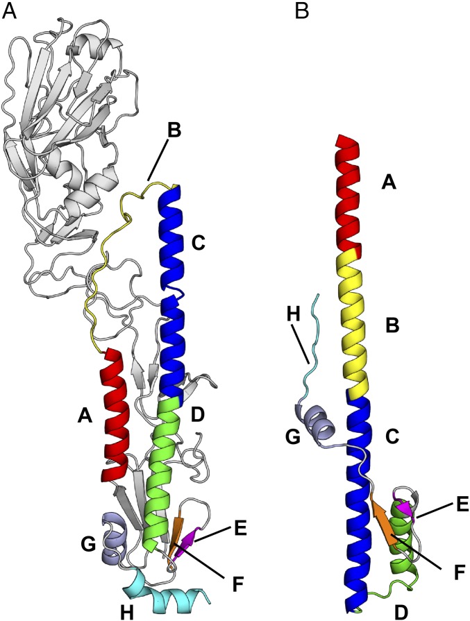
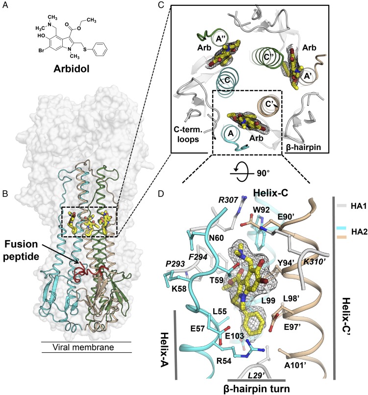
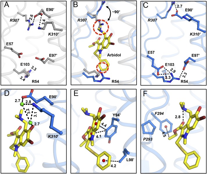
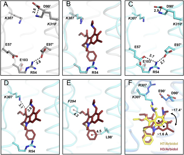
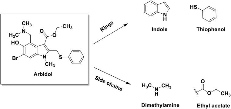
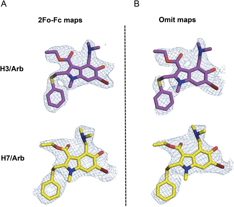
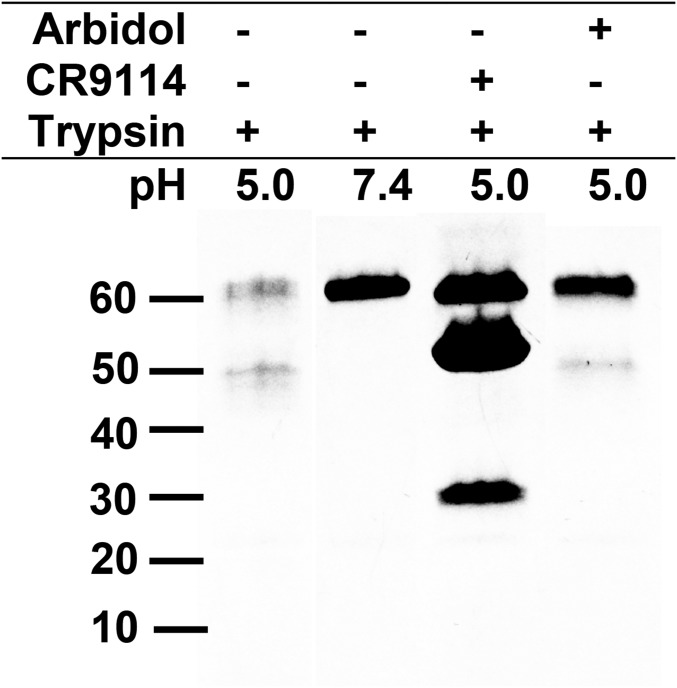
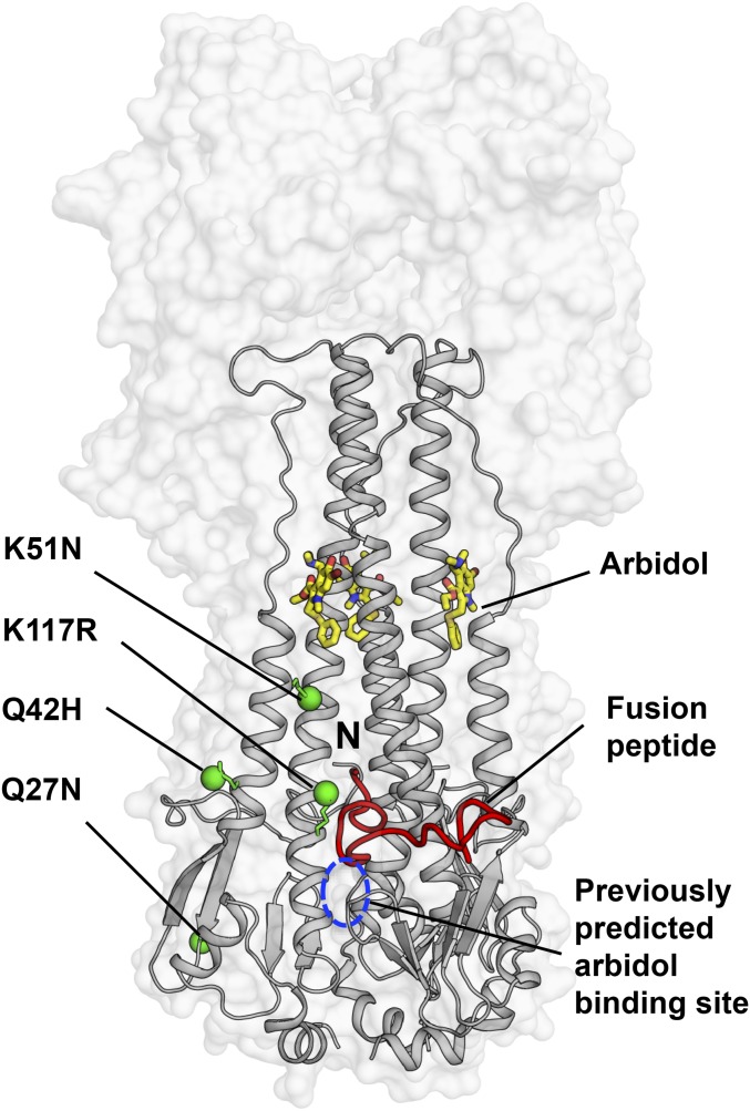
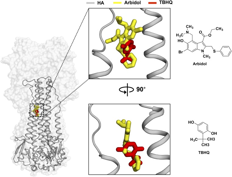
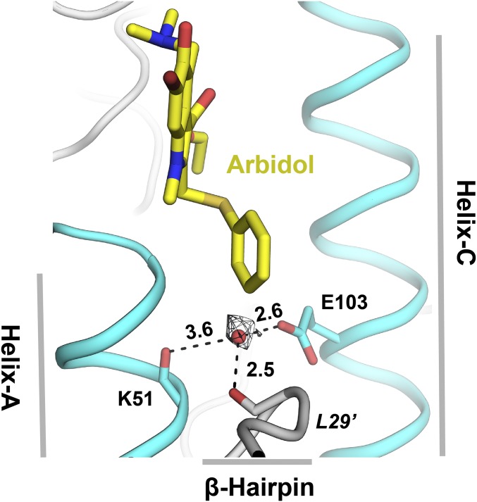
The broad-spectrum antiviral drug Arbidol shows efficacy against influenza viruses by targeting the hemagglutinin (HA) fusion machinery. However, the structural basis of the mechanism underlying fusion inhibition by Arbidol has remained obscure, thereby hindering its further development as a specific and optimized influenza therapeutic. We determined crystal structures of Arbidol in complex with influenza virus HA from pandemic 1968 H3N2 and recent 2013 H7N9 viruses. Arbidol binds in a hydrophobic cavity in the HA trimer stem at the interface between two protomers. This cavity is distal to the conserved epitope targeted by broadly neutralizing stem antibodies and is ∼16 Å from the fusion peptide. Arbidol primarily makes hydrophobic interactions with the binding site but also induces some conformational rearrangements to form a network of inter- and intraprotomer salt bridges. By functioning as molecular glue, Arbidol stabilizes the prefusion conformation of HA that inhibits the large conformational rearrangements associated with membrane fusion in the low pH of the endosome. This unique binding mode compared with the small-molecule inhibitors of other class I fusion proteins enhances our understanding of how small molecules can function as fusion inhibitors and guides the development of broad-spectrum therapeutics against influenza virus.

Keywords: Arbidol; X-ray crystallography; fusion inhibitor; hemagglutinin; influenza virus.


Conflict of interest statement

The authors declare no conflict of interest.

Figures

Fig. 1.
Fig. 1.
Prefusion and postfusion conformations of influenza HA. (A) Prefusion conformation of H7N9 A/Shanghai/2/2013 (H7/SH2; PDB 4LN6) HA at neutral pH. (B) Postfusion conformation of TBHA (PDB 1HTM) at fusion pH 5.0. The secondary structure elements that are involved in the large conformational rearrangements in going from the pre- to postfusion form of the HA mainly involve α-helices, but also loops and β-sheets. These elements are labeled from A to H in different colors.
Fig. 2.
Fig. 2.
The Arbidol-binding site on influenza virus HA. (A) Molecular structure of Arbidol. (B) Crystal structure of Arbidol in complex with H7N9 A/Shanghai/2/2013 (H7/SH2) HA. The HA trimer is shown as a transparent gray surface, HA2 is shown in cyan, beige, and green secondary structure backbone traces, and Arbidol is shown as yellow sticks. One of the three N-terminal fusion peptides in HA2 in the trimer has been highlighted as a red ribbon. (C) Three identical Arbidol-binding sites are shown viewed along the threefold symmetry axis of the trimer. HA1 lateral short loops and the bottom β-hairpin turn are represented in the gray cartoons. (D) The Arbidol-binding site consists of helix-A and helix-C from protomer 1 (cyan) and helix-C′ from protomer 2 (beige). A 2Fo-Fc electron density map (black mesh) is contoured at 1σ around Arbidol.
Fig. 3.
Fig. 3.
Molecular interactions in the Arbidol–H7/SH2 complex. (A) Charged residues in the Arbidol-binding site of the apo structure of H7N9 A/Shanghai/2/2013 (H7/SH2; PDB ID code 4LN6) (gray sticks). (B) Superimposition of apo and Arbidol-bound structures of H7/SH2 HA. Arg54 and Arg307 in the apo conformation (gray sticks) would clash (red dashed circles) with Arbidol (yellow sticks), whereas in the Arbidol-bound conformation, Arg54 and Arg307 (blue sticks) move away from the binding pocket and form alternative intra- and interprotomer salt bridges. (C) Charged residues in the Arbidol-binding site of the Arbidol–H7/SH2 complex (blue sticks). (DF) Noncovalent interactions in the Arbidol–H7/SH2 complex. Arbidol is represented by yellow sticks, HA residues as blue ball-and-stick models, centroids of rings as red spheres, and water molecules as green spheres. Noncovalent interaction mediated through salt-bridge networks (A and C), water molecules (D), and CH–π bonds (E and F) are highlighted using black dashed lines with distances in Ångstroms.
Fig. 4.
Fig. 4.
Molecular interactions in the Arbidol–H3/HK68 complex. (A) Charged residues in the Arbidol-binding site of the apo structure of H3N2 A/Hong Kong/1/1968 (H3/HK68; PDB ID code 4FNK) (gray sticks). (B) Superimposition of apo and Arbidol bound structures of H3/HK68. (C) Charged residues in the Arbidol-binding site of the Arbidol–H3/HK68 complex are shown as cyan sticks. (D and E) Noncovalent intramolecular interactions in the Arbidol–H3/HK68 complex. Arbidol is shown as reddish-brown sticks and HA residues are shown as cyan ball-and-sticks. Noncovalent intramolecular salt-bridge interactions in the Arbidol-binding site (A and C), inter- and intramolecular H-bonds made by Arbidol (D), and intramolecular CH–π bonds (E) are indicated by black dashed lines with distances in Ångstroms. (F) Superimposition of Arbidol-bound structures with H7/SH2 and H3/HK68 HAs. Arbidol bound to H7 is shown as yellow sticks, and Arbidol bound to H3 is shown as brown sticks. The distance between centroids of the two indole rings of Arbidol is ∼1.6 Å and is represented by a black dashed line; the torsional angle between the planes of the two different conformations of Arbidol is ∼17.4°.
Fig. S1.
Fig. S1.
Arbidol structure and its components. The structure of Arbidol and its component rings and side chains.
Fig. S2.
Fig. S2.
Electron density maps for the crystal structures of Arbidol in complex with H3 and H7 HAs. (A and B) 2Fo-Fc (A) and simulated annealing omit (B) maps, contoured at 1σ. For Arbidol in complex with H3/HK68, C, O, N, and Br are represented as magenta, red, blue, and brown sticks, respectively. For Arbidol in complex with H7/SH2, C, O, N, and Br are represented as yellow, red, blue, and brown sticks, respectively. Electron density maps are represented as a blue mesh.
Fig. 5.
Fig. 5.
Trypsin-susceptibility assay of Arbidol. SDS/PAGE analysis of the trypsin-susceptibility assay of Arbidol. Lane 1, acidified PR8 H1 HA treated with trypsin. Lane 2, PR8 HA treated with trypsin without prior acidification. Lane 3, acidified PR8-CR9114 Fab complex treated with trypsin (additional bands correspond to the Fab (∼50 kDa) and the heavy and light chains of the Fab (∼30 kDa). Lane 4, acidified PR8–Arbidol complex treated with trypsin.
Fig. 6.
Fig. 6.
Mapping of the Arbidol-resistance mutations on HA. The crystal structure of Arbidol in complex with H7N9 A/Shanghai/2/2013 (H7/SH2) HA is represented with the HA shown as a transparent gray surface. The HA2 is also shown in gray secondary structure backbone traces, and Arbidol is shown as yellow sticks. One of the three N-terminal fusion peptides in the trimer has been highlighted as a red ribbon, and its N terminus is labeled “N.” Previously reported Arbidol-resistance mutations K51N, K117R, Q42H, and Q27N in HA2 are shown as green spheres, and the previously predicted binding site for Arbidol from docking studies is marked by a blue dashed circle.
Fig. S3.
Fig. S3.
Binding modes of Arbidol and TBHQ on HA. (Left) Crystal structure of Arbidol in complex with H7N9 A/Shanghai/2/2013 (H7/SH2). The HA is shown as a transparent gray surface, HA2 is shown as gray secondary structure backbone traces, and Arbidol is shown as yellow sticks. One of the three binding sites of Arbidol is illustrated. TBHQ in complex with H14N5 A/mallard/Astrakhan/263/1982 (PDB ID code 3EYK) is superimposed on the H7/SH2 HA and is shown as red sticks. (Center) The same complex rotated by 90°. (Right) The chemical structures of Arbidol (Upper) and TBHQ (Lower).
Fig. 7.
Fig. 7.
Group-specific binding mode of Arbidol. (AC) Comparison of the location of the Arbidol-binding site in the apo structures of group 1 H1 A/Puerto Rico/8/1934 HA (PDB ID code 1RU7; red) (A) and in group 2 H7N9 A/Shanghai/2/2013 (H7/SH2; PDB ID code 4LN6; blue) (B) and H3N2 A/Hong Kong/1/1968 (H3/HK68; PDB ID code 4FNK; cyan) (C) HAs. The extra helical turn that blocks the Arbidol-binding pocket in group 1 H1 HAs is formed by residues 57–60, whereas the Arbidol-binding site (indicated by black dots) is accessible in group 2 HAs.
Fig. S4.
Fig. S4.
Group-specific binding of Arbidol. (A and B) Superimposition of group 1 H1 A/Puerto Rico/8/1934 (PR8) HA (PDB 1RU7; red) onto group 2 Arbidol-bound structures of H7N9 A/Shanghai/2/2013 (H7/SH2; blue) (A) and H3N2 A/Hong Kong/1/1968 (H3/HK68; cyan) (B) HAs. Arbidol is shown as a gray molecular surface. The extra helical turn that blocks the Arbidol-binding pocket in group 1 H1 is formed by HA2 residues 57–60. In H1 HA, Lys58 forms a salt bridge with Glu97′, further blocking the Arbidol-binding site, unlike the Glu97′–Arg54 salt bridge, which is more accommodating for Arbidol binding, in group 2 HAs. Hydrophobic residues from H1/PR8 HA that clash with the conformation of Arbidol in group 2 HAs are shown as red sticks.
Fig. 8.
Fig. 8.
Location of small-molecule inhibitors in class I fusion proteins. Prefusion structures of RSV F (PDB ID code 5EA3) (A), Ebola GP (PDB ID code 5JQ7) (B), and HA (C) are shown as transparent molecular surfaces with the small molecules JNJ2408068, toremifene, and Arbidol, respectively. The subunits in the glycoproteins RSV F (F1 and F2), Ebola GP (GP1 and GP2), and Influenza HA (HA1 and HA2) are represented by dark and light gray surfaces, respectively. Small molecules are shown as a yellow molecular surface, and fusion peptide is shown as a red ribbon. The top panels show the chemical structures of JNJ2408068, toremifene, and Arbidol, respectively. The middle and bottom panels represent the same complexes, rotated by 90° with a side view (middle panels) and a top view (bottom panels).
Fig. S5.
Fig. S5.
Ordered water molecule in the Arbidol-binding site in H7/SH2 HA. The ordered water molecule (shown as a red sphere) mediates interactions (indicated by black dashed lines) between HA2 Lys51 of helix-A, Glu103 of helix-C (cyan), and the β-hairpin Leu29′ carbonyl (gray). Arbidol is shown as yellow sticks. Distances are in Ångstroms.

References

    1. Beveridge WI. The chronicle of influenza epidemics. Hist Philos Life Sci. 1991;13(2):223–234. - PubMed
    1. Nguyen-Van-Tam JS, Hampson AW. The epidemiology and clinical impact of pandemic influenza. Vaccine. 2003;21(16):1762–1768. - PubMed
    1. Salzberg S. The contents of the syringe. Nature. 2008;454(7201):160–161. - PMC - PubMed
    1. Davlin SL, et al. Influenza activity - United States, 2015-16 season and composition of the 2016-17 influenza vaccine. MMWR Morb Mortal Wkly Rep. 2016;65(22):567–575. - PubMed
    1. Hensley SE. Challenges of selecting seasonal influenza vaccine strains for humans with diverse pre-exposure histories. Curr Opin Virol. 2014;8:85–89. - PMC - PubMed

Publication types

MeSH terms