. 2017 Feb 14;91(5):e02199-16.
doi: 10.1128/JVI.02199-16. Print 2017 Mar 1.

Genesis and Dissemination of Highly Pathogenic H5N6 Avian Influenza Viruses

Affiliations

Genesis and Dissemination of Highly Pathogenic H5N6 Avian Influenza Viruses

Lei Yang et al. J Virol. .

Abstract

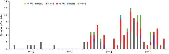
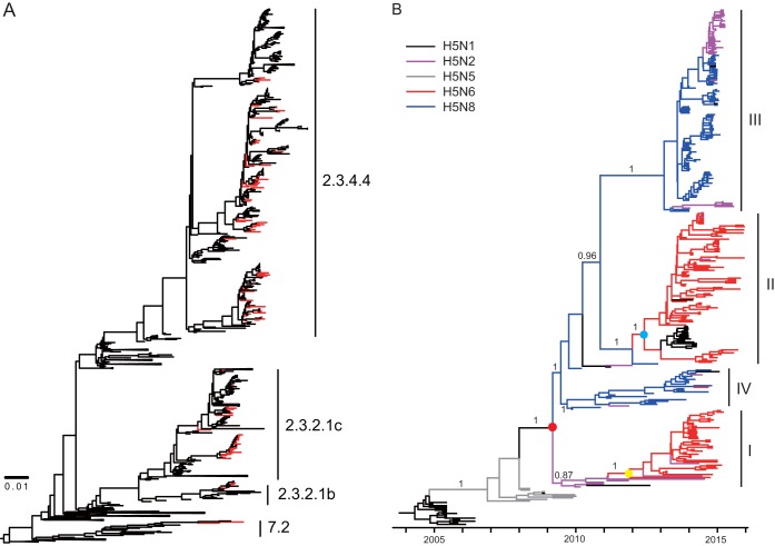
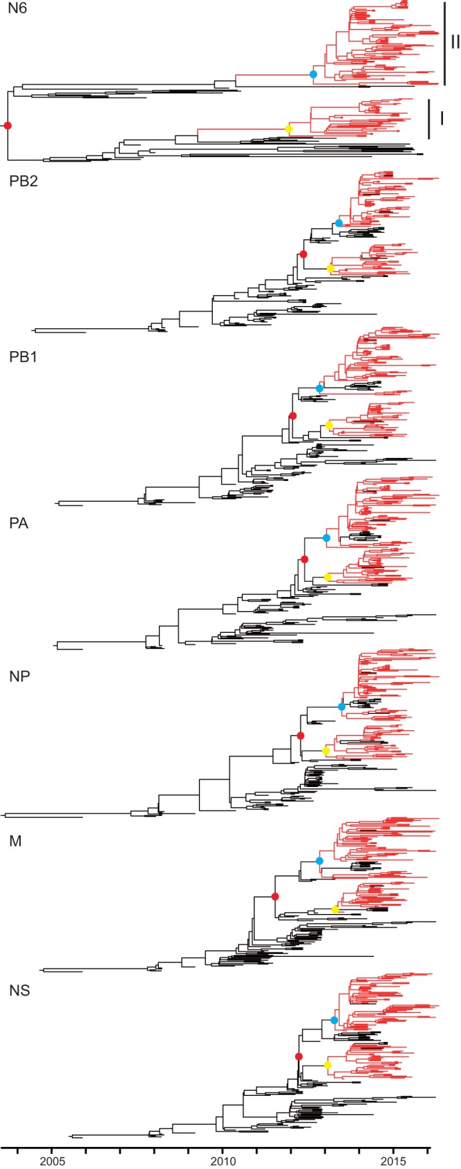
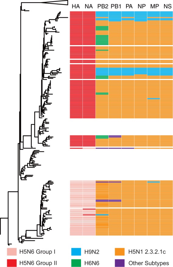
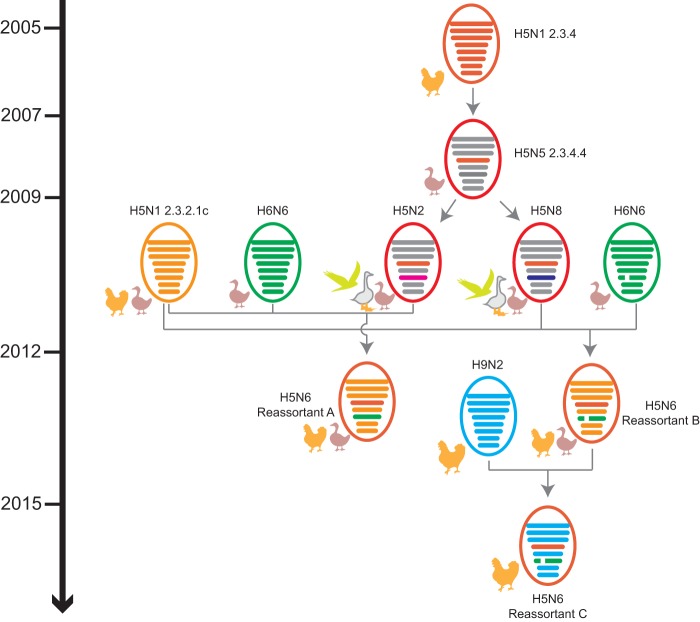
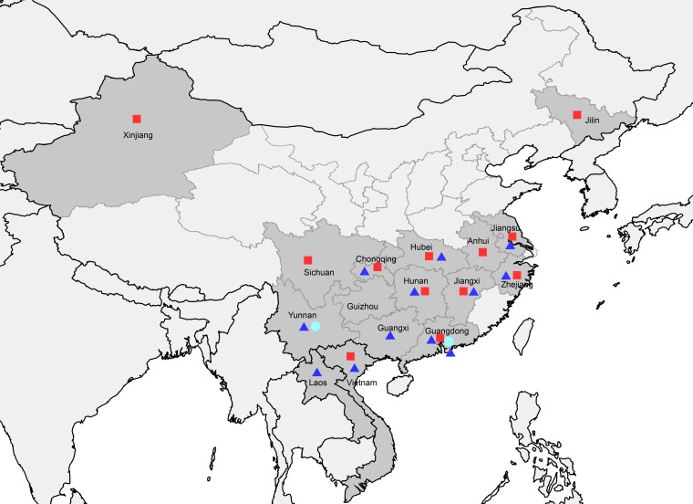
Clade 2.3.4.4 highly pathogenic avian influenza viruses (H5Nx) have spread from Asia to other parts of the world. Since 2014, human infections with clade 2.3.4.4 highly pathogenic avian influenza H5N6 viruses have been continuously reported in China. To investigate the genesis of the virus, we analyzed 123 H5 or N6 environmental viruses sampled from live-poultry markets or farms from 2012 to 2015 in Mainland China. Our results indicated that clade 2.3.4.4 H5N2/N6/N8 viruses shared the same hemagglutinin gene as originated in early 2009. From 2012 to 2015, the genesis of highly pathogenic avian influenza H5N6 viruses occurred via two independent pathways. Three major reassortant H5N6 viruses (reassortants A, B, and C) were generated. Internal genes of reassortant A and B viruses and reassortant C viruses derived from clade 2.3.2.1c H5N1 and H9N2 viruses, respectively. Many mammalian adaption mutations and antigenic variations were detected among the three reassortant viruses. Considering their wide circulation and dynamic reassortment in poultry, we highly recommend close monitoring of the viruses in poultry and humans. IMPORTANCE Since 2014, clade 2.3.4.4 highly pathogenic avian influenza (H5Nx) viruses have caused many outbreaks in both wild and domestic birds globally. Severe human cases with novel H5N6 viruses in this group were also reported in China in 2014 and 2015. To investigate the genesis of the genetic diversity of these H5N6 viruses, we sequenced 123 H5 or N6 environmental viruses sampled from 2012 to 2015 in China. Sequence analysis indicated that three major reassortants of these H5N6 viruses had been generated by two independent evolutionary pathways. The H5N6 reassortant viruses had been detected in most provinces of southern China and neighboring countries. Considering the mammalian adaption mutations and antigenic variation detected, the spread of these viruses should be monitored carefully due to their pandemic potential.

Keywords: H5N6; highly pathogenic avian influenza; reassortment.


Figures

FIG 1
FIG 1
Time distribution of H5 or N6 viruses isolated in this study. The number of isolates of each subtype was accumulated by month.
FIG 2
FIG 2
Evolutionary analysis of H5 gene sequences. (A) Maximum likelihood phylogenetic analysis of HA genes of 108 HPAI H5 viruses. The viruses isolated in our study are highlighted in red. (B) Bayesian maximum clade credibility (MCC) phylogeny of clade 2.3.4.4 HPAI H5Nx viruses. The phylogenetic clades that included clade 2.3.4.4 HPAI H5N2/N6/N8 viruses were obtained from the dated phylogeny of HA gene segments constructed by molecular phylogenetic analysis and are further aligned onto the same time scale. The branches in purple, blue, red, gray, and black represent clade 2.3.4.4 HPAI H5N2, H5N8, H5N6, H5N5, and H5N1 viruses, respectively. The tMRCAs of the clade HPAI H5Nx viruses and the two H5N6 subgroups are indicated by red, blue, and yellow dots. The posterior probabilities of the main branches are shown as numbers. For details of the phylogeny, see Fig. S1 in the supplemental material.
FIG 3
FIG 3
Genomic divergence of NA and six internal genes of clade 2.3.4.4 HPAI H5N6 viruses. The red and black branches represent the clade 2.3.4.4 HPAI H5N6 viruses and other related viruses, respectively. The tMRCAs of the two subgroups and each subgroup of H5N6 viruses are highlighted in solid red, blue, and yellow, respectively. For details of the phylogeny, see Fig. S2 to S8 in the supplemental material.
FIG 4
FIG 4
Reassortant patterns of the clade 2.3.4.4 HPAI H5N6 viruses and their potential donor-like viruses. A phylogenetic tree demonstrating the similar evolutionary pattern of clade 2.3.4.4 HPAI H5Nx HA gene in Fig. 2 is shown on the left. The unrooted tree was based on the full-length HA gene sequences. Reassortant patterns of the H5N6 viruses are listed on the right. Eight gene segments are indicated at the top of each bar. The colors of the bars represent the potential donor-like viruses listed at the bottom.
FIG 5
FIG 5
Possible evolutionary pathways toward the generation of clade 2.3.4.4 HPAI H5N2/N6/N8 viruses and the diverse genotypes of HPAI H5N6 viruses. Virus particles are represented by colored ovals containing horizontal bars that represent the eight gene segments (from top to bottom: PB2, PB1, PA, HA, NP, NA, M, and NS). Segments in descendant viruses are colored according to their corresponding source viruses (top) to illustrate gene ancestry through reassortment events. Source viruses for a reassortment are adjacent to arrow tails; arrowheads point to the resulting reassortants. A broken bar in segment 6 (NA) indicates a stalk region deletion. The timeline on the left indicates the possible time of virus emergence or reassortment events.
FIG 6
FIG 6
Geographic distributions of three reassortant HPAI H5N6 viruses in Mainland China and neighboring Laos and Vietnam. Provinces in which clade 2.3.4.4 HPAI H5N6 viruses were isolated in Mainland China, Laos, and Vietnam are in dark gray. Solid red squares, blue triangles, and cyan circles indicate the reassortant A, B, and C H5N6 viruses, respectively. This map was drawn using ArcGIS (ESRI) software version 9.

References

    1. Duan L, Campitelli L, Fan XH, Leung YH, Vijaykrishna D, Zhang JX, Donatelli I, Delogu M, Li KS, Foni E, Chiapponi C, Wu WL, Kai H, Webster RG, Shortridge KF, Peiris JS, Smith GJ, Chen H, Guan Y. 2007. Characterization of low-pathogenic H5 subtype influenza viruses from Eurasia: implications for the origin of highly pathogenic H5N1 viruses. J Virol 81:7529–7539. doi: 10.1128/JVI.00327-07. - DOI - PMC - PubMed
    1. Duan L, Bahl J, Smith GJ, Wang J, Vijaykrishna D, Zhang LJ, Zhang JX, Li KS, Fan XH, Cheung CL, Huang K, Poon LL, Shortridge KF, Webster RG, Peiris JS, Chen H, Guan Y. 2008. The development and genetic diversity of H5N1 influenza virus in China, 1996–2006. Virology 380:243–254. doi: 10.1016/j.virol.2008.07.038. - DOI - PMC - PubMed
    1. WHO/OIE/FAO H5N1 Evolution Working Group. 29 October 2011. Continued evolution of highly pathogenic avian influenza A (H5N1): updated nomenclature. Influenza Other Respir Viruses doi: 10.1111/j.1750-2659.2011.00298.x. - DOI - PMC - PubMed
    1. Liu J, Xiao H, Lei F, Zhu Q, Qin K, Zhang XW, Zhang XL, Zhao D, Wang G, Feng Y, Ma J, Liu W, Wang J, Gao GF. 2005. Highly pathogenic H5N1 influenza virus infection in migratory birds. Science 309:1206. doi: 10.1126/science.1115273. - DOI - PubMed
    1. Hill SC, Lee YJ, Song BM, Kang HM, Lee EK, Hanna A, Gilbert M, Brown IH, Pybus OG. 2015. Wild waterfowl migration and domestic duck density shape the epidemiology of highly pathogenic H5N8 influenza in the Republic of Korea. Infect Genet Evol 34:267–277. doi: 10.1016/j.meegid.2015.06.014. - DOI - PMC - PubMed

Publication types

MeSH terms