. 2016 Dec 12:12:3161-3173.
doi: 10.2147/NDT.S121779. eCollection 2016.

Treadmill exercise promotes neuroprotection against cerebral ischemia-reperfusion injury via downregulation of pro-inflammatory mediators

Affiliations

Treadmill exercise promotes neuroprotection against cerebral ischemia-reperfusion injury via downregulation of pro-inflammatory mediators

Ying Zhang et al. Neuropsychiatr Dis Treat. .

Abstract

Background: Stroke is one of the major causes of morbidity and mortality worldwide, which is associated with serious physical deficits that affect daily living and quality of life and produces immense public health and economic burdens. Both clinical and experimental data suggest that early physical training after ischemic brain injury may reduce the extent of motor dysfunction. However, the exact mechanisms have not been fully elucidated. The aim of this study was to investigate the effects of aerobic exercise on neuroprotection and understand the underlying mechanisms.

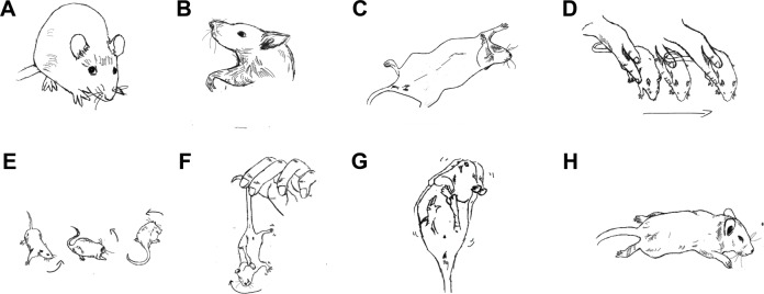
Materials and methods: Middle cerebral artery occlusion (MCAO) was conducted to establish a rat model of cerebral ischemia-reperfusion injury to mimic ischemic stroke. Experimental animals were divided into the following three groups: sham (n=34), MCAO (n=39), and MCAO plus treadmill exercise (n=28). The effects of aerobic exercise intervention on ischemic brain injury were evaluated using functional scoring, histological analysis, and Bio-Plex Protein Assays.

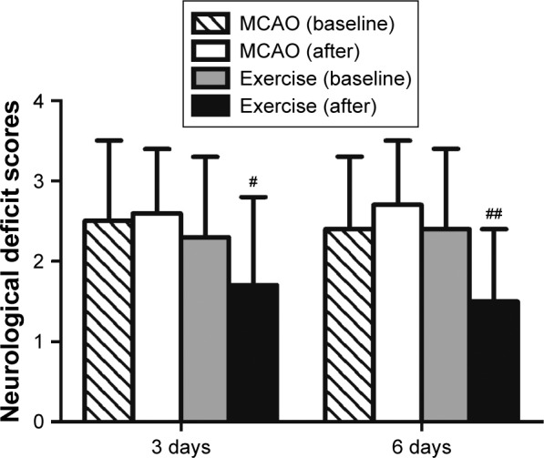
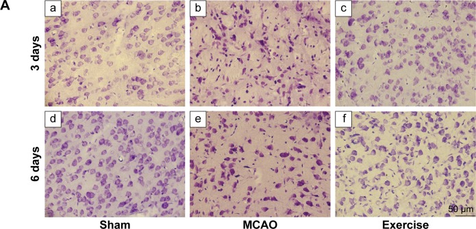
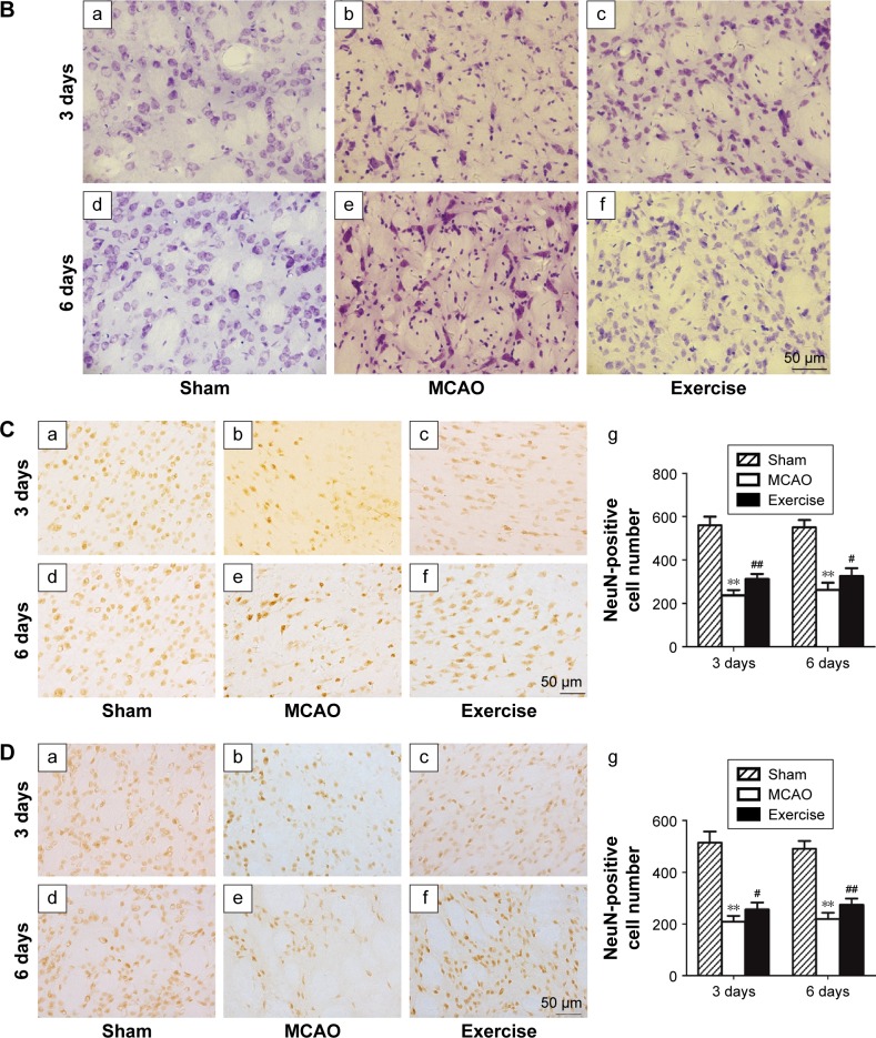
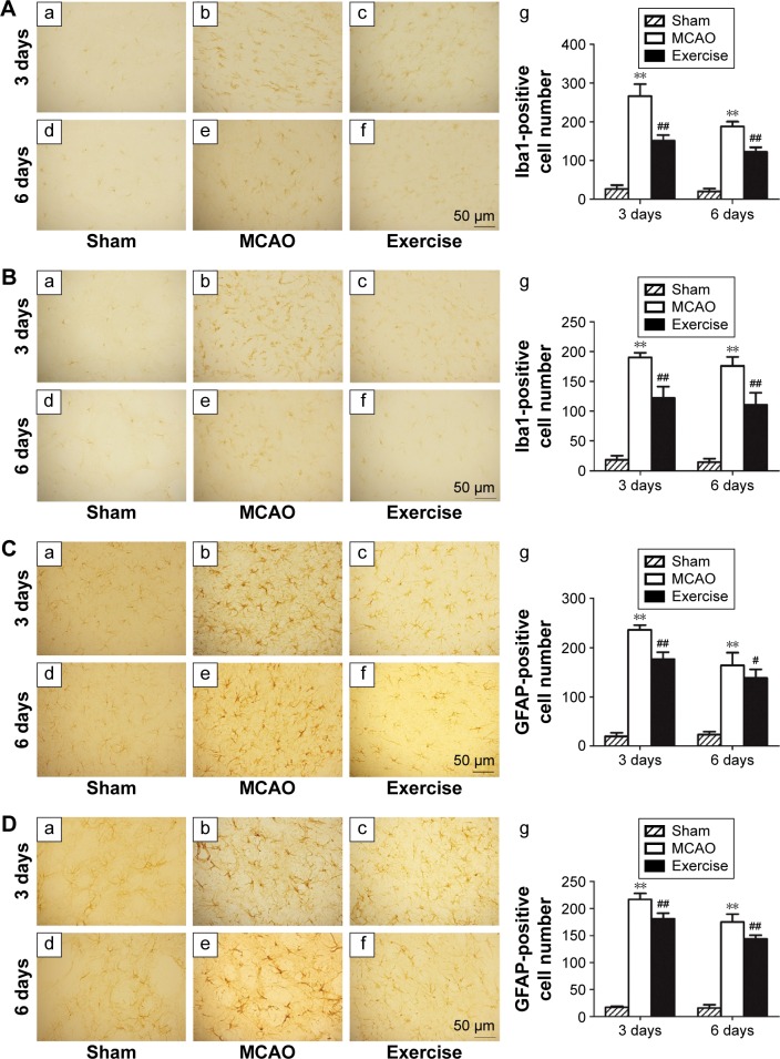
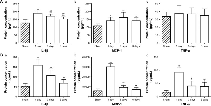
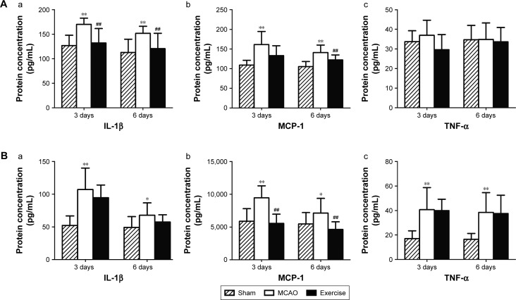
Results: Early aerobic exercise intervention was found to improve motor function, prevent death of neuronal cells, and suppress the activation of microglial cells and astrocytes. Furthermore, it was observed that aerobic exercise downregulated the expression of the cytokine interleukin-1β and the chemokine monocyte chemotactic protein-1 after transient MCAO in experimental rats.

Conclusion: This study demonstrates that treadmill exercise rehabilitation promotes neuroprotection against cerebral ischemia-reperfusion injury via the downregulation of proinflammatory mediators.

Keywords: chemokine; cytokine; rat model; rehabilitation; stroke.


Conflict of interest statement

The authors report no conflicts of interest in this work.

Figures

Figure 1
Figure 1
Graphical representation of motor function scores. Notes: Rats with no deficit were scored 0 (A); rats with forelimb flexion influencing wrist (B) or shoulder (C) were scored 1; rats with forelimb flexion plus decreased resistance to lateral push were scored 2 (D); rats with unidirectional circling to the deficit side were scored 3 (E); rats with longitudinal spinning to the deficit side (F) or seizure activity (G) were scored 4; rats with no movement were scored 5 (H).
Figure 2
Figure 2
Exercise improved motor function after ischemic brain injury. Notes: Rat neurological behavior was evaluated based on a modified Bederson scoring method. Treadmill exercise was started 1 day after surgery and lasted for 3-day and 6-day periods. Behavioral scores were measured before the exercise intervention and at the end of the two time points. The neurological deficit scores did not show statistical difference in the MCAO groups after 3-day and 6-day periods, respectively. The neurological deficit scores of the baseline did not show statistical difference between the MCAO group and exercise group either. Behavioral scores improved after 3-day (#P<0.05) and 6-day exercises (##P<0.01) in comparison with the MCAO groups, respectively. Abbreviation: MCAO, middle cerebral artery occlusion.
Figure 3
Figure 3
Exercise prevented neuronal cell death in rat model of ischemic brain injury. Notes: (A) Nissl staining in cortex: a. sham group 3 days after surgery, b. MCAO group 3 days after surgery, c. exercise group 3 days after surgery, d. sham group 6 days after surgery, e. MCAO group 6 days after surgery, f. exercise group 6 days after surgery. (B) Nissl staining in caudatum: a. sham group 3 days after surgery, b. MCAO group 3 days after surgery, c. exercise group 3 days after surgery, d. sham group 6 days after surgery, e. MCAO group 6 days after surgery, f. exercise group 6 days after surgery. (C) Immunohistochemistry staining using anti-NeuN antibody in cortex: a. sham group 3 days after surgery, b. MCAO group 3 days after surgery, c. exercise group 3 days after surgery, d. sham group 6 days after surgery, e. MCAO group 6 days after surgery, f. exercise group 6 days after surgery, g. quantification of stained surviving neuronal cells. (D) Immunohistochemistry staining using anti-NeuN antibody in caudatum: a. sham group 3 days after surgery, b. MCAO group 3 days after surgery, c. exercise group 3 days after surgery, d. sham group 6 days after surgery, e. MCAO group 6 days after surgery, f. exercise group 6 days after surgery, g. quantification of stained surviving neuronal cells. Bar =50 μm for all images; **P<0.01 in comparison with the sham group; #P<0.05 and ##P<0.01 in comparison with the MCAO group. Abbreviation: MCAO, middle cerebral artery occlusion.
Figure 3
Figure 3
Exercise prevented neuronal cell death in rat model of ischemic brain injury. Notes: (A) Nissl staining in cortex: a. sham group 3 days after surgery, b. MCAO group 3 days after surgery, c. exercise group 3 days after surgery, d. sham group 6 days after surgery, e. MCAO group 6 days after surgery, f. exercise group 6 days after surgery. (B) Nissl staining in caudatum: a. sham group 3 days after surgery, b. MCAO group 3 days after surgery, c. exercise group 3 days after surgery, d. sham group 6 days after surgery, e. MCAO group 6 days after surgery, f. exercise group 6 days after surgery. (C) Immunohistochemistry staining using anti-NeuN antibody in cortex: a. sham group 3 days after surgery, b. MCAO group 3 days after surgery, c. exercise group 3 days after surgery, d. sham group 6 days after surgery, e. MCAO group 6 days after surgery, f. exercise group 6 days after surgery, g. quantification of stained surviving neuronal cells. (D) Immunohistochemistry staining using anti-NeuN antibody in caudatum: a. sham group 3 days after surgery, b. MCAO group 3 days after surgery, c. exercise group 3 days after surgery, d. sham group 6 days after surgery, e. MCAO group 6 days after surgery, f. exercise group 6 days after surgery, g. quantification of stained surviving neuronal cells. Bar =50 μm for all images; **P<0.01 in comparison with the sham group; #P<0.05 and ##P<0.01 in comparison with the MCAO group. Abbreviation: MCAO, middle cerebral artery occlusion.
Figure 4
Figure 4
Exercise inhibited the activation of microglial cells and astrocytes. Notes: (A) Immunohistochemistry staining using anti-Iba1 antibody in cortex: a. sham group 3 days after surgery, b. MCAO group 3 days after surgery, c. exercise group 3 days after surgery, d. sham group 6 days after surgery, e. MCAO group 6 days after surgery, f. exercise group 6 days after surgery, g. quantification of stained microglial cells. (B) Immunohistochemistry staining using anti-Iba1 antibody in caudatum: a. sham group 3 days after surgery, b. MCAO group 3 days after surgery, c. exercise group 3 days after surgery, d. sham group 6 days after surgery, e. MCAO group 6 days after surgery, f. exercise group 6 days after surgery, g. quantification of stained microglial cells. (C) Immunohistochemistry staining using anti-GFAP antibody in cortex: a. sham group 3 days after surgery, b. MCAO group 3 days after surgery, c. exercise group 3 days after surgery, d. sham group 6 days after surgery, e. MCAO group 6 days after surgery, f. exercise group 6 days after surgery, g. quantification of stained astrocytes. (D) Immunohistochemistry staining using anti-GFAP antibody in caudatum: a. sham group 3 days after surgery, b. MCAO group 3 days after surgery, c. exercise group 3 days after surgery, d. sham group 6 days after surgery, e. MCAO group 6 days after surgery, f. exercise group 6 days after surgery, g. quantification of stained astrocytes. Bar =50 μm for all images; **P<0.01 in comparison with the sham group; #P<0.05 and ##P<0.01 in comparison with the MCAO group. Abbreviations: GFAP, glial fibrillary acidic protein; MCAO, middle cerebral artery occlusion.
Figure 5
Figure 5
Changes of cytokine levels in the brain and blood after MCAO. Notes: Protein levels of cytokines after 1, 3, and 6 days post surgery were measured by Bio-Plex Multiplex Cytokine Assays (A) in the brain a. IL-1β protein concentration, b. MCP-1 protein concentration, c. TNF-α protein concentration and (B) in peripheral blood a. IL-1β protein concentration, b. MCP-1 protein concentration, c. TNF-α, protein concentration. (sham group: n=10 for baseline; MCAO group: n=12 for 1-day, n=9 for 3-day, n=10 for 6-day time points). *P<0.05 and **P<0.01 in comparison with the sham group; #P<0.05 and ##P<0.01 in comparison with the level on 1 day post MCAO. Abbreviations: IL-1β, interleukin-1β; MCAO, middle cerebral artery occlusion; MCP-1, monocyte chemotactic protein-1; TNF-α, tumor necrosis factor-α.
Figure 6
Figure 6
Effects of exercise on cytokine/chemokine changes after MCAO. Notes: Bio-Plex Multiplex Cytokine Assays were performed to measure protein levels of IL-1β, MCP-1, and TNF-α at 3-day and 6-day time points among the groups of sham, MCAO, and exercise (A) in the brain a. IL-1β protein concentration, b. MCP-1 protein concentration, c. TNF-α protein concentration and (B) In peripheral blood a. IL-1β protein concentration, b. MCP-1 protein concentration, c. TNF-α protein concentration. (sham group: n=8 for 3-day and n=8 for 6-day time points; MCAO group: n=9 for 3-day and n=10 for 6-day time points; exercise group: n=11 for 3-day and n=9 for 6-day time points). *P<0.05 and **P<0.01 in comparison with the sham group. #P<0.05 and ##P<0.01 in comparison with the MCAO group. Abbreviations: IL-1β, interleukin-1β; MCAO, middle cerebral artery occlusion; MCP-1, monocyte chemotactic protein-1; TNF-α, tumor necrosis factor-α.

Similar articles

Cited by

References

    1. GBD 2013 Mortality and Causes of Death Collaborators Global, regional, and national age-sex specific all-cause and cause-specific mortality for 240 causes of death, 1990–2013: a systematic analysis for the Global Burden of Disease Study 2013. Lancet. 2015;385(9963):117–171. - PMC - PubMed
    1. Saxena SK, Ng TP, Koh G, Yong D, Fong NP. Is improvement in impaired cognition and depressive symptoms in post-stroke patients associated with recovery in activities of daily living? Acta Neurol Scand. 2007;115(5):339–346. - PubMed
    1. Mozaffarian D, Benjamin EJ, Go AS, et al. Heart disease and stroke statistics – 2015 update: a report from the American Heart Association. Circulation. 2015;131(4):434–441. - PubMed
    1. Koziol JA, Feng AC. On the analysis and interpretation of outcome measures in stroke clinical trials. Stoke. 2006;37(10):2644–2647. - PubMed
    1. Rizos EC, Ntzani EE, Bika E, Kostapanos MS, Elisaf MS. Association between omega-3 fatty acid supplementation and risk of major cardiovascular disease events: a systematic review and meta-analysis. JAMA. 2012;308(10):1024–1033. - PubMed