

Transcatheter Closure of Ruptured Sinus of Valsalva Aneurysm: Report of two cases
- PMID: 28003903
- PMCID: PMC5135468
- DOI: 10.18295/squmj.2016.16.04.020
Transcatheter Closure of Ruptured Sinus of Valsalva Aneurysm: Report of two cases
Abstract
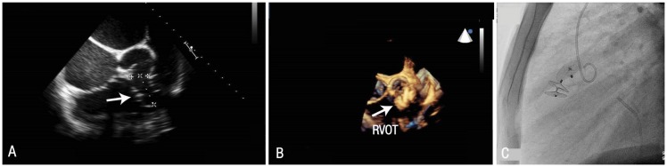
A ruptured sinus of Valsalva aneurysm (RSVA) is a rare cardiac anomaly. Traditionally, RSVAs were repaired surgically; however, percutaneous transcatheter closure is the current treatment of choice. We report two cases of RSVA which were closed using this approach. The first case was a 45-year-old female who presented to the Royal Hospital, Muscat, Oman, in 2014 with a RSVA in the right ventricle. The second case was a 39-year-old male who was admitted to the Sultan Qaboos University Hospital, Muscat, in 2015 with a large multifenestrated RSVA extending into the right ventricle outflow tract. Each patient underwent transcutaneous cardiac catheterisation using three-dimensional echocardiography. Both interventions were technically successful; however, the second patient required a subsequent surgery due to the continuing presence of a significant shunt. Transcatheter closure of RSVAs is an effective alternative to surgical repair, although large multifenestrated RSVAs should be repaired surgically to ensure complete closure.
Keywords: Cardiac Catheterization; Case Report; Oman; Ruptured Aneurysm; Sinus of Valsalva; Three-Dimensional Echocardiography.
Figures

References
-
- Perloff JK. The Clinical Recognition of Congenital Heart Disease. 5th ed. Philadelphia, Pennsylvania, USA: Saunders; 2003. pp. 457–70.
-
- Chu SH, Hung CR, How SS, Chang H, Wang SS, Tsai CH, et al. Ruptured aneurysms of the sinus of Valsalva in Oriental patients. J Thorac Cardiovasc Surg. 1990;99:288–98. - PubMed
-
- Shah RP, Ding ZP, Ng AS, Quek SS. A ten-year review of ruptured sinus of Valsalva: Clinico-pathological and echo-Doppler features. Singapore Med J. 2001;42:473–6. - PubMed
LinkOut - more resources
Full Text Sources
Other Literature Sources