

Thymic Epidermoid Cyst: Clinical and Imaging Manifestations of This Rare Anterior Mediastinal Mass
- PMID: 28003927
- PMCID: PMC5143738
- DOI: 10.1155/2016/5789321
Thymic Epidermoid Cyst: Clinical and Imaging Manifestations of This Rare Anterior Mediastinal Mass
Abstract
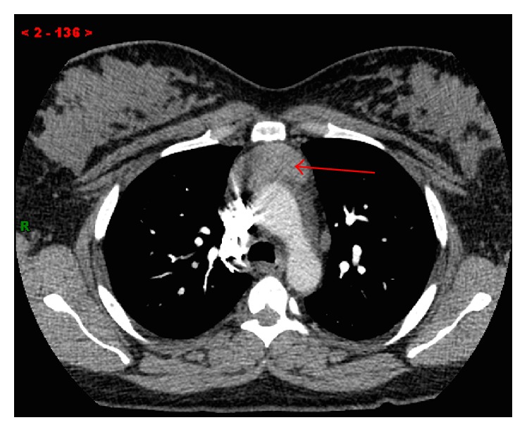
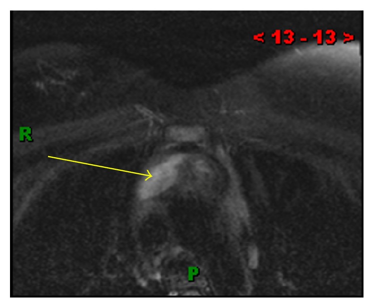
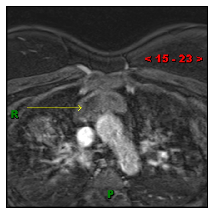
Thymic epidermoid cysts are an extremely rare entity. These arise from epidermal cells that migrate to the thymus. The radiologic diagnosis of this rare lesion is challenging. We describe a case of an otherwise healthy 35-year-old woman who presented with an acute onset of chest pain and shortness of breath. She was found to have an anterior mediastinal mass. The imaging findings were, however, not characteristic for any single diagnostic entity. Since the imaging was inconclusive, surgical resection was performed for definitive diagnosis. The mass was found to be a thymic epidermoid cyst. This case underlines the significance for radiologists to be aware that epidermoid cysts can occur in the thymus and should be considered in the differential diagnosis for a heterogeneous anterior mediastinal mass.
Conflict of interest statement
The authors declare that there is no conflict of interests regarding the publication of this paper.
Figures


Similar articles
-
CT Findings of Thymic Epidermoid Cyst in the Anterior Mediastinum: A Case Report and Literature Review.Taehan Yongsang Uihakhoe Chi. 2022 Jan;83(1):212-217. doi: 10.3348/jksr.2021.0014. Epub 2021 Oct 18. Taehan Yongsang Uihakhoe Chi. 2022. PMID: 36237357 Free PMC article.
-
Epidermoid Thymic Cyst - A Very Rare Mediastinal Mass.Rev Port Cir Cardiotorac Vasc. 2019 Oct-Dec;26(4):267-268. Rev Port Cir Cardiotorac Vasc. 2019. PMID: 32006449
-
[Rare Case of Thymic Epidermoid Cyst;Report of a Case].Kyobu Geka. 2019 Dec;72(13):1119-1122. Kyobu Geka. 2019. PMID: 31879391 Japanese.
-
Thymic adenocarcinoma associated with thymic cyst: a case report and review of literature.Int J Clin Exp Pathol. 2015 May 1;8(5):5890-5. eCollection 2015. Int J Clin Exp Pathol. 2015. PMID: 26191314 Free PMC article. Review.
-
Complete recovery of an isolated left vocal fold palsy associated with a benign mediastinal thymic cyst.J Laryngol Otol. 2008 Feb;122(2):e4. doi: 10.1017/S0022215107001326. Epub 2008 Jan 10. J Laryngol Otol. 2008. PMID: 18184448 Review.
Cited by
-
CT Findings of Thymic Epidermoid Cyst in the Anterior Mediastinum: A Case Report and Literature Review.Taehan Yongsang Uihakhoe Chi. 2022 Jan;83(1):212-217. doi: 10.3348/jksr.2021.0014. Epub 2021 Oct 18. Taehan Yongsang Uihakhoe Chi. 2022. PMID: 36237357 Free PMC article.
-
Mediastinal Epidermoid Cyst in a 5-Year-Old Girl.European J Pediatr Surg Rep. 2018 Jan;6(1):e24-e26. doi: 10.1055/s-0037-1621707. Epub 2018 Mar 22. European J Pediatr Surg Rep. 2018. PMID: 29577001 Free PMC article.
References
-
- Delamarre J., Dupas J. L., Muir J. F., Deschepper B., Sevestre H., Capron J. P. Gardner's syndrome and epidermoid cyst of the thymus. Gastroenterologie Clinique et Biologique. 1987;11(5):421–423. - PubMed
LinkOut - more resources
Full Text Sources
Other Literature Sources
