. 2017 Mar 15;595(6):2043-2064.
doi: 10.1113/JP273335. Epub 2017 Feb 1.

Non-chemosensitive parafacial neurons simultaneously regulate active expiration and airway patency under hypercapnia in rats

Affiliations

Non-chemosensitive parafacial neurons simultaneously regulate active expiration and airway patency under hypercapnia in rats

Alan A de Britto et al. J Physiol. .

Abstract

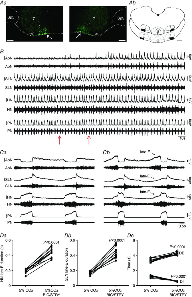
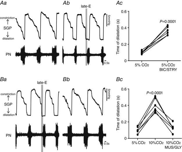
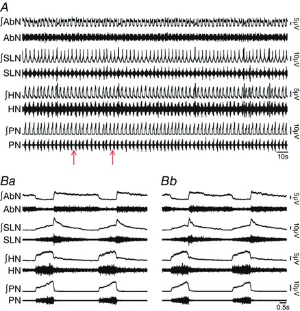
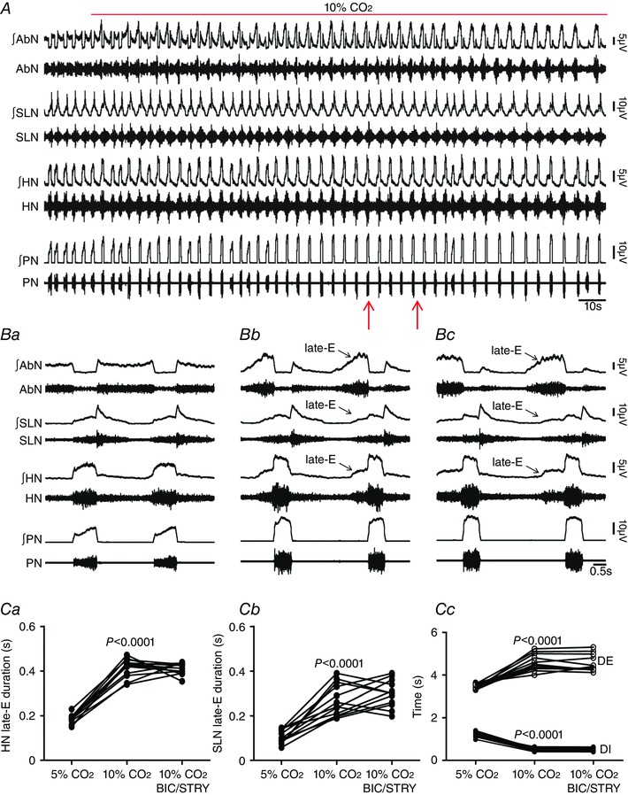
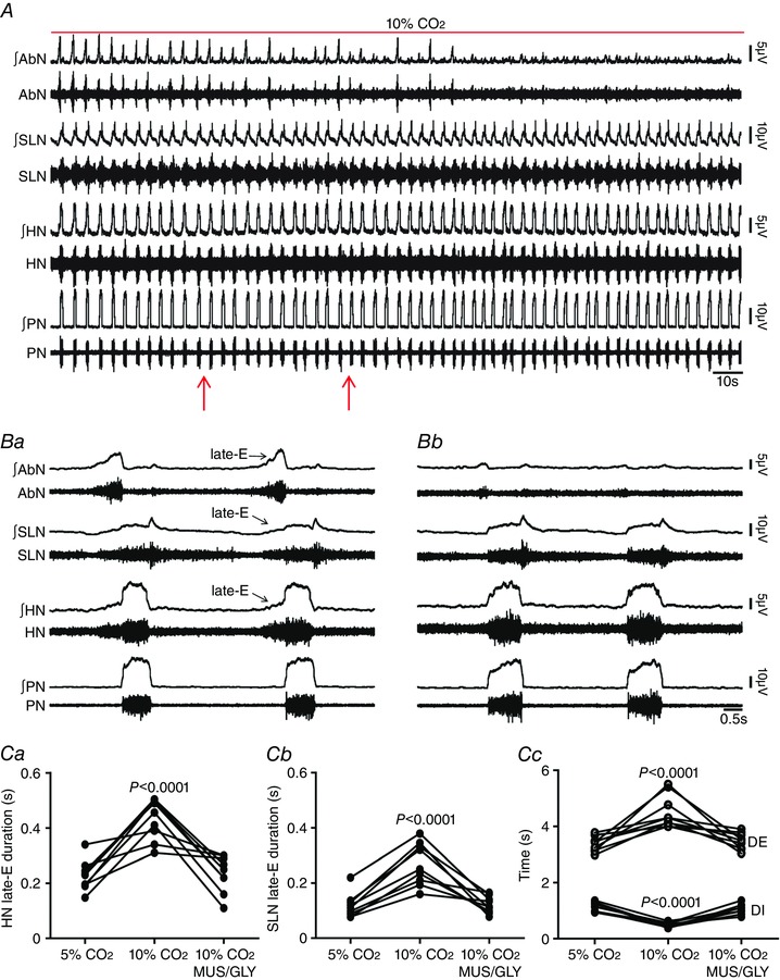
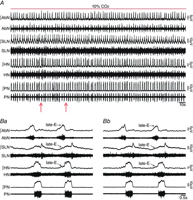
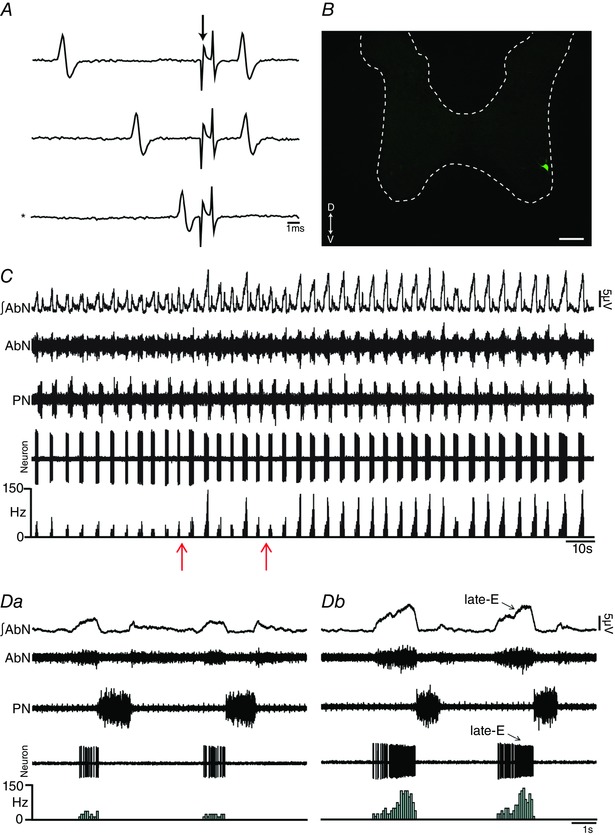
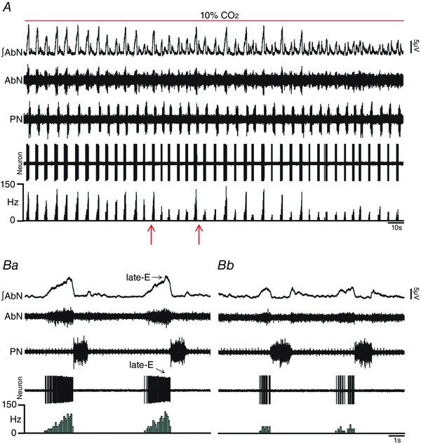
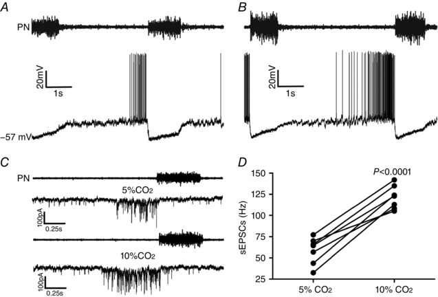
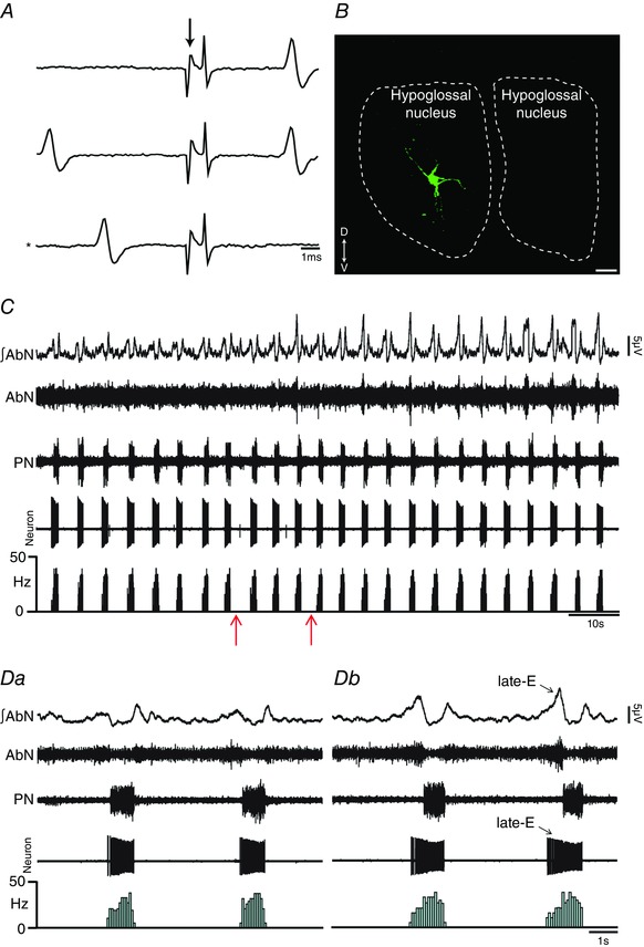
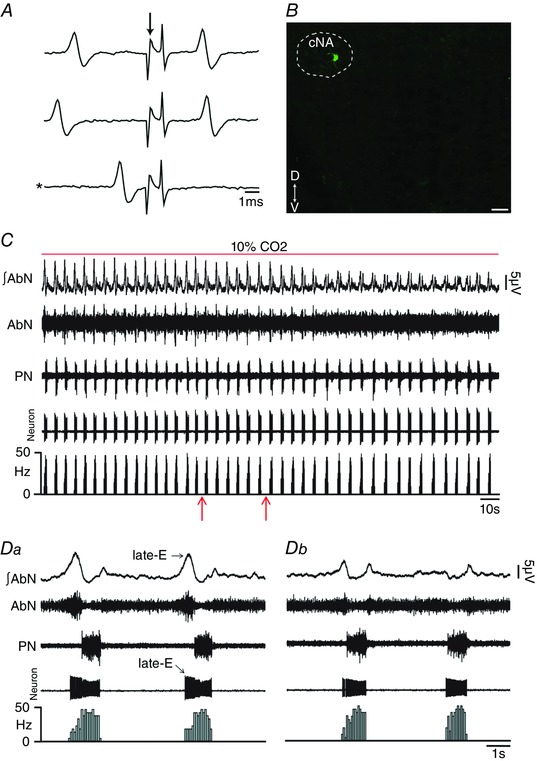
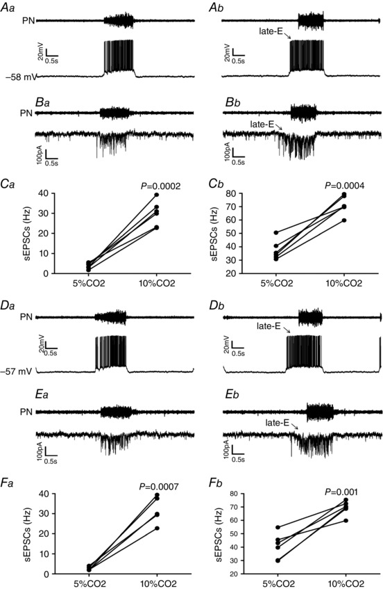
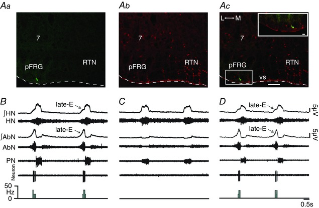
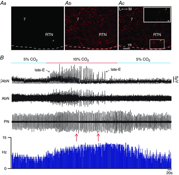
Key points: Hypercapnia or parafacial respiratory group (pFRG) disinhibition at normocapnia evokes active expiration in rats by recruitment of pFRG late-expiratory (late-E) neurons. We show that hypercapnia simultaneously evoked active expiration and exaggerated glottal dilatation by late-E synaptic excitation of abdominal, hypoglossal and laryngeal motoneurons. Simultaneous rhythmic expiratory activity in previously silent pFRG late-E neurons, which did not express the marker of ventral medullary CO2 -sensitive neurons (transcription factor Phox2b), was also evoked by hypercapnia. Hypercapnia-evoked active expiration, neural and neuronal late-E activities were eliminated by pFRG inhibition, but not after blockade of synaptic excitation. Hypercapnia produces disinhibition of non-chemosensitive pFRG late-E neurons to evoke active expiration and concomitant cranial motor respiratory responses controlling the oropharyngeal and upper airway patency.

Abstract: Hypercapnia produces active expiration in rats and the recruitment of late-expiratory (late-E) neurons located in the parafacial respiratory group (pFRG) of the ventral medullary brainstem. We tested the hypothesis that hypercapnia produces active expiration and concomitant cranial respiratory motor responses controlling the oropharyngeal and upper airway patency by disinhibition of pFRG late-E neurons, but not via synaptic excitation. Phrenic nerve, abdominal nerve (AbN), cranial respiratory motor nerves, subglottal pressure, and medullary and spinal neurons/motoneurons were recorded in in situ preparations of juvenile rats. Hypercapnia evoked AbN active expiration, exaggerated late-E discharges in cranial respiratory motor outflows, and glottal dilatation via late-E synaptic excitation of abdominal, hypoglossal and laryngeal motoneurons. Simultaneous rhythmic late-E activity in previously silent pFRG neurons, which did not express the marker of ventral medullary CO2 -sensitive neurons (transcription factor Phox2b), was also evoked by hypercapnia. In addition, hypercapnia-evoked AbN active expiration, neural and neuronal late-E activities were eliminated by pFRG inhibition, but not after blockade of synaptic excitation. On the other hand, pFRG inhibition did not affect either hypercapnia-induced inspiratory increases in respiratory motor outflows or CO2 sensitivity of the more medial Phox2b-positive neurons in the retrotrapezoid nucleus (RTN). Our data suggest that neither RTN Phox2b-positive nor other CO2 -sensitive brainstem neurons activate Phox2b-negative pFRG late-E neurons under hypercapnia to produce AbN active expiration and concomitant cranial motor respiratory responses controlling the oropharyngeal and upper airway patency. Hypercapnia produces disinhibition of non-chemosensitive pFRG late-E neurons in in situ preparations of juvenile rats to activate abdominal, hypoglossal and laryngeal motoneurons.

Keywords: abdominal active expiration; airway patency; late-expiratory neurons; parafacial respiratory group; respiratory motoneurons.


Figures

Figure 1
Figure 1. pFRG disinhibition evoked active expiration and concomitant responses in cranial and spinal respiratory motor outflows in in situ preparations of rats
Aa, photomicrographs showing typical sites of bilateral microinjections in the pFRG (at bregma level −11.40 mm; 200 μm rostral to the caudal pole of facial nucleus). The fluorescent microbeads are located in the ventral medullary surface (vs; arrows) ∼ 500 μm medial to the spinal trigeminal tract (Sp5). Scale bars: 100 μm; 7, motor facial nucleus. Ab, schematic drawings of coronal sections of the brainstem showing the sites of bilateral microinjections into the pFRG of all in situ preparations of rats used in the present study (at bregma level between –11.30 mm and −11.50 mm; 100–300 μm rostral to the caudal pole of facial nucleus). Scale bar: 500 μm. B, raw and integrated (∫) records of AbN, SLN, HN and PN activities from one in situ preparation of rat under normocapnia before and after bilateral microinjections (arrows) of BIC/STRY into pFRG. C, magnification of two respiratory cycles from the same in situ preparation of rat before (Ca) and after (Cb) bilateral microinjections of BIC/STRY into pFRG. Note that pFRG disinhibition evoked AbN active expiration, HN and SLN late‐E activities, decreased DI, increased DE, but did not affect the PN frequency. D, grouped data comparing the duration of HN (Da) and SLN (Db) late‐E activity, DI and DE (Dc) under normocapnia before and after bilateral microinjections of BIC/STRY into pFRG. [Color figure can be viewed at wileyonlinelibrary.com]
Figure 2
Figure 2. Hypercapnia or pFRG disinhibition exaggerated late‐E glottal dilatation in in situ preparations of rats
Representative tracings of dynamic changes in glottis during the respiratory cycle, including dilatation during inspiration (decrease in SGP) and constriction following inspiration (increase in SGP) from one in situ preparation of rat under normocapnia before (Aa) and after bilateral microinjections of BIC/STRY into pFRG (Ab). Ac, grouped data comparing the time of glottal dilatation in relation to inspiratory activity (PN) before and after bilateral microinjections of BIC/STRY into pFRG of rats under normocapnia. Note that pFRG disinhibition exaggerated late‐E glottal dilatation. B, representative tracings of dynamic changes in glottal resistance during the respiratory cycle from one in situ preparation of rat under hypercapnia before (Ba) and after bilateral microinjections of MUS/GLY into pFRG (Bb). Bc, grouped data comparing the time of glottal dilatation in relation to inspiratory activity (PN) before and after bilateral microinjections of MUS/GLY into pFRG of rats under hypercapnia. Note that pFRG inhibition normalized the exaggerated late‐E glottal dilatation evoked by hypercapnia.
Figure 3
Figure 3. Bilateral microinjections of vehicle into pFRG did not affect the respiratory pattern in in situ preparations of rats
A, raw and integrated (∫) records of AbN, SLN, HN and PN activities from one in situ preparation of rat under normocapnia before and after bilateral microinjections (arrows) of vehicle into pFRG. B, magnification of two respiratory cycles from the same in situ preparation of rat before (Ba) and after (Bb) bilateral microinjections of vehicle into pFRG. Note the absence of changes in the respiratory pattern after vehicle microinjections into pFRG. [Color figure can be viewed at wileyonlinelibrary.com]
Figure 4
Figure 4. pFRG disinhibition did not affect hypercapnia‐evoked active expiration and concomitant responses in cranial and spinal respiratory motor outflows in in situ preparations of rats
A, raw and integrated (∫) records of AbN, SLN, HN and PN activities from one in situ preparation of rat under normocapnia, hypercapnia and after bilateral microinjections (arrows) of BIC/STRY into pFRG during hypercapnia. B, magnification of two respiratory cycles from the same in situ preparation of rat under normocapnia (Ba), hypercapnia (Bb) and after bilateral microinjections of BIC/STRY into pFRG during hypercapnia (Bc). Note that hypercapnia evoked AbN active expiration, HN and SLN late‐E activities, decreased DI, increased DE, but did not affect the PN frequency. pFRG disinhibition was not able to produce any additional change in the AbN and in cranial and spinal respiratory motor outflows during hypercapnia. C, grouped data comparing the duration of HN (Ca) and SLN (Cb) late‐E activity, DI and DE (Cc) under normocapnia, hypercapnia and after bilateral microinjections of BIC/STRY into pFRG during hypercapnia. [Color figure can be viewed at wileyonlinelibrary.com]
Figure 5
Figure 5. pFRG inhibition eliminated hypercapnia‐evoked active expiration and concomitant responses in cranial and spinal respiratory motor outflows in in situ preparations of rats
A, raw and integrated (∫) records of AbN, SLN, HN and PN activities from one in situ preparation of rat under hypercapnia before and after bilateral microinjections (arrows) of MUS/GLY into pFRG. B, magnification of two respiratory cycles from the same in situ preparation of rat under hypercapnia before (Ba) and after (Bc) bilateral microinjections of MUS/GLY into pFRG. Note that hypercapnia evoked AbN active expiration, HN and SLN late‐E activities, decreased DI and increased DE. pFRG inhibition eliminated hypercapnia‐evoked active expiration and concomitant responses in cranial and spinal respiratory motor outflows. C, grouped data comparing the duration of HN (Ca) and SLN (Cb) late‐E activity, DI and DE (Cc) under hypercapnia before and after bilateral microinjections of MUS/GLY into pFRG. [Color figure can be viewed at wileyonlinelibrary.com]
Figure 6
Figure 6. Blockade of ionotropic glutamatergic receptors in pFRG did not affect hypercapnia‐evoked active expiration and concomitant responses in cranial and spinal respiratory motor outflows in in situ preparations of rats
A, raw and integrated (∫) records of AbN, SLN, HN and PN activities from one in situ preparation of rat under hypercapnia before and after bilateral microinjections (arrows) of KYN into pFRG. B, magnification of two respiratory cycles from the same in situ preparation of rat under hypercapnia before (Ba) and after (Bc) bilateral microinjections of KYN into pFRG. Note that hypercapnia evoked AbN active expiration, HN and SLN late‐E activity, decreased DI and increased DE. Blockade of synaptic excitation in the pFRG did not affect these responses. [Color figure can be viewed at wileyonlinelibrary.com]
Figure 7
Figure 7. pFRG disinhibition increased the firing frequency of spinal abdominal aug‐E motoneurons in in situ preparations of rats
A, representative spinal abdominal aug‐E motoneuron that was antidromically activated from the T12 AbN. Asterisk indicates sweep when the antidromic spike was absent as the result of a collision with a spontaneous spike used to trigger the stimulus (stimulus artifact at arrow). B, spinal abdominal aug‐E motoneuron labelled in situ with biocytin (Alexa 488, green) located in the ventral aspect of spinal cord. Scale bar: 100 μm. C, raw and integrated (∫) extracellular records of AbN, PN and spinal abdominal aug‐E motoneuron from one in situ preparation of rat under normocapnia before and after bilateral microinjections (arrows) of BIC/STRY into pFRG. D, magnification of two respiratory cycles from the same in situ preparation of rat under normocapnia before (Da) and after (Dc) bilateral microinjections of BIC/STRY into pFRG. Note that pFRG disinhibition evoked AbN active expiration and increased the firing frequency of spinal abdominal aug‐E motoneurons at the end of E2 phase. [Color figure can be viewed at wileyonlinelibrary.com]
Figure 8
Figure 8. pFRG inhibition blockade the effects of hypercapnia on the firing frequency of spinal abdominal aug‐E motoneurons in in situ preparations of rats
A, raw and integrated (∫) extracellular records of AbN, PN and spinal abdominal aug‐E motoneurons from one in situ preparation of rat under hypercapnia before and after bilateral microinjections (arrows) of MUS/GLY into pFRG. B, magnification of two respiratory cycles from the same in situ preparation of rat under hypercapnia before (Ba) and after (Bc) bilateral microinjections of MUS/GLY into pFRG. Note that pFRG inhibition eliminated AbN active expiration and normalized the increased firing frequency of spinal abdominal aug‐E motoneurons, at the end of E2 phase, induced by hypercapnia. [Color figure can be viewed at wileyonlinelibrary.com]
Figure 9
Figure 9. Hypercapnia increases synaptic excitation to spinal abdominal aug‐E motoneurons in in situ preparations of rats
A and B, raw records of PN and whole cell patch current clamp records of spinal abdominal aug‐E motoneurons from one in situ preparation of rat under normocapnia (A) and hypercapnia (B). C, raw records of PN and whole cell patch voltage clamp (−70 mV) records of spinal abdominal aug‐E motoneurons from one in situ preparation of rat under normocapnia and hypercapnia. Note that hypercapnia increased the spinal abdominal aug‐E motoneuron firing frequency and the frequency, but not the amplitude, of sEPSCs at the end of E2 phase. D, grouped data comparing the frequency of sEPSCs to spinal abdominal aug‐E motoneurons at the end of E2 phase under normocapnia and hypercapnia.
Figure 10
Figure 10. pFRG disinhibition increased the firing frequency of hypoglossal inspiratory motoneurons in in situ preparations of rats
A, representative hypoglossal inspiratory motoneuron that was antidromically activated from HN. Asterisk indicates sweep when the antidromic spike was absent as the result of a collision with a spontaneous spike used to trigger the stimulus (stimulus artifact at arrow). B, hypoglossal inspiratory motoneuron labelled in situ with biocytin (Alexa 488, green) located in the hypoglossal nucleus. Scale bar: 25 μm. C, raw and integrated (∫) extracellular records of AbN, PN and hypoglossal inspiratory motoneurons from one in situ preparation of rat under normocapnia before and after bilateral microinjections (arrows) of BIC/STRY into pFRG. D, magnification of two respiratory cycles from the same in situ preparation of rat under normocapnia before (Da) and after (Db) bilateral microinjections of BIC/STRY into pFRG. Note that pFRG disinhibition evoked AbN active expiration and increased the firing frequency of hypoglossal inspiratory motoneurons at the end of E2 phase. [Color figure can be viewed at wileyonlinelibrary.com]
Figure 11
Figure 11. pFRG inhibition blockade the effects of hypercapnia on the firing frequency of laryngeal motoneurons in in situ preparations of rats
A, representative laryngeal inspiratory motoneuron that was antidromically activated from RLN. Asterisk indicates sweep when the antidromic spike was absent as the result of a collision with a spontaneous spike used to trigger the stimulus (stimulus artifact at arrow). B, laryngeal inspiratory motoneuron labelled in situ with biocytin (Alexa 488, green) located in the cNA. Scale bar: 50 μm. C, raw and integrated (∫) extracellular records of AbN, PN and laryngeal inspiratory motoneurons from one in situ preparation of rat under hypercapnia before and after bilateral microinjections (arrows) of MUS/GLY into pFRG. D, magnification of two respiratory cycles from the same in situ preparation of rat under hypercapnia before (Da) and after (Db) bilateral microinjections of MUS/GLY into pFRG. Note that pFRG inhibition eliminated AbN active expiration and normalized the increased firing frequency of laryngeal inspiratory motoneurons at the end of E2 phase induced by hypercapnia. [Color figure can be viewed at wileyonlinelibrary.com]
Figure 12
Figure 12. Hypercapnia increases synaptic excitation to hypoglossal and laryngeal inspiratory motoneurons in in situ preparations of rats
A, raw records of PN and whole cell patch current clamp records of hypoglossal inspiratory motoneurons from one in situ preparation of rat under normocapnia (Aa) and hypercapnia (Ab). B, raw records of PN and whole cell patch voltage clamp (−70 mV) records of hypoglossal inspiratory motoneurons from one in situ preparation of rat under normocapnia (Ba) and hypercapnia (Bb). C, grouped data comparing the frequency of sEPSCs to hypoglossal inspiratory motoneurons under normocapnia and hypercapnia at the end of E2 phase (Ca) and during inspiration (Cb). D, raw records of PN and whole cell patch current clamp records of laryngeal inspiratory motoneurons from one in situ preparation of rat under normocapnia (Da) and hypercapnia (Db). E, raw records of PN and whole cell patch voltage clamp (−70 mV) records of laryngeal inspiratory motoneurons from one in situ preparation of rat under normocapnia (Ea) and hypercapnia (Eb). F, grouped data comparing the frequency of sEPSCs to laryngeal inspiratory motoneurons under normocapnia and hypercapnia at the end of E2 phase (Fa) and during inspiration (Fb). Note that hypercapnia increased the firing frequency of hypoglossal and laryngeal inspiratory motoneurons, and the frequency, but not the amplitude, of sEPSCs not only at the end of E2 phase but also during inspiration.
Figure 13
Figure 13. Hypercapnia or pFRG disinhibition evoked Phox2b‐negative late‐E neuronal activity in in situ preparations of rats
A, late‐E neuron labelled in situ with biocytin (Alexa 488, green; at bregma level −11.40 mm) located in the ventral medullary surface (vs) in pFRG more laterally to the RTN (Aa), Phox2b immunofluorescence revealed with Alexa 647 (red) (Ab) and the overlay (Ac). Scale bar: 100 μm; 7, motor facial nucleus; L, lateral; M, medial. Note the absence of Phox2b immunostaining in the labelled pFRG late‐E neuron in the enlargement of the corresponding box in Ac. Scale bar: 20 μm. B, raw and integrated (∫) extracellular records of HN, AbN, PN and pFRG late‐E neurons from one in situ preparation of rat under hypercapnia. Note that hypercapnia evoked AbN active expiration and simultaneous late‐E activity in HN and in pFRG neurons. C, normocapnia simultaneously eliminated AbN active expiration, HN and pFRG late‐E activity. D, bilateral microinjections of BIC/STRY into pFRG again evoked pFRG late‐E neuronal activity in the same in situ preparation of rat. [Color figure can be viewed at wileyonlinelibrary.com]
Figure 14
Figure 14. pFRG inhibition, but not blockade of synaptic excitation, eliminated hypercapnia‐evoked pFRG late‐E neuronal activity in in situ preparations of rats
A, raw extracellular records of PN and pFRG late‐E neurons from one in situ preparation of rat under hypercapnia before and after bilateral microinjections of MUS/GLY (arrows) into pFRG. B, magnification of one respiratory cycle from the same in situ preparation under hypercapnia before (Ba) and after (Bb) bilateral microinjections of MUS/GLY into pFRG. Note that pFRG inhibition eliminated pFRG late‐E neuronal activity induced by hypercapnia. C, raw extracellular records of PN and pFRG late‐E neurons from another in situ preparation of rat under hypercapnia before and after bilateral microinjections of KYN (arrows) into pFRG. D, magnification of one respiratory cycle from the same in situ preparation under hypercapnia before (Da) and after (Db) bilateral microinjections of KYN into pFRG. Note the absence of changes in late‐E neuronal activity after blockade of ionotropic glutamatergic receptors in the pFRG. [Color figure can be viewed at wileyonlinelibrary.com]
Figure 15
Figure 15. pFRG inhibition did not affect CO2 sensitivity of Phox2b‐positive RTN neurons in in situ preparations of rats
Aa, RTN CO2‐sensitive neuron labelled in situ with biocytin (Alexa 488, green; at bregma level −11.60 mm), Phox2b immunofluorescence (Ab) revealed with Alexa 647 (red) and the overlay (Ac). Scale bar: 100 μm; 7, motor facial nucleus; L, lateral; M, medial. Note the Phox2b immunostaining in the nucleus of a labelled RTN neuron in the enlargement of the corresponding box in Ac. Scale bar: 20 μm. B, raw and integrated (∫) extracellular records of AbN, PN and RTN Phox2b‐positive neurons from one in situ preparation of rat under normocapnia, hypercapnia, before and after bilateral microinjections of MUS/GLY (arrows) into pFRG. Note that hypercapnia evoked AbN active expiration and increased the Phox2b‐positive RTN neuron firing frequency. Bilateral inhibition of pFRG did not affect the CO2 sensitivity of Phox2b‐positive RTN neurons, despite elimination of AbN active expiration. Normocapnia reduced the RTN Phox2b‐positive neuron firing frequency. [Color figure can be viewed at wileyonlinelibrary.com]

Similar articles

Cited by

References

    1. Abbott SB, Stornetta RL, Coates MB & Guyenet PG (2011). Phox2b‐expressing neurons of the parafacial region regulate breathing rate, inspiration, and expiration in conscious rats. J Neurosci 31, 16410–16422. - PMC - PubMed
    1. Abdala AP, Paton JF & Smith JC (2015). Defining inhibitory neurone function in respiratory circuits: opportunities with optogenetics? J Physiol 593, 3033–3046. - PMC - PubMed
    1. Abdala AP, Rybak IA, Smith JC & Paton JF (2009). Abdominal expiratory activity in the rat brainstem‐spinal cord in situ: patterns, origins and implications for respiratory rhythm generation. J Physiol 587, 3539–3559. - PMC - PubMed
    1. Abraham KA, Feingold H, Fuller DD, Jenkins M, Mateika JH & Fregosi RF (2002). Respiratory‐related activation of human abdominal muscles during exercise. J Physiol 541, 653–663. - PMC - PubMed
    1. Andrews CG & Pagliardini S (2015). Expiratory activation of abdominal muscle is associated with improved respiratory stability and an increase in minute ventilation in REM epochs of adult rats. J Appl Physiol (1985) 119, 968–974. - PMC - PubMed

Publication types

LinkOut - more resources