. 2016 Dec 22:6:38846.
doi: 10.1038/srep38846.

Investigating the Neuroprotective Effects of Turmeric Extract: Structural Interactions of β-Amyloid Peptide with Single Curcuminoids

Affiliations

Investigating the Neuroprotective Effects of Turmeric Extract: Structural Interactions of β-Amyloid Peptide with Single Curcuminoids

Rosario Randino et al. Sci Rep. .

Abstract

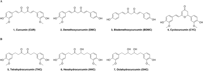
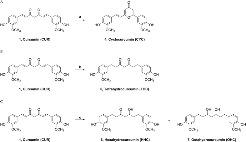
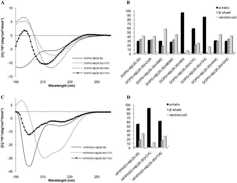
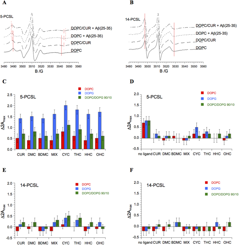
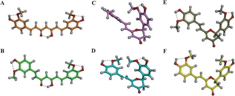
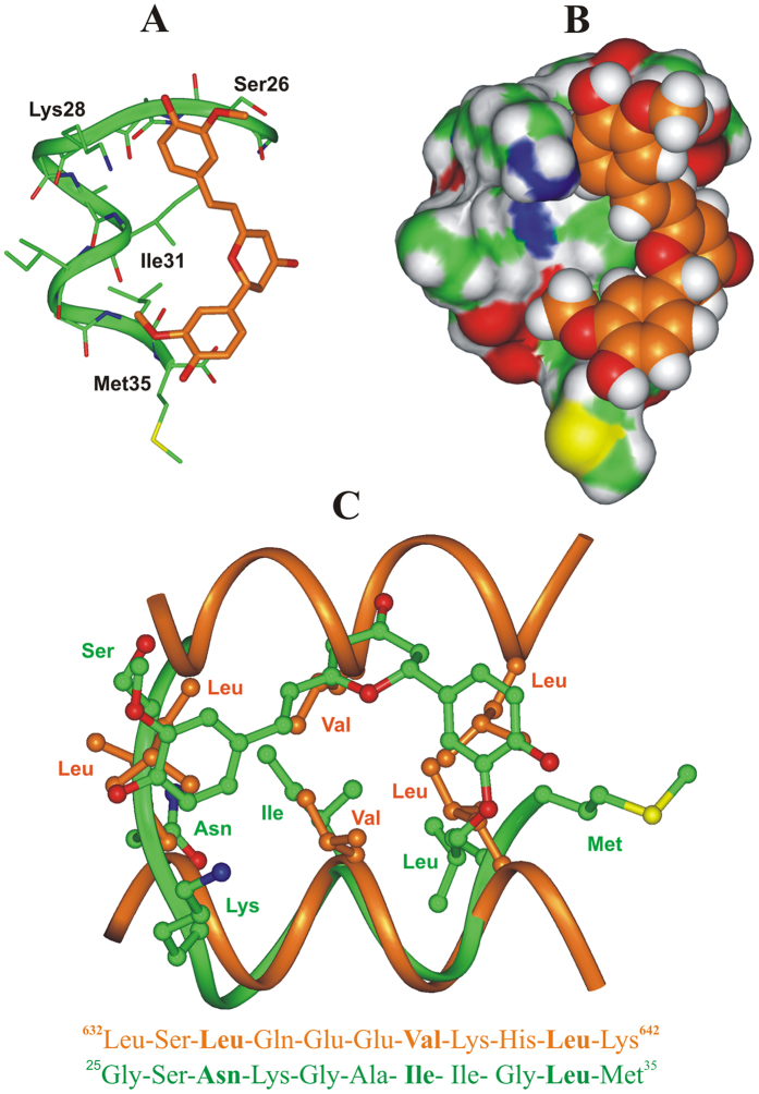
A broad biophysical analysis was performed to investigate the molecular basis of the neuroprotective action of Curcuma longa extracts in Alzheimer's disease. By combining circular dichroism and electron paramagnetic resonance experiments with molecular modeling calculations, the minor components of Curcuma longa extracts, such as demethoxycurcumin (2, DMC), bisdemethoxycurcumin (3, BDMC) and cyclocurcumin (4, CYC), were analyzed in a membrane environment mimicking the phospholipid bilayer. Our study provides the first evidence on the relative role of single curcuminoids interacting with Aβ-peptide. When the CYC and curcumin metabolite tetrahydrocurcumin (5, THC) were inserted into an anionic lipid solution, a significant modification of the Aβ CD curves was detected. These data were implemented by EPR experiments, demonstrating that CYC reaches the inner part of the bilayer, while the other curcuminoids are localized close to the membrane interface. Computational studies provided a model for the curcuminoid-Aβ interaction, highlighting the importance of a constrained "semi-folded" conformation to interact with Aβ analogously to the pattern observed in α-helical coiled-coil peptide structures. This combined approach led to a better understanding of the intriguing in vitro and in vivo activity of curcuminoids as anti-Alzheimer agents, paving a new path for the rational design of optimized druggable analogues.


Figures

Figure 1
Figure 1
(A) Structures of natural curcumin derivatives, curcuminoids, (1–4). (B) Structures of main curcumin metabolites (5–7).
Figure 2
Figure 2
(A) Synthesis of cyclocurcumin, CYC (4); reagents and conditions: a) TFA, MW, 100 °C, 4 min, 10%. (B) Synthesis of THC (5); reagents and conditions: b) H2 (1 atm), Pt/C, MeOH, r.t., 1 h, 77%. (C) Synthesis of HHC (6) and OHC (7); reagents and conditions: c) H2 (6.8 atm), Pt/C, MeOH, r.t., 13 h; 6, HHC (37%); 7, OHC (55%).
Figure 3
Figure 3
(A) CD spectra of Aβ(25–35) in DOPG vesicles in the absence and the presence of ligands MIX, (B) Secondary structure quantification of Aβ(25–35) in DOPG vesicles in the presence of curcuminoid compounds. (C) CD spectra of Aβ(25–35) in HFIP/water mixture in the absence and the presence of ligands MIX, (D) Secondary structure quantification of Aβ(25–35) in HFIP/water mixture in the presence of curcuminoid compounds.
Figure 4
Figure 4
EPR spectra of 5-PCSL (A) and 14-PCSL (B) in lipid bilayers composed of DOPC or DOPC and CUR, in the absence and in the presence of Aβ(25–35). Red lines are guides for the eye, highlighting the shifts of the low-field maximum and high-field minimum. Increase in outer hyperfine splitting of 5-PCSL and 14-PCSL in DOPC, DOPG and DOPC/DOPG 90/10 bilayers upon inclusion of ligands (CUR, DMC, BDMC, MIX, CYC, THC, HHC and OHC) in the lipid formulation (panels C,E) and upon addition of Aβ(25/35) to the system (panels D,F).
Figure 5
Figure 5
PM7 lowest energy conformers of i) keto-enol CUR conf I (A; orange) and conf II (B; green) and ii) CYC conf I (C; pink), conf II (D; cyan), conf III (E; gray), and conf IV (F; yellow). The conformers are displayed as ball & stick models and colored by atom type: O, red; H, white. Hydrogen bonds are highlighted with a black dashed line.
Figure 6
Figure 6
(A,B) Docked structure of CYC (orange) in complex with Aβ(25–35) peptide (green). (A) The complex is displayed as ribbon and sticks and peptide interacting residues are labeled; hydrogens are omitted for sake of clarity. (B) The Connolly surface of the peptide and the vdW volume of CYC are displayed. (C) X-ray structure of the coiled-coil dimerization motif of human ROCK I protein (carbon atoms: orange; PDB ID: 3O0Z) superimposed on the docked complex of CYC: Aβ(25–35) peptide (carbon atoms: green). Hydrogens are omitted for clarity. The corresponding 2-D sequence of human ROCK I protein and Aβ(25–35) are reported and the conserved residues of the heptad repeat motif are highlighted in bold. Molecules are colored by atom type (O: red; N: blue; S: yellow and H: white).

Similar articles

Cited by

References

    1. Selkoe D. J. Alzheimer’s disease: Genes, proteins, and therapy. Physiol Rev 81, 741–766 (2001). - PubMed
    1. Walsh D. M. & Selkoe D. J. Deciphering the molecular basis of memory failure in Alzheimer’s disease. Neuron 44, 181–193 (2004). - PubMed
    1. Sipe J. D. & Cohen A. S. Review: History of the amyloid fibril. J Struct Biol 130, 88–98 (2000). - PubMed
    1. Hardy J. & Selkoe D. J. Medicine - The amyloid hypothesis of Alzheimer’s disease: Progress and problems on the road to therapeutics. Science 297, 353–356 (2002). - PubMed
    1. Iversen L. L., Mortishiresmith R. J., Pollack S. J. & Shearman M. S. The Toxicity in-Vitro of Beta-Amyloid Protein. Biochem J 311, 1–16 (1995). - PMC - PubMed

Publication types