. 2016 Dec 22:6:39628.
doi: 10.1038/srep39628.

Passive immunization with anti-ActA and anti-listeriolysin O antibodies protects against Listeria monocytogenes infection in mice

Affiliations

Passive immunization with anti-ActA and anti-listeriolysin O antibodies protects against Listeria monocytogenes infection in mice

Krisana Asano et al. Sci Rep. .

Abstract

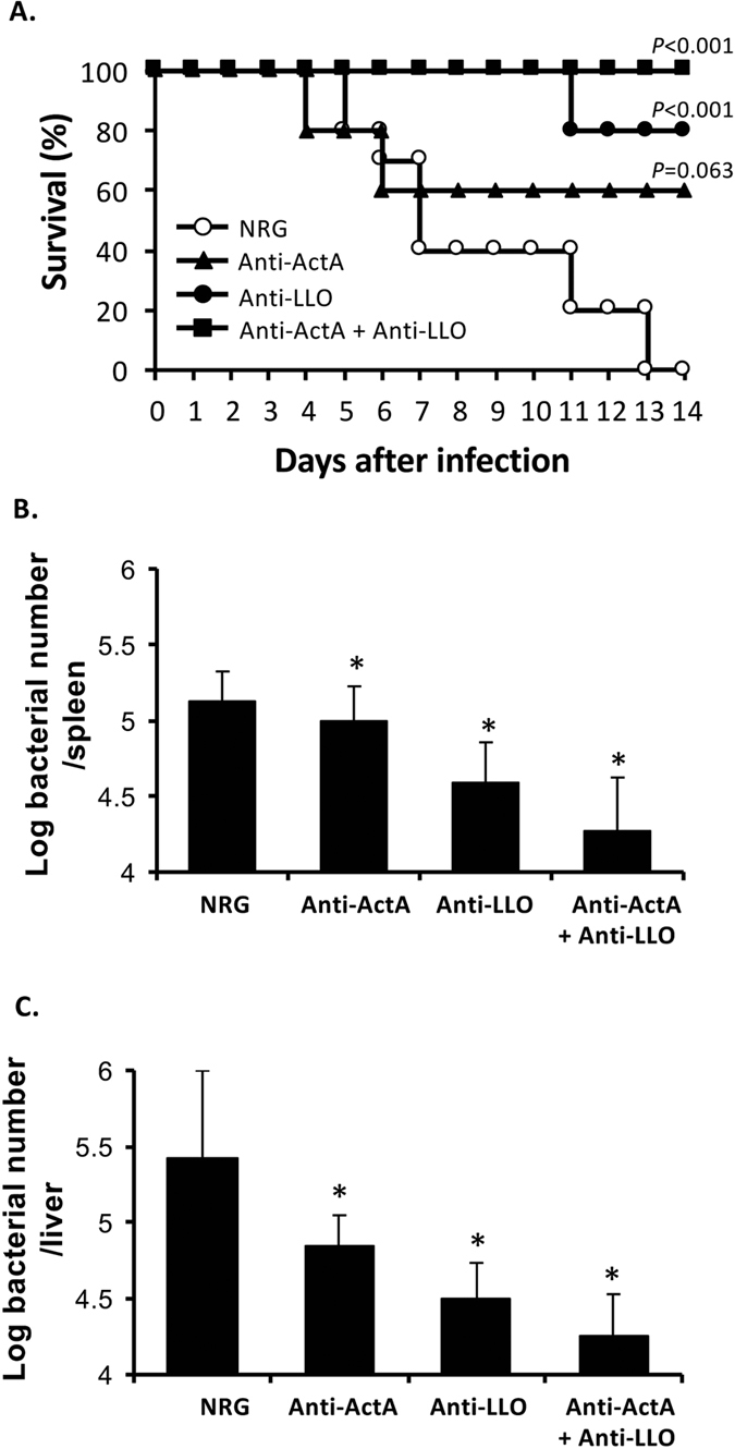
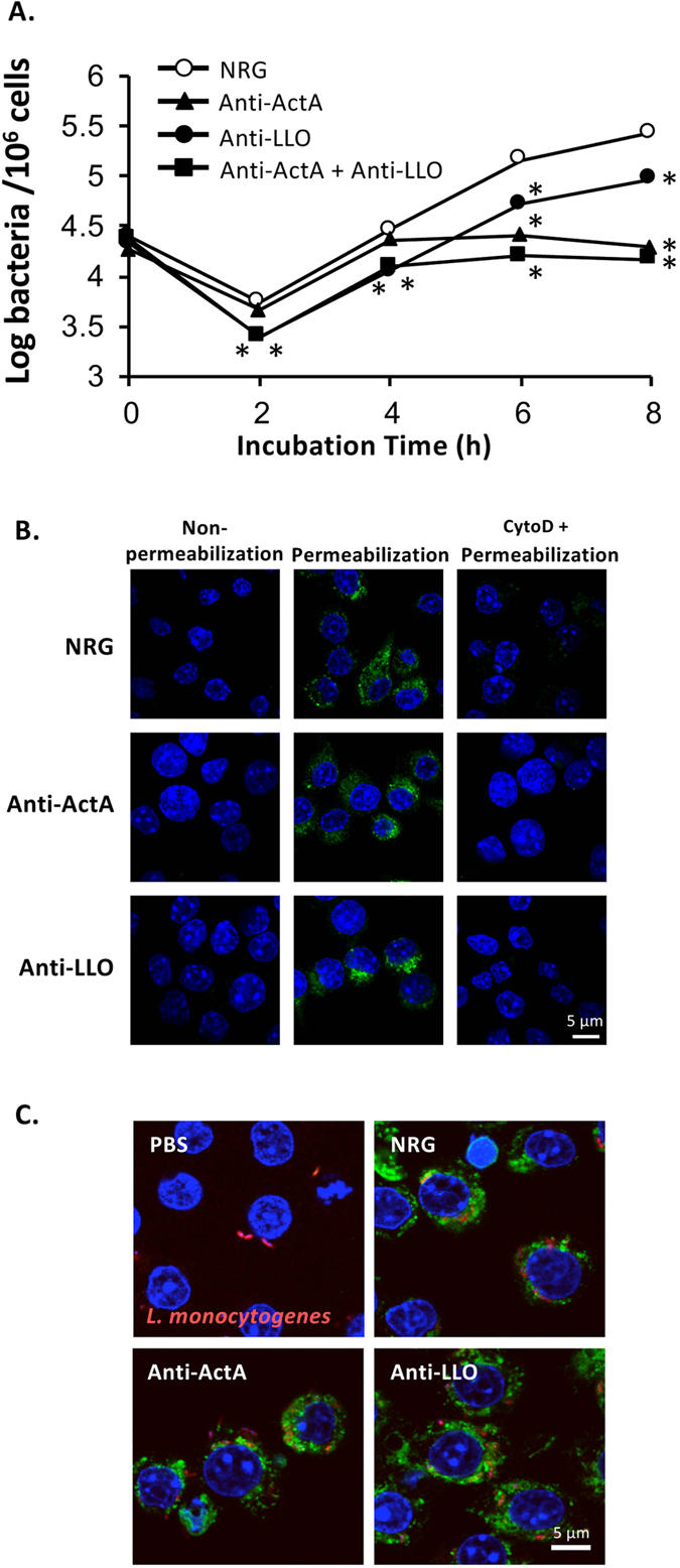
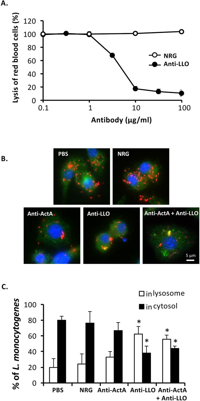
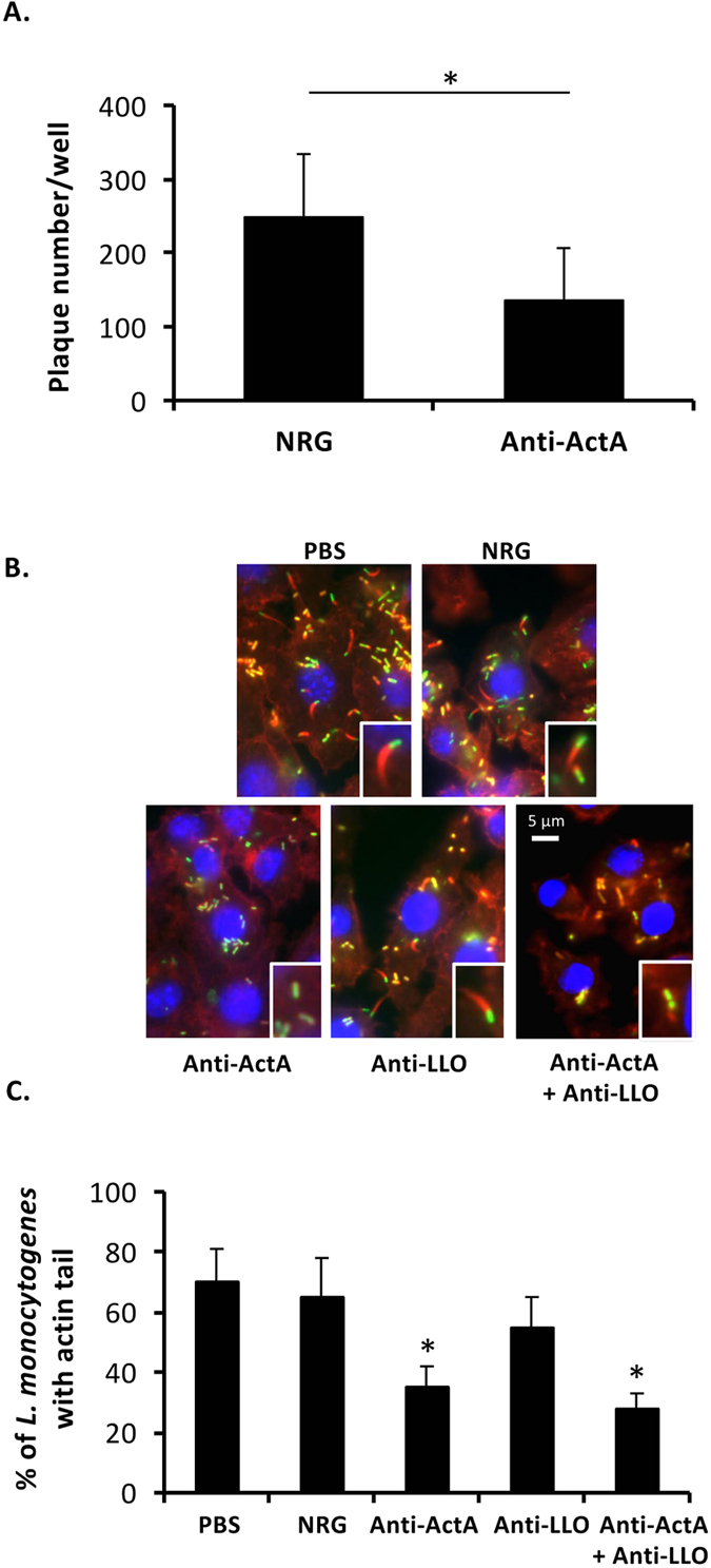
Listeria monocytogenes is an intracellular pathogen that causes listeriosis. Due to its intracellular niche, L. monocytogenes has evolved to limit immune recognition and response to infection. Antibodies that are slightly induced by listerial infection are completely unable to protect re-infection of L. monocytogenes. Thus, a role of antibody on the protective effect against L. monocytogenes infection has been neglected for a long time. In the present study, we reported that passive immunization with an excessive amount of antibodies against ActA and listeriolysin O (LLO) attenuates severity of L. monocytogenes infection. Combination of these antibodies improved survival of L. monocytogenes infected mice. Bacterial load in spleen and liver of listerial infected mice and infected RAW264.7 cells were significantly reduced by administration of anti-ActA and anti-LLO antibodies. In addition, anti-LLO antibody neutralized LLO activity and inhibited the bacterial escape from the lysosomal compartments. Moreover, anti-ActA antibody neutralized ActA activity and suppressed actin tail formation and cell-to-cell spread. Thus, our studies reveal that passive immunization with the excessive amount of anti-ActA and -LLO antibodies has potential to provide the protective effect against listerial infection.


Figures

Figure 1
Figure 1. Passive immunization of anti-ActA and anti-LLO antibodies protects mice from listerial infection.
Mice were administered intravenously with the antibody or NRG 1 mg/mouse. Mice were infected intravenously with 1 × 106 CFU L. monocytogenes 24 h later. (A) Survival was observed for 14 days (n = 10). P-values calculated by log rank test are indicated. (B,C) On day 3 after infection, the bacterial load in the spleens (B) and livers (C) were enumerated (mean ± S.D. n = 9). *P < 0.05.
Figure 2
Figure 2. Antibodies internalize into murine macrophages and reduce intracellular number of L. monocytogenes.
RAW264.7 cells were plated on 24-well culture plates at 1 × 106 cell/well. (A) The cells were treated with 100 μg of the antibody or NRG and infected simultaneously with L. monocytogenes at MOI 10. After incubation for 30 min, the extracellular bacteria were eliminated with 50 μg/ml gentamicin. At each time point, intracellular bacterial number was enumerated (mean ± S.D., n = 9). *P < 0.01. (B) The cells were treated with 100 μg of the antibody or NRG for 2 h. After fixing, immunostaining was performed using AlexaFluor 488-conjugated donkey anti-rabbit IgG. DAPI was used to stain cell nucleus. Left panel: the cells were not permeabilized with Triton X-100, middle and right panels: the cells were permeabilized with Triton X-100, right panel: endocytosis was blocked by cytochalasin D before treatment with the antibody. (C) The cells were treated 100 μg of the antibody or NRG or PBS and infected simultaneously with DsRedEx-labeled L. monocytogenes at MOI 10. After incubation for 30 min, the extracellular bacteria were washed and eliminated with 50 μg/ml gentamicin. After fixation and permeabilization, immunostaining was performed using AlexaFluor 488-conjugated donkey anti-rabbit IgG. DAPI was used to stain cell nucleus.
Figure 3
Figure 3. Neutralizing activity of the anti-LLO antibody.
(A) rLLO (1.0 μg/ml) was pre-incubated with anti-LLO antibody or NRG for 1 h at 37 °C prior to incubation with RBC at 37 °C for 45 min. Lysis of RBC was determined at absorbance 541 nm (mean ± S.D., n = 3). Neutralization of hemolytic activity of LLO by the antibody was determined from a reduction of lysis of RBC. (B) RAW264.7 cells were cultivated on sterilized glass slides. The cells were treated with the antibody, NRG or PBS and infected simultaneously with DsRedEx-expressing L. monocytogenes. After incubation for 30 min, the extracellular bacteria were eliminated by gentamicin. At 4 h after infection, the cells were fixed and permeabilized. The cells were stained using mouse monoclonal anti-LAMP1 antibody and AlexaFluor 488-conjugated donkey anti-mouse IgG. DAPI was used to stain cell nucleus. Fluorescent signals were observed under fluorescence microscope. (C) Percentages of L. monocytogenes localized in lysosome and cytoplasm were quantified from at least 100 areas of 3-independent experiments (mean ± S.D.). *P < 0.01.
Figure 4
Figure 4. Neutralizing activity of the anti-ActA antibody.
(A) NMuLi cells were treated with the antibody or NRG and infected with simultaneously with L. monocytogenes at MOI 10. Plaques under noble agar were enumerated on day 3 after infection. NRG was used as a control (mean ± S.D., n = 6). *P < 0.05. (B) RAW264.7 cells were cultivated on sterilized glass slides. The cells were treated with the antibody, NRG or PBS and infected simultaneously with YFP-expressing L. monocytogenes. After incubation for 30 min, the extracellular bacteria were eliminated by gentamicin. At 6 h after infection, actin tail was stained using rhodamine-conjugated phalloidin. DAPI was used to stain cell nucleus. (C) Percentage of L. monocytogenes containing actin tail was quantified from at least 100 areas of 3-independent experiments (mean ± S.D.). *P < 0.01.

References

    1. Farber J. M. & Peterkin P. I. Listeria monocytogenes, a food-borne pathogen. Microbiol Rev 55, 476–511 (1991). - PMC - PubMed
    1. Vázquez-Boland J. A. et al.. Listeria pathogenesis and molecular virulence determinants. Clin Microbiol Rev 14, 584–640, doi: 10.1128/CMR.14.3.584-640.2001 (2001). - DOI - PMC - PubMed
    1. Tilney L. G. & Portnoy D. A. Actin filaments and the growth, movement, and spread of the intracellular bacterial parasite, Listeria monocytogenes. J Cell Biol 109, 1597–1608, doi: 10.1083/jcb.109.4.1597 (1989). - DOI - PMC - PubMed
    1. Osanai A. et al.. Fibronectin-binding protein, FbpA, is the adhesin responsible for pathogenesis of Listeria monocytogenes infection. Microbiol Immunol 57, 253–262, doi: 10.1111/1348-0421.12030 (2013). - DOI - PubMed
    1. Bubert A., Kuhn M., Goebel W. & Köhler S. Structural and functional properties of the p60 proteins from different Listeria species. J Bacteriol 174, 8166–8171 (1992). - PMC - PubMed

Publication types

MeSH terms